国际口腔医学杂志 ›› 2024, Vol. 51 ›› Issue (4): 456-466.doi: 10.7518/gjkq.2024058
• 论著 • 上一篇
韩婧文1( ),王蕾1,任诗琦1,王红宇1,黄颖怡1,李佳敏1,郑艳1,2()
Jingwen Han1(),Lei Wang1,Shiqi Ren1,Hongyu Wang1,Yingyi Huang1,Jiamin Li1,Yan Zheng1,2()
摘要:
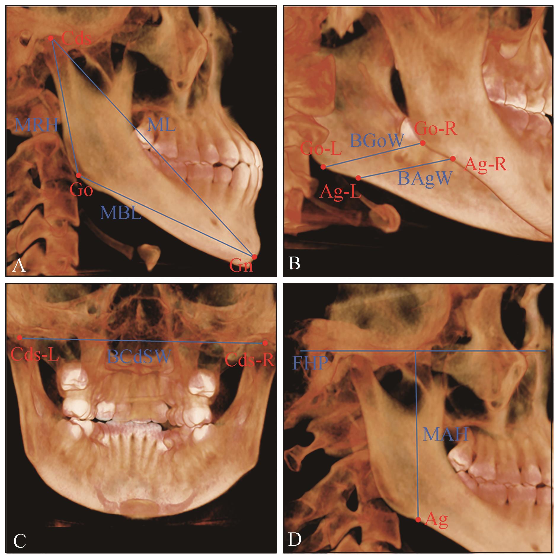
目的 通过测量具有不同颞下颌关节形态特征的青少年生长高峰期前后下颌骨在三维方向的生长量,探索颞下颌关节形态特征与下颌骨生长潜力的关系。 方法 选取生长发育正常、未经正畸治疗的青少年横向样本226例,收集各样本的锥形束计算机断层扫描(CBCT)资料,根据蝶枕软骨联合融合程度分为生长期前组及生长期后组,测量并计算髁突位置、髁突高颈比、髁突角、髁头直立角,根据结果分为具有不同颞下颌关节形态特征的各组,测量分析各组下颌骨在三维方向的生长量。 结果 无论髁突在关节窝中处于何种位置,生长高峰期前后,均可观察到下颌长度及高度显著增长,仅在髁突非正中位时,观察到下颌宽度增长差异具有统计学意义;髁突短而粗,可观察到下颌在三维方向生长均显著,髁突长而细,可观察到下颌骨长度与高度显著增长而宽度增长差异无统计学意义;髁突角较大,可观察到下颌在三维方向生长均显著,髁突角较小,可观察到下颌长度与高度增长显著而宽度增长差异无统计学意义;髁头直立角较大,可观察到下颌的长度及高度增长显著而宽度增长差异无统计学意义,髁头直立角较小,可观察到下颌长度与宽度增长显著而高度增长差异无统计学意义。 结论 颞下颌关节形态特征可作为预测下颌骨生长潜力的参考指标,具有不同颞下颌关节形态特征的下颌骨在生长高峰期间在各方向的生长量存在差异。
中图分类号:
| 1 | 刘纯, 贾莹, 杨世榕, 等. 大鼠髁突软骨下骨骨微结构生长发育的特征[J]. 中国组织工程研究, 2022, 26(32): 5162-5166. |
| Liu C, Jia Y, Yang SR, et al. Characteristics of the growth, development and microarchitecture of condyle subchondral bone in rats[J]. Chin J Tissue Eng Res, 2022, 26(32): 5162-5166. | |
| 2 | Saccucci M, Polimeni A, Festa F, et al. Do skeletal cephalometric characteristics correlate with condylar volume, surface and shape? A 3D analysis[J]. Head Face Med, 2012, 8: 15. |
| 3 | Al-Gumaei WS, Al-Attab R, Alhammadi MS, et al. Evaluation of spheno-occipital synchondrosis fusion in Chinese population using CBCT: a cross-sectio-nal study[J]. J Contemp Dent Pract, 2022, 23(1): 8-13. |
| 4 | 傅开元, 胡敏, 余强, 等. 颞下颌关节紊乱病锥形束CT检查规范及诊断标准的专家共识[J]. 中华口腔医学杂志, 2020, 55(9): 613-616. |
| Fu KY, Hu M, Yu Q, et al. Experts consensus on cone-beam CT examination specification and diagnostic criteria of temporomandibular disorders[J]. Chin J Stomatol, 2020, 55(9): 613-616. | |
| 5 | Tun Oo L, Miyamoto JJ, Takada JI, et al. Three-dimensional characteristics of temporomandibular joint morphology and condylar movement in patients with mandibular asymmetry[J]. Prog Orthod, 2022, 23(1): 50. |
| 6 | Ikeda M, Miyamoto JJ, Takada JI, et al. Association between 3-dimensional mandibular morphology and condylar movement in subjects with mandibular asymmetry[J]. Am J Orthod Dentofacial Orthop, 2017, 151(2): 324-334. |
| 7 | Franklin D, Flavel A. Brief communication: timing of spheno-occipital closure in modern Western Australians[J]. Am J Phys Anthropol, 2014, 153(1): 132-138. |
| 8 | Al-Gumaei WS, Al-Attab R, Al-Tayar B, et al. Comparison of spheno-occipital synchondrosis maturation stages with three-dimensional assessment of mandibular growth[J]. BMC Oral Health, 2022, 22(1): 654. |
| 9 | Okamoto K, Ito J, Tokiguchi S, et al. High-resolution CT findings in the development of the sphenooccipital synchondrosis[J]. AJNR Am J Neurora-diol, 1996, 17(1): 117-120. |
| 10 | Pullinger AG, Solberg WK, Hollender L, et al. Relationship of mandibular condylar position to dental occlusion factors in an asymptomatic population[J]. Am J Orthod Dentofacial Orthop, 1987, 91(3): 200-206. |
| 11 | Berraquero R, Palacios J, Rodríguez JI. The role of the condylar cartilage in mandibular growth. A study in thanatophoric dysplasia[J]. Am J Orthod Dentofac Orthop, 1992, 102(3): 220-226. |
| 12 | Kajikawa A, Hirabayashi S, Harii K. An experimental study on the growth of condylar cartilage, using a new vascularized mandible heterotopic transplant model[J]. J Oral Maxillofac Surg, 2003, 61(2): 239-245. |
| 13 | Shen G, Darendeliler MA. The adaptive remodeling of condylar cartilage-a transition from chondrogenesis to osteogenesis[J]. J Dent Res, 2005, 84(8): 691-699. |
| 14 | Bjork A. Facial growth in man, studied with the aid of metallic implants[J]. Acta Odontol Scand, 1955, 13(1): 9-34. |
| 15 | Chen YX, Li LF, Li Y, et al. Comprehensive positional and morphological assessments of the temporomandibular joint in adolescents with skeletal Class Ⅲ malocclusion: a retrospective CBCT study[J]. BMC Oral Health, 2023, 23(1): 78. |
| 16 | Burke G, Major P, Glover K, et al. Correlations between condylar characteristics and facial morphology in Class Ⅱ preadolescent patients[J]. Am J Orthod Dentofacial Orthop, 1998, 114(3): 328-336. |
| 17 | Ma QL, Bimal P, Mei L, et al. Temporomandibular condylar morphology in diverse maxillary-mandibular skeletal patterns: a 3-dimensional cone-beam computed tomography study[J]. J Am Dent Assoc, 2018, 149(7): 589-598. |
| 18 | 戴微微, 王秀颖, 潘思思, 等. 替牙期功能性Ⅲ类错𬌗髁突位置的CBCT研究[J]. 实用口腔医学杂志, 2018, 34(4): 548-551. |
| Dai WW, Wang XY, Pan SS, et al. The characteristic of condylar postion in the subjects with mixed dentition and Pseudo-Class Ⅲ malocclusion: a CBCT study[J]. J Pract Stomatol, 2018, 34(4): 548-551. | |
| 19 | 胡敏, 毕长青, 周丹, 等. 安氏Ⅲ类错𬌗正畸前后颞下颌关节形态变化的研究[J]. 现代口腔医学杂志, 2000, 14(5): 317-319. |
| Hu M, Bi CQ, Zhou D, et al. The study on TMJ morphological changes in patients with Angle Class Ⅲ malocclusion pretreatment and after treatment[J]. J Mod Stomatol, 2000, 14(5): 317-319. | |
| 20 | Pullinger AG, Solberg WK, Hollender L, et al. Tomographic analysis of mandibular condyle position in diagnostic subgroups of temporomandibular disorders[J]. J Prosthet Dent, 1986, 55(6): 723-729. |
| 21 | Shokri A, Zarch HH, Hafezmaleki F, et al. Comparative assessment of condylar position in patients with temporomandibular disorder (TMD) and asympto-matic patients using cone-beam computed tomography[J]. Dent Med Probl, 2019, 56(1): 81-87. |
| 22 | Cohlmia JT, Ghosh J, Sinha PK, et al. Tomographic assessment of temporomandibular joints in patients with malocclusion[J]. Angle Orthod, 1996, 66(1): 27-35. |
| 23 | Paknahad M, Shahidi S. Association between condylar position and vertical skeletal craniofacial morphology: a cone beam computed tomography study[J]. Int Orthod, 2017, 15(4): 740-751. |
| 24 | Akahane Y, Deguchi T, Hunt NP. Morphology of the temporomandibular joint in skeletal Class Ⅲ symmetrical and asymmetrical cases: a study by cephalometric laminography[J]. J Orthod, 2001, 28(2): 119-128. |
| 25 | 韩婧文, 任诗琦, 刘星宇, 等. 成人不同垂直及矢状骨面型髁突特征的研究[J]. 国际口腔医学杂志, 2022, 49(2): 153-162. |
| Han JW, Ren SQ, Liu XY, et al. Features of condyles of adult patients with different vertical and sagittal skeletal facial types[J]. Int J Stomatol, 2022, 49(2): 153-162. | |
| 26 | Doraczynska-Kowalik A, Nelke KH, Pawlak W, et al. Genetic factors involved in mandibular prognathism[J]. J Craniofac Surg, 2017, 28(5): e422-e431. |
| 27 | Moreno Uribe LM, Howe SC, Kummet C, et al. Phenotypic diversity in white adults with moderate to severe Class Ⅱ malocclusion[J]. Am J Orthod Dentofacial Orthop, 2014, 145(3): 305-316. |
| 28 | Arnett GW, Bergman RT. Facial keys to orthodontic diagnosis and treatment planning. Part Ⅰ [J]. Am J Orthod Dentofacial Orthop, 1993, 103(4): 299-312. |
| 29 | Arnett GW, Bergman RT. Facial keys to orthodontic diagnosis and treatment planning. Part Ⅱ[J]. Am J Orthod Dentofacial Orthop, 1993, 103(5): 395-411. |
| 30 | Katsavrias EG, Halazonetis DJ. Condyle and fossa shape in Class Ⅱ and Class Ⅲ skeletal patterns: a morphometric tomographic study[J]. Am J Orthod Dentofacial Orthop, 2005, 128(3): 337-346. |
| 31 | 柳汀. 不同矢状骨面型高角错𬌗畸形患者颞下颌关节的CBCT研究[D]. 天津: 天津医科大学, 2017. |
| Liu T. Morphological study on temporomandibular joint in high-angle patients with different sagittal skeletal pattern by CBCT[D]. Tianjin: Tianjin Medical University, 2017. | |
| 32 | Lv WX, Nie Q, Gu Y. Three-dimensional analysis of mandibular characteristics in patients with skeletal Class Ⅱ malocclusion and chin deviation[J]. Am J Orthod Dentofac Orthop, 2021, 160(3): 392-400. |
| 33 | Liu W, Wang Y, Zhang Y, et al. Study of condylar asymmetry in Angle Class Ⅲ malocclusion with mandibular deviation[J]. J Craniofac Surg, 2015, 26(3): e264-e268. |
| 34 | Kim HO, Lee W, Kook YA, et al. Comparison of the condyle-fossa relationship between skeletal Class Ⅲ malocclusion patients with and without asymmetry: a retrospective three-dimensional cone-beam computed tomograpy study[J]. Korean J Orthod, 2013, 43(5): 209-217. |
| 35 | Obwegeser HL, Makek MS. Hemimandibular hyperplasia: hemimandibular elongation[J]. J Maxillofac Surg, 1986, 14(4): 183-208. |
| 36 | Santander P, Quast A, Olbrisch C, et al. Comprehensive 3D analysis of condylar morphology in adults with different skeletal patterns-a cross-sectional study[J]. Head Face Med, 2020, 16(1): 33. |
| 37 | Björk A. Prediction of mandibular growth rotation[J]. Am J Orthod, 1969, 55(6): 585-599. |
| 38 | Shirley NR, Jantz RL. Spheno-occipital synchondrosis fusion in modern Americans[J]. J Forensic Sci, 2011, 56(3): 580-585. |
| 39 | Alhazmi A, Vargas E, Palomo JM, et al. Timing and rate of spheno-occipital synchondrosis closure and its relationship to puberty[J]. PLoS One, 2017, 12(8): e0183305. |
| 40 | Ferrillo M, Curci C, Roccuzzo A, et al. Reliability of cervical vertebral maturation compared to hand-wrist for skeletal maturation assessment in growing subjects: a systematic review[J]. J Back Musculos-kelet Rehabil, 2021, 34(6): 925-936. |
| 41 | Gabriel DB, Southard KA, Qian F, et al. Cervical vertebrae maturation method: poor reproducibility[J]. Am J Orthod Dentofacial Orthop, 2009, 136(4): 478.e1-478.e7, 478-480. |
| 42 | Zhao XG, Lin JX, Jiang JH, et al. Validity and re-liability of a method for assessment of cervical vertebral maturation[J]. Angle Orthod, 2012, 82(2): 229-234. |
| 43 | Scarfe WC, Farman AG, Sukovic P. Clinical applications of cone-beam computed tomography in dental practice[J]. J Can Dent Assoc, 2006, 72(1): 75-80. |
| 44 | Schlueter B, Kim KB, Oliver D, et al. Cone beam computed tomography 3D reconstruction of the mandibular condyle[J]. Angle Orthod, 2008, 78(5): 880-888. |
| 45 | Diwakar R, Bucci R, Kaushik A, et al. Three-dimensional assessment of temporomandibular joint morphology and facial asymmetry in individuals with different vertical skeletal growth patterns[J]. Int J Environ Res Public Health, 2023, 20(2): 1437. |
| 46 | Lim YS, Chung DH, Lee JW, et al. Reliability and validity of mandibular posterior vertical asymmetry index in panoramic radiography compared with cone-beam computed tomography[J]. Am J Orthod Dentofacial Orthop, 2018, 153(4): 558-567. |
| 47 | Al-Gumaei WS, Long H, Al-Attab R, et al. Compa-rison of three-dimensional maxillary growth across spheno-occipital synchondrosis maturation stages[J]. BMC Oral Health, 2023, 23(1): 100. |
| 48 | Manabe A, Ishida T, Kanda E, et al. Evaluation of maxillary and mandibular growth patterns with cephalometric analysis based on cervical vertebral maturation: a Japanese cross-sectional study[J]. PLoS One, 2022, 17(4): e0265272. |
| 49 | 陶珂金, 刘光俊, 冯剑颖. 颞下颌关节间隙改变与关节盘移位及程度的关系[J]. 口腔颌面修复学杂志, 2022, 23(3): 196-200. |
| Tao KJ, Liu GJ, Feng JY. Relationship of temporomandibular joint space to disc displacement and degree[J]. Chin J Prosthodont, 2022, 23(3): 196-200. |
| [1] | 张静,钟亦思,郑耘昊,张莉,熊鑫. 不同类型颞下颌关节紊乱病患者焦虑抑郁情绪的差异[J]. 国际口腔医学杂志, 2024, 51(3): 296-302. |
| [2] | 胡雅瑄,马子涵,王将凌,汪永跃. 可降解新型聚乳酸膜在引导骨组织再生中的应用[J]. 国际口腔医学杂志, 2024, 51(2): 187-192. |
| [3] | 薛晴,齐慧川,胡敏. 机械应力下初级纤毛在骨和颞下颌关节软骨改建中力学感知作用的研究进展[J]. 国际口腔医学杂志, 2024, 51(2): 201-207. |
| [4] | 徐书奎,张珊,谢新宇,马文盛. 上颌前方牵引矫治骨性Ⅲ类错 畸形远期疗效稳定性的研究进展[J]. 国际口腔医学杂志, 2023, 50(6): 646-652. 畸形远期疗效稳定性的研究进展[J]. 国际口腔医学杂志, 2023, 50(6): 646-652. |
| [5] | 石佳鑫,王淳艺,李精韬. Pierre Robin序列征患者腭裂临床治疗的研究进展[J]. 国际口腔医学杂志, 2023, 50(2): 237-242. |
| [6] | 李佩桐,时彬冕,许春梅,谢旭东,王骏. Gli1阳性间充质干细胞在牙及牙周组织中的分布及作用[J]. 国际口腔医学杂志, 2023, 50(1): 37-42. |
| [7] | 张宇宁,曾妮,张焙,石冰,郑谦. 咽后壁瓣咽成形术对腭裂术后患者颌面部生长影响的初步研究[J]. 国际口腔医学杂志, 2023, 50(1): 66-71. |
| [8] | 张珊,葛晓磊,李杰,谢新宇,常维维,马文盛. 上颌前方牵引矫治对颌骨生长发育长期影响的Meta分析[J]. 国际口腔医学杂志, 2022, 49(5): 548-555. |
| [9] | 黎静文,周力. 颈椎成熟法评估下颌骨骨龄的研究进展[J]. 国际口腔医学杂志, 2022, 49(3): 337-342. |
| [10] | 韩婧文,任诗琦,刘星宇,郎鑫,储梦诗,Waseem Saleh Abdo Kaid Algumaei,郑艳. 成人不同垂直及矢状骨面型髁突特征的研究[J]. 国际口腔医学杂志, 2022, 49(2): 153-162. |
| [11] | 张哲,刘进,王卫红,陈志强,杨春,刘丽. 焦磷酸钙沉积症继发颞下颌关节脱位1例[J]. 国际口腔医学杂志, 2021, 48(6): 664-667. |
| [12] | 许琳,王如意,勾薪瑞,王晓莉,李宇. 甲状旁腺激素相关蛋白调控下颌髁突软骨的研究进展[J]. 国际口腔医学杂志, 2021, 48(5): 549-555. |
| [13] | 刘嘉程,孟昭松,李宏捷,隋磊. 卵泡抑素在口腔颌面部发育中的作用及其治疗应用前景[J]. 国际口腔医学杂志, 2021, 48(5): 556-562. |
| [14] | 方苓力,谭玺,叶雨丝,黄兰,何瑶. 颞下颌关节退行性变早期髁突软骨细胞行为改变的实验研究[J]. 国际口腔医学杂志, 2021, 48(4): 417-425. |
| [15] | 金作林. 颅面部生长发育与早期生长改良[J]. 国际口腔医学杂志, 2021, 48(1): 7-11. |
|
||