国际口腔医学杂志 ›› 2024, Vol. 51 ›› Issue (2): 187-192.doi: 10.7518/gjkq.2024030
胡雅瑄( ),马子涵,王将凌,汪永跃()
Yaxuan Hu(),Zihan Ma,Jiangling Wang,Yongyue Wang()
摘要:
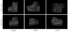
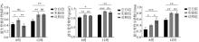
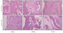
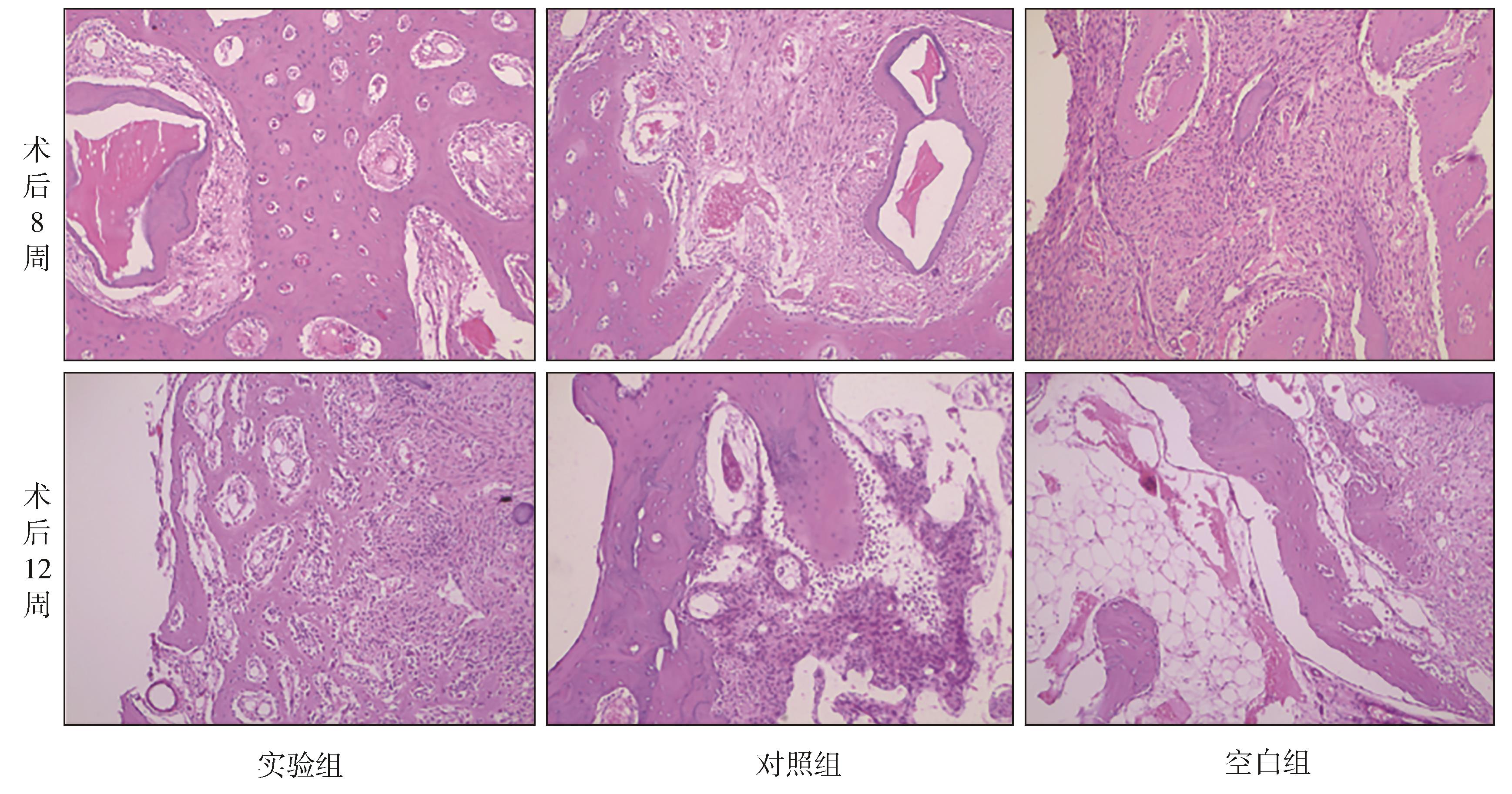
目的 探索一种可降解新型聚乳酸膜(PDLLA/PLLA)在引导骨组织再生中的应用效果。 方法 新西兰大白兔24只,体重2.5~3.0 kg,在动物一侧下颌骨体部近下颌骨下缘处制备10 mm×5 mm×3 mm箱状骨缺损,然后将动物随机分为实验组、对照组和空白组,每组8只。实验组动物骨缺损处填Bio-oss骨粉后将PDLLA/PLLA覆盖于缺损表面,对照组动物骨缺损处填Bio-oss骨粉后将Guidor聚乳酸可吸收膜覆盖于缺损表面,空白组动物不作处理。术后8、12周采集缺损处标本,进行大体观察、Micro-CT检查和组织病理学观察。 结果 实验期间各组实验动物均未发生炎症和排异反应,各组创口愈合良好,成骨活跃。大体观察显示,术后8周实验组动物成骨量较多,材料降解较少,对照组动物成骨量较实验组少,材料降解完全;术后12周实验组动物和对照组动物成骨量相当,实验组材料进一步降解,空白组动物成骨量少于实验组和对照组。术后8、12周,Micro-CT可以观察到实验组和对照组缺损区域新生骨明显多于空白组。术后8、12周,实验组动物和对照组动物新生骨相对骨体积分数(BV/TV)、骨密度(BMD)和骨小梁数量(Tb.N)均显著高于空白组动物(P<0.05),且术后8周实验组动物新生骨BV/TV高于对照组(P<0.05);但在术后12周时实验组与对照组新生骨BV/TV、BMD和Tb.N比较,差异无统计学意义(P>0.05)。组织切片观察显示,术后8周实验组动物新生骨小梁周边细胞生长活跃,并可见少量成骨细胞及破骨细胞;术后12周实验组动物骨小梁周围可见大量成骨细胞及破骨细胞,骨缺损部位骨组织密度接近周边正常骨组织。 结论 与对照组和空白组相比,PDLLA/PLLA呈现出了良好的生物相容性和骨传导性,可以明显促进缺损处愈合。
中图分类号:
| 1 | Gentile P, Chiono V, Tonda-Turo C, et al. Polymeric membranes for guided bone regeneration[J]. Biotechnol J, 2011, 6(10): 1187-1197. |
| 2 | Singhvi MS, Zinjarde SS, Gokhale DV. Polylactic acid: synthesis and biomedical applications[J]. J Appl Microbiol, 2019, 127(6): 1612-1626. |
| 3 | de França JOC, da Silva Valadares D, Paiva MF, et al. Polymers based on PLA from synthesis using D, L-lactic acid (or racemic lactide) and some biomedical applications: a short review[J]. Polymers (Basel), 2022, 14(12): 2317. |
| 4 | Ilyas RA, Sapuan SM, Harussani MM, et al. Polylactic acid (PLA) biocomposite: processing, additive manufacturing and advanced applications[J]. Polymers (Basel), 2021, 13(8): 1326. |
| 5 | Zaaba NF, Jaafar M. A review on degradation mecha-nisms of polylactic acid: hydrolytic, photodegradative, microbial, and enzymatic degradation[J]. Polym Eng Sci, 2020, 60(9): 2061-2075. |
| 6 | Annunziata M, Nastri L, Borgonovo A, et al. Poly-D-L-lactic acid membranes for bone regeneration[J]. J Craniofac Surg, 2015, 26(5): 1691-1696. |
| 7 | Yan WJ, Yang FH, Liu ZN, et al. Anti-inflammatory and mineralization effects of an ASP/PLGA-ASP/ACP/PLLA-PLGA composite membrane as a dental pulp capping agent[J]. J Funct Biomater, 2022, 13(3): 106. |
| 8 | Scantlebury TV. 1982-1992: a decade of technology development for guided tissue regeneration[J]. J Periodontol, 1993, 64(): 1129-1137. |
| 9 | Stoecklin-Wasmer C, Rutjes AWS, da Costa BR, et al. Absorbable collagen membranes for periodontal regeneration[J]. J Dent Res, 2013, 92(9): 773-781. |
| 10 | Ramires GAD, Helena JT, Oliveira JCS, et al. Eva-luation of guided bone regeneration in critical defects using bovine and porcine collagen membranes: histomorphometric and immunohistochemical analyses[J]. Int J Biomater, 2021, 2021: 8828194. |
| 11 | Annunziata M, Nastri L, Cecoro G, et al. The use of poly-d, l-lactic acid (PDLLA) devices for bone augmentation techniques: a systematic review[J]. Molecules, 2017, 22(12): 2214. |
| 12 | Castro-Aguirre E, Iñiguez-Franco F, Samsudin H, et al. Poly(lactic acid)-mass production, processing, industrial applications, and end of life[J]. Adv Drug Deliv Rev, 2016, 107: 333-366. |
| 13 | 廖凯荣, 全大萍, 高建文, 等. PLLA/PDLLA共混物的力学性能及体外降解特性研究[J].中山大学学报(自然科学版), 2002, 41(1): 51-54. |
| Liao KR, Quan DP, Gao JW, et al. The mechanical properties and degradation behavior in vitro of PLLA/PDLLA blends[J]. Acta Sci Natur Univ Sunyatseni, 2002, 41(1): 51-54. | |
| 14 | 徐高祥, 张鲁鲁, 高华丽, 等. 不同比例PLLA/PDLLA/5% HA复合物体外降解性能的研究[J]. 中国实验诊断学, 2017, 21(6): 1067-1071. |
| Xu GX, Zhang LL, Gao LH, et al. Study on in vitro degradation performance of PLLA/PDLLA/5% HA complex with different proportions[J]. Chin J Lab Diagn, 2017, 21(6): 1067-1071. | |
| 15 | Sitompul JP, Setyawan D, Nabila AG, et al. Synthesis of nanocomposite materials for biodegradable food packaging[J]. J Oil Palm Res, 2019, 2(1): 33-45. |
| 16 | Friedmann A, Stavropoulos A, Bilhan H. GTR treatment in furcation grade Ⅱ periodontal defects with the recently reintroduced guidor PLA matrix barrier: a case series with chronological step-by-step illustrations[J]. Case Rep Dent, 2020, 2020: 8856049. |
| [1] | 马瑜鸿,赵蕾. 微创非手术牙周治疗技术的临床研究进展[J]. 国际口腔医学杂志, 2024, 51(2): 227-232. |
| [2] | 徐彦雪,付丽. 功能等级引导骨再生膜的研究进展[J]. 国际口腔医学杂志, 2023, 50(3): 353-358. |
| [3] | 李春洁, 毕小琴, 朱桂全. 口腔颌面部肿瘤患者游离皮瓣修复术的并发症预防及处理[J]. 国际口腔医学杂志, 2023, 50(2): 127-137. |
| [4] | 木合森·牙生江,买买提吐逊·吐尔地. 带线锚钉在口腔颌面外科的应用[J]. 国际口腔医学杂志, 2023, 50(1): 114-119. |
| [5] | 黎静文,周力. 颈椎成熟法评估下颌骨骨龄的研究进展[J]. 国际口腔医学杂志, 2022, 49(3): 337-342. |
| [6] | 李嫣斐,张新春. 牙本质作为骨修复材料的研究进展[J]. 国际口腔医学杂志, 2022, 49(2): 197-203. |
| [7] | 郭雨婷,吕学超. 药物调控牙髓干细胞成骨分化的研究进展[J]. 国际口腔医学杂志, 2021, 48(6): 737-744. |
| [8] | 王剑. 浅谈嵌体和高嵌体修复的临床应用[J]. 国际口腔医学杂志, 2021, 48(5): 497-505. |
| [9] | 陈宸,田雨婷,程立,王国松,胡涛. 儿童第一恒磨牙大面积缺损永久修复时机的考量和展望[J]. 国际口腔医学杂志, 2021, 48(2): 129-134. |
| [10] | 张心驰,吴炜. 颌面骨再生领域3D打印技术及应用材料的研究进展[J]. 国际口腔医学杂志, 2020, 47(6): 677-685. |
| [11] | 付世锦,曾刊,李鑫,杨静,汪成林,叶玲. 骨保护素/核因子κB受体活化因子配体影响肺癌细胞下颌骨与股骨转移差异的初步研究[J]. 国际口腔医学杂志, 2020, 47(5): 538-546. |
| [12] | 刘育豪,张陶. 形状记忆高分子材料在骨缺损修复再生领域的研究进展[J]. 国际口腔医学杂志, 2020, 47(2): 219-224. |
| [13] | 林阳阳,侯敏. 双侧下颌支矢状骨劈开术对下颌近心骨段位移变化的影响[J]. 国际口腔医学杂志, 2019, 46(6): 718-723. |
| [14] | 王小萌,王晓,史册,孙宏晨,黄洋. 骨形态发生蛋白信号通路及其交叉对话对下颌骨发育的影响[J]. 国际口腔医学杂志, 2019, 46(3): 258-262. |
| [15] | 韩雨亭,吴燕茹. 应用龈壁提升术修复牙体缺损的研究进展[J]. 国际口腔医学杂志, 2019, 46(3): 349-355. |
|