国际口腔医学杂志 ›› 2026, Vol. 53 ›› Issue (2): 257-265.doi: 10.7518/gjkq.2026210
• 综述 • 上一篇
林子妍1( ),许来俊2()
Ziyan Lin1(),Laijun Xu2()
摘要:
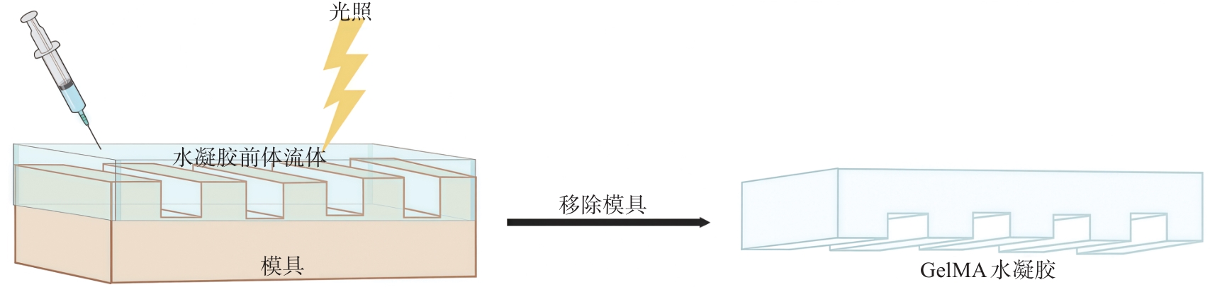
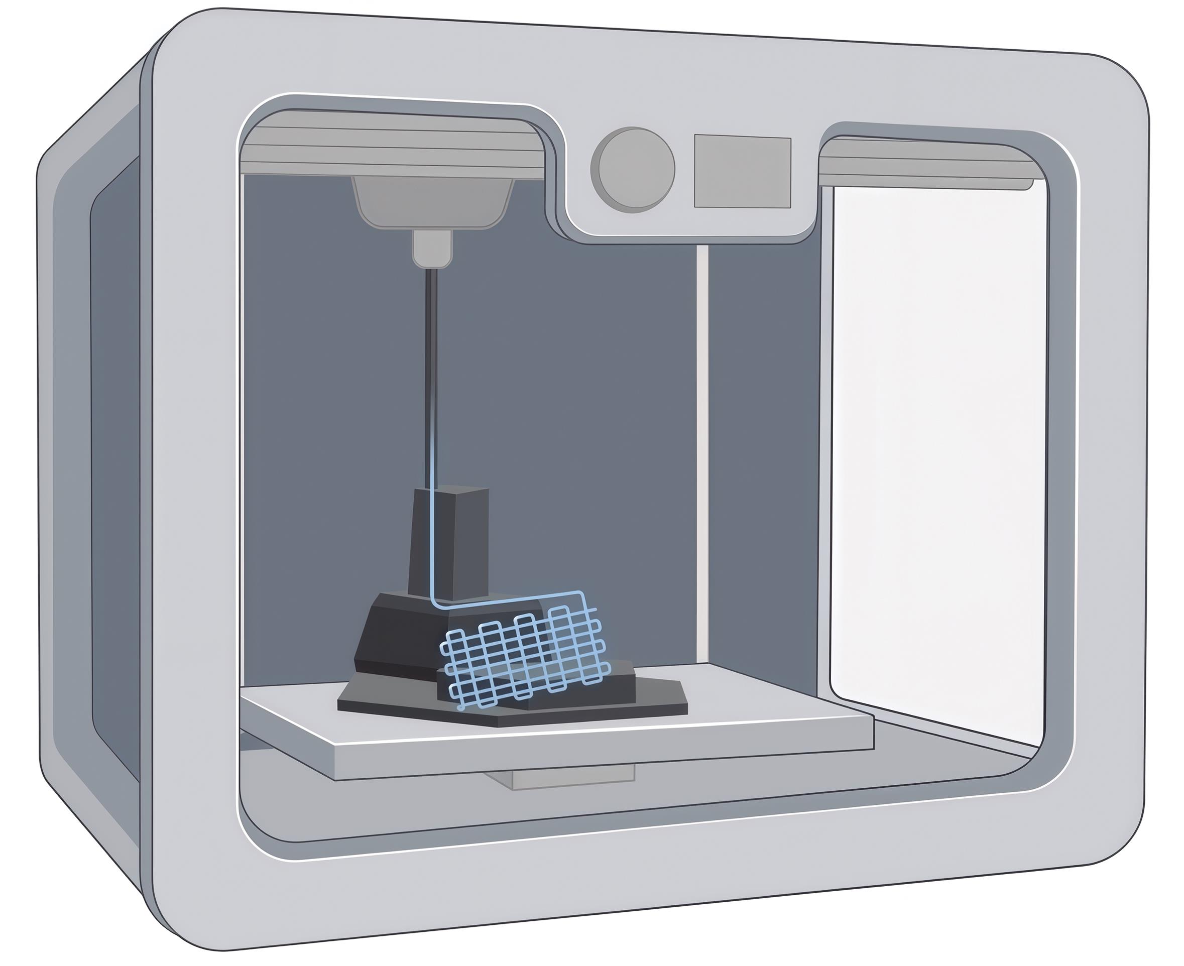
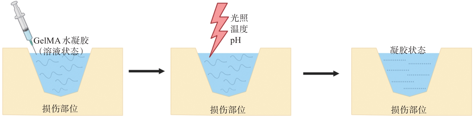
甲基丙烯酰化明胶(GelMA)水凝胶作为热门生物材料,以其适于细胞生长和分化的三维结构、优异的生物相容性等特性,在骨组织工程、皮肤组织缺损修复等领域已有广泛研究。近年来,GelMA水凝胶及其复合材料在神经组织修复领域的应用逐渐兴起,本文旨在探讨GelMA水凝胶应用于神经组织修复的研究进展,为神经组织再生提供新的见解与思路。
中图分类号:
| [1] | Lopes B, Sousa P, Alvites R, et al. Peripheral nerve injury treatments and advances: one health perspective[J]. Int J Mol Sci, 2022, 23(2): 918. |
| [2] | Luo LH, He Y, Jin L, et al. Application of bioactive hydrogels combined with dental pulp stem cells for the repair of large gap peripheral nerve injuries[J]. Bioact Mater, 2021, 6(3): 638-654. |
| [3] | van Hoorick J, Tytgat L, Dobos A, et al. (Photo-) crosslinkable gelatin derivatives for biofabrication applications[J]. Acta Biomater, 2019, 97: 46-73. |
| [4] | Chen SY, Zhao YX, Yan XL, et al. PAM/GO/gel/SA composite hydrogel conduit with bioactivity for repairing peripheral nerve injury[J]. J Biomed Mater Res A, 2019, 107(6): 1273-1283. |
| [5] | Zhou PH, Xu PP, Guan JJ, et al. Promoting 3D neuronal differentiation in hydrogel for spinal cord regeneration[J]. Colloids Surf B Biointerfaces, 2020, 194: 111214. |
| [6] | He WH, Zhang XX, Li XZ, et al. A decellularized spinal cord extracellular matrix-gel/GelMA hydrogel three-dimensional composite scaffold promotes recovery from spinal cord injury via synergism with human menstrual blood-derived stem cells[J]. J Mater Chem B, 2022, 10(30): 5753-5764. |
| [7] | Tan MH, Xu WZ, Yan G, et al. Oriented artificial ni-che provides physical-biochemical stimulations for rapid nerve regeneration[J]. Mater Today Bio, 2023, 22: 100736. |
| [8] | Xu HL, Gao ZH, Wang ZY, et al. Electrospun PCL nerve conduit filled with GelMA gel for CNTF and IGF-1 delivery in promoting sciatic nerve regeneration in rat[J]. ACS Biomater Sci Eng, 2023, 9(11): 6309-6321. |
| [9] | Dong Q, Yang XD, Liang X, et al. Composite hydrogel conduit incorporated with platelet-rich plasma improved the regenerative microenvironment for peripheral nerve repair[J]. ACS Appl Mater Interfaces, 2023, 15(20): 24120-24133. |
| [10] | Dey M, Ozbolat IT. 3D bioprinting of cells, tissues and organs[J]. Sci Rep, 2020, 10(1): 14023. |
| [11] | Das S, Thimukonda Jegadeesan J, Basu B. Advancing peripheral nerve regeneration: 3D bioprinting of GelMA-based cell-laden electroactive bioinks for nerve conduits[J]. ACS Biomater Sci Eng, 2024, 10(3): 1620-1645. |
| [12] | Zhang LM, Zhang H, Wang HR, et al. Fabrication of multi-channel nerve guidance conduits contai-ning schwann cells based on multi-material 3D bioprinting[J]. 3D Print Addit Manuf, 2023, 10(5): 1046-1054. |
| [13] | Wu WB, Dong YC, Liu HF, et al. 3D printed elastic hydrogel conduits with 7, 8-dihydroxyflavone relea-se for peripheral nerve repair[J]. Mater Today Bio, 2023, 20: 100652. |
| [14] | Tao J, Zhang JM, Du T, et al. Rapid 3D printing of functional nanoparticle-enhanced conduits for effective nerve repair[J]. Acta Biomater, 2019, 90: 49-59. |
| [15] | Wu ZX, Xie S, Kang YF, et al. Biocompatibility evaluation of a 3D-bioprinted alginate-GelMA-bacteria nanocellulose (BNC) scaffold laden with oriented-growth RSC96 cells[J]. Mater Sci Eng C Mater Biol Appl, 2021, 129: 112393. |
| [16] | Hamid OA, Eltaher HM, Sottile V, et al. 3D bioprinting of a stem cell-laden, multi-material tubular composite: an approach for spinal cord repair[J]. Mater Sci Eng C Mater Biol Appl, 2021, 120: 111707. |
| [17] | Ning LQ, Zhu N, Mohabatpour F, et al. Bioprinting Schwann cell-laden scaffolds from low-viscosity hydrogel compositions[J]. J Mater Chem B, 2019, 7(29): 4538-4551. |
| [18] | Zhou H, Jing SL, Xiong W, et al. Metal-organic framework materials promote neural differentiation of dental pulp stem cells in spinal cord injury[J]. J Nanobiotechnology, 2023, 21(1): 316. |
| [19] | Wang QC, Ge L, Guo JL, et al. Acid neutralization by composite lysine nanoparticles for spinal cord injury recovery through mitigating mitochondrial dysfunction[J]. ACS Biomater Sci Eng, 2024, 10(7): 4480-4495. |
| [20] | Wang H, Tang Q, Lu Y, et al. Berberine-loaded MSC-derived sEVs encapsulated in injectable GelMA hydrogel for spinal cord injury repair[J]. Int J Pharm, 2023, 643: 123283. |
| [21] | Qi ZP, Pan S, Yang XY, et al. Injectable hydrogel loaded with CDs and FTY720 combined with neural stem cells for the treatment of spinal cord injury[J]. Int J Nanomedicine, 2024, 19: 4081-4101. |
| [22] | Tsai EC, Dalton PD, Shoichet MS, et al. Matrix inclusion within synthetic hydrogel guidance channels improves specific supraspinal and local axonal regeneration after complete spinal cord transection[J]. Biomaterials, 2006, 27(3): 519-533. |
| [23] | Zhuang H, Bu SS, Hua L, et al. Gelatin-methacry-lamide gel loaded with microspheres to deliver GD-NF in bilayer collagen conduit promoting sciatic nerve growth[J]. Int J Nanomedicine, 2016, 11: 1383-1394. |
| [24] | Hu YN, Chen ZY, Wang HY, et al. Conductive nerve guidance conduits based on morpho butterfly wings for peripheral nerve repair[J]. ACS Nano, 2022, 16(2): 1868-1879. |
| [25] | Mendes AX, Caballero Aguilar L, do Nascimento AT, et al. Integrating graphene oxide-hydrogels and electrical stimulation for controlled neurotrophic factor encapsulation: a promising approach for efficient nerve tissue regeneration[J]. ACS Appl Bio Mater, 2024, 7(6): 4175-4192. |
| [26] | Cai YT, Huang Q, Wang PH, et al. Conductive hydrogel conduits with growth factor gradients for peripheral nerve repair in diabetics with non-suture ta-pe[J]. Adv Healthc Mater, 2022, 11(16): e2200755. |
| [27] | Wu W, Jia S, Xu H, et al. Supramolecular hydrogel microspheres of platelet-derived growth factor mimetic peptide promote recovery from spinal cord injury[J]. ACS Nano, 2023, 17(4): 3818-3837. |
| [28] | Heo DN, Lee SJ, Timsina R, et al. Development of 3D printable conductive hydrogel with crystallized PEDOT: PSS for neural tissue engineering[J]. Mater Sci Eng C Mater Biol Appl, 2019, 99: 582-590. |
| [29] | Liu Y, Yu H, Yu P, et al. Gelatin methacryloyl hydrogel scaffold loaded with activated Schwann cells attenuates apoptosis and promotes functional reco-very following spinal cord injury[J]. Exp Ther Med, 2023, 25(4): 144. |
| [30] | Pepelanova I, Kruppa K, Scheper T, et al. Gelatin-methacryloyl (GelMA) hydrogels with defined degree of functionalization as a versatile toolkit for 3D cell culture and extrusion bioprinting[J]. Bioengineering, 2018, 5(3): 55. |
| [31] | Zhao HB, Liu M, Zhang YJ, et al. Nanocomposite hydrogels for tissue engineering applications[J]. Nanoscale, 2020, 12(28): 14976-14995. |
| [32] | Zhang XW, Zhang H, Zhang Y, et al. 3D printed reduced graphene oxide-GelMA hybrid hydrogel scaffolds for potential neuralized bone regeneration[J]. J Mater Chem B, 2023, 11(6): 1288-1301. |
| [33] | Park J, Jeon J, Kim B, et al. Electrically conductive hydrogel nerve guidance conduits for peripheral ner-ve regeneration[J]. Adv Funct Mater, 2020, 30(39): 2003759. |
| [34] | Zhao FY, Cheng J, Sun MY, et al. Digestion degree is a key factor to regulate the printability of pure tendon decellularized extracellular matrix bio-ink in extrusion-based 3D cell printing[J]. Biofabrication, 2020, 12(4): 045011. |
| [35] | Yu C, Ma XY, Zhu W, et al. Scanningless and continuous 3D bioprinting of human tissues with decellularized extracellular matrix[J]. Biomaterials, 2019, 194: 1-13. |
| [36] | Wang T, Han Y, Wu ZJ, et al. Tissue-specific hydrogels for three-dimensional printing and potential application in peripheral nerve regeneration[J]. Tissue Eng Part A, 2022, 28(3/4): 161-174. |
| [37] | Xu YW, Zhou J, Liu CC, et al. Understanding the role of tissue-specific decellularized spinal cord matrix hydrogel for neural stem/progenitor cell microenvironment reconstruction and spinal cord injury[J]. Biomaterials, 2021, 268: 120596. |
| [1] | 李甜,李丽洁. 基于支架的牙髓组织预血管化技术的研究进展[J]. 国际口腔医学杂志, 2025, 52(5): 594-605. |
| [2] | 黄启航,王航,王耀钟,李德超. 静电纺丝纳米纤维在颌面部组织修复中的应用[J]. 国际口腔医学杂志, 2025, 52(4): 526-533. |
| [3] | 高丽钞,刘畅,刘云通,罗瑜雪,曹钰彬,华成舸. 拔牙术后舌神经功能障碍的诊治进展[J]. 国际口腔医学杂志, 2024, 51(4): 489-497. |
| [4] | 陈润智,张文涛,陈枫,杨帆. 丝素蛋白水凝胶的改性方法及其在骨组织工程中的应用[J]. 国际口腔医学杂志, 2023, 50(6): 739-746. |
| [5] | 吴嘉馨,程兴群,吴红崑. 透明质酸在修复龈乳头退缩中的临床应用进展[J]. 国际口腔医学杂志, 2023, 50(3): 347-352. |
| [6] | 蔡超莹,陈学鹏,胡济安. 外泌体复合支架用于口腔组织工程的研究进展[J]. 国际口腔医学杂志, 2022, 49(4): 489-496. |
| [7] | 施培磊,于晨浩,谢旭东,吴亚菲,王骏. 牙源性间充质干细胞应用于牙周组织缺损修复的研究进展[J]. 国际口腔医学杂志, 2021, 48(6): 690-695. |
| [8] | 巩靖蕾,黄艳梅,王军. 多相支架在牙周再生领域的研究进展[J]. 国际口腔医学杂志, 2021, 48(5): 563-569. |
| [9] | 曹春玲,韩冰,王晓燕. 水凝胶用于牙髓再生的研究进展[J]. 国际口腔医学杂志, 2021, 48(2): 192-197. |
| [10] | 李佩仪,张新春. 微环境酸碱度在组织工程骨再生中作用的研究进展[J]. 国际口腔医学杂志, 2021, 48(1): 64-70. |
| [11] | 刘育豪,张陶. 形状记忆高分子材料在骨缺损修复再生领域的研究进展[J]. 国际口腔医学杂志, 2020, 47(2): 219-224. |
| [12] | 邹俊东,刘定坤,杨楠,王谜,刘志辉. 生物活性玻璃/壳聚糖复合材料在生物医学领域的应用[J]. 国际口腔医学杂志, 2020, 47(1): 90-94. |
| [13] | 梅宏翔,张懿丹,张城浩,刘恩言,陈昊,赵志河,廖文. 表没食子儿茶素没食子酸酯在干细胞增殖及成骨分化作用中的研究现状[J]. 国际口腔医学杂志, 2019, 46(4): 431-436. |
| [14] | 董正谋,刘锐,刘鲁川,温秀杰. 种子细胞在牙周组织再生治疗中的研究进展[J]. 国际口腔医学杂志, 2019, 46(1): 48-54. |
| [15] | 李龙飚,汪成林,叶玲. 天然支架材料在牙髓组织工程再生中的研究进展[J]. 国际口腔医学杂志, 2018, 45(6): 666-672. |
|
||