国际口腔医学杂志 ›› 2021, Vol. 48 ›› Issue (6): 635-639.doi: 10.7518/gjkq.2021111
熊梦琳1,2( ),吴龙1,2,马丽1,2,赵今1,2()
Xiong Menglin1,2(),Wu Long1,2,Ma Li1,2,Zhao Jin1,2()
摘要:
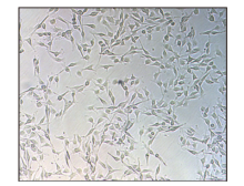
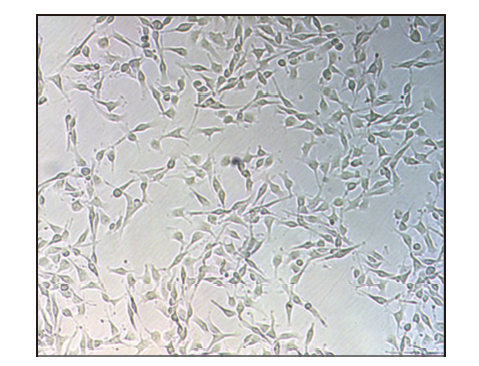
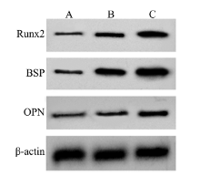
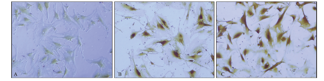
目的 探讨转化生长因子-β2(TGF-β2)对牙髓干细胞(DPSCs)增殖和分化的影响。方法 分离、培养DPSCs,分为阴性对照组、阳性对照组和TGF-β2组。四甲基偶氮唑盐比色法(MTT)检测细胞活力,碱性磷酸酶(ALP)活性测定试剂盒检测细胞内ALP活性,蛋白质印迹法检测Runt相关转录因子2(Runx2)、骨涎蛋白(BSP)和骨桥蛋白(OPN)的表达,免疫组织化学染色检测Runx2阳性表达。结果 与阳性对照组相比,TGF-β2作用3、7、10 d后DPSCs的活力不断升高,细胞内ALP活性上调,Runx2、BSP和OPN蛋白表达水平升高,Runx2阳性表达程度升高(P<0.05)。结论 TGF-β2可促进DPSCs增殖及成骨分化。
中图分类号:
| [1] |
Yusof MFH, Zahari W, Hashim SNM, et al. Angiogenic and osteogenic potentials of dental stem cells in bone tissue engineering[J]. J Oral Biol Craniofacial Res, 2018, 8(1):48-53.
doi: 10.1016/j.jobcr.2017.10.003 |
| [2] | 仵韩, 木合塔尔·霍加 . 转化生长因子-β诱导牙髓干细胞成骨向分化的研究进展[J]. 中华实用诊断与治疗杂志, 2017, 31(11):1128-1130. |
| Wu H, Muhetaer HJ. Advances in osteogenic dif-ferentiation of dental pulp stem cells induced by transforming growth factor-β[J]. J Chin Pract Diagn Ther, 2017, 31(11):1128-1130. | |
| [3] |
Salkın H, Gönen ZB, Ergen E, et al. Effects of TGF-β1 overexpression on biological characteristics of hu-man dental pulp-derived mesenchymal stromal cells[J]. Int J Stem Cells, 2019, 12(1):170-182.
doi: 10.15283/ijsc18051 pmid: 30595006 |
| [4] |
Hara Y, Ghazizadeh M, Shimizu H, et al. Delayed expression of circulating TGF-β1 and BMP-2 levels in human nonunion long bone fracture healing[J]. J Nippon Med Sch, 2017, 84(1):12-18.
doi: 10.1272/jnms.84.12 |
| [5] |
Verrecchia F, Rédini F. Transforming growth factor-β signaling plays a pivotal role in the interplay between osteosarcoma cells and their microenvironment[J]. Front Oncol, 2018, 8:133.
doi: 10.3389/fonc.2018.00133 pmid: 29761075 |
| [6] |
Deng MY, Mei TN, Hou TY, et al. TGFβ3 recruits endogenous mesenchymal stem cells to initiate bone regeneration[J]. Stem Cell Res Ther, 2017, 8:258.
doi: 10.1186/s13287-017-0693-0 |
| [7] | Huang LJ, Yi LX, Zhang CL, et al. Synergistic effects of FGF-18 and TGF-β3 on the chondrogenesis of human adipose-derived mesenchymal stem cells in the pellet culture[J]. Stem Cells Int, 2018, 2018:1-10. |
| [8] |
Kalinichenko SG, Matveeva NY, Kostiv RE, et al. Role of vascular endothelial growth factor and transforming growth factor-β2 in rat bone tissue after bone fracture and placement of titanium implants wi-th bioactive bioresorbable coatings[J]. Bull Exp Biol Med, 2017, 162(5):671-675.
doi: 10.1007/s10517-017-3684-3 |
| [9] | 卢金金, 刘欣辰, 周怡君, 等. 牙髓干细胞在软组织再生和修复中的研究进展[J]. 海南医学, 2019, 30(13):1752-1755. |
| Lu JJ, Liu XC, Zhou YJ, et al. Research progress of dental pulp stem cells in soft tissue regeneration and repair[J]. Hainan Med J, 2019, 30(13):1752-1755. | |
| [10] |
Dole NS, Mazur CM, Acevedo C, et al. Osteocyte-intrinsic TGF-β signaling regulates bone quality through perilacunar/canalicular remodeling[J]. Cell Rep, 2017, 21(9):2585-2596.
doi: 10.1016/j.celrep.2017.10.115 |
| [11] | 王腾, 木合塔尔·霍加, 李军 . 转化生长因子β3联合牙髓干细胞在种植体骨结合中作用的实验研究[J]. 中华口腔医学杂志, 2017, 52(6):367-373. |
| Wang T, Muhetaer HJ, Li J. Experimental study of transforming growth factor-β3 combined with den-tal pulp stem cells in promoting the implant’s os-seointegration[J]. Chin J Stomatol, 2017, 52(6):367-373. | |
| [12] | 胡正雄, 李彪, 蓝天, 等. TGF-β2和geneX对BrdU标记骨髓间充质干细胞增殖与成骨分化的作用[J]. 昆明医科大学学报, 2016, 37(2):10-14. |
| Hu ZX, Li B, Lan T, et al. Effect of TGF- β2 and geneX on the proliferation and osteogenic differen-tiation of Brd U-labeled bone mesenchymal stem cells[J]. J Kunming Med Univ, 2016, 37(2):10-14. | |
| [13] |
Nakamura T, Nakamura-Takahashi A, Kasahara M, et al. Tissue-nonspecific alkaline phosphatase promotes the osteogenic differentiation of osteoprogenitor cells[J]. Biochem Biophys Res Commun, 2020, 524(3):702-709.
doi: 10.1016/j.bbrc.2020.01.136 |
| [14] | Hou Z, Wang Z, Tao Y, et al. KLF2 regulates osteoblast differentiation by targeting of Runx2[J]. Lab Invest, 2019, 99(2):271-280. |
| [15] | 胡胜涛. 自体PRP对成骨细胞骨涎蛋白表达的影响[D]. 石家庄: 河北医科大学, 2014. |
| Hu ST. Effect of autologous PRP on the expression of bone sialoprotein in osteoblasts[D]. Shijiazhuang: Hebei Medical University, 2014. | |
| [16] |
Wang Y, Yao J, Yuan M, et al. Osteoblasts can induce dental pulp stem cells to undergo osteogenic differentiation[J]. Cytotechnology, 2013, 65(2):223-231.
doi: 10.1007/s10616-012-9479-5 |
| [17] |
Carvalho MS, Cabral JM, da Silva CL, et al. Synergistic effect of extracellularly supplemented osteopontin and osteocalcin on stem cell proliferation, osteogenic differentiation, and angiogenic properties[J]. J Cell Biochem, 2019, 120(4):6555-6569.
doi: 10.1002/jcb.27948 pmid: 30362184 |
| [1] | 周金阔,张晋弘,史晓晶,刘广顺,姜磊,刘倩峰. 长链非编码RNA小核仁RNA宿主基因22调控微小RNA-27b-3p对口腔鳞状细胞癌细胞增殖、侵袭和迁移的影响[J]. 国际口腔医学杂志, 2024, 51(1): 52-59. |
| [2] | 古丽其合热·阿布来提,秦旭,朱光勋. 线粒体自噬在牙周炎发生发展过程中的研究进展[J]. 国际口腔医学杂志, 2024, 51(1): 68-73. |
| [3] | 于乐蓉,李祥伟,艾虹. 牙髓干细胞干性维持的研究进展[J]. 国际口腔医学杂志, 2023, 50(4): 463-471. |
| [4] | 刘体倩,梁星,刘蔚晴,李晓虹,朱睿. 咬合创伤在牙周炎发生发展中的作用及机制的研究进展[J]. 国际口腔医学杂志, 2023, 50(1): 19-24. |
| [5] | 张静怡,李丹薇,孙宇,雷雅燕,刘涛,龚瑜. 复合树脂及复合体对成骨细胞毒性及成骨向分化的影响[J]. 国际口腔医学杂志, 2022, 49(4): 412-419. |
| [6] | 李佩,林凌,赵玮. 乳牙牙髓干细胞在口腔组织再生修复中的研究进展[J]. 国际口腔医学杂志, 2022, 49(4): 483-488. |
| [7] | 洪娅娅,陈学鹏,姒蜜思. 非编码RNA调控牙囊干细胞成骨分化的研究进展[J]. 国际口腔医学杂志, 2022, 49(3): 263-271. |
| [8] | 付恒怡,汪成林. 人牙髓干细胞无血清培养方法的研究进展[J]. 国际口腔医学杂志, 2022, 49(2): 220-226. |
| [9] | 郭雨婷,吕学超. 药物调控牙髓干细胞成骨分化的研究进展[J]. 国际口腔医学杂志, 2021, 48(6): 737-744. |
| [10] | 刘娟,陈斌,闫福华. 富血小板血浆和浓缩生长因子对人牙周膜细胞增殖和成骨分化影响的研究[J]. 国际口腔医学杂志, 2021, 48(5): 520-527. |
| [11] | 李静雅,税钰森,郭永文. 循环牵张应力影响人牙周膜细胞成骨分化机制的研究进展[J]. 国际口腔医学杂志, 2020, 47(6): 652-660. |
| [12] | 杨叶青,陈明,吴补领. 环状非编码RNA在间充质干细胞成骨向分化中作用的研究进展[J]. 国际口腔医学杂志, 2020, 47(3): 257-262. |
| [13] | 刘俊圻,陈艺尹,杨文宾. RNA腺嘌呤6-甲基化修饰调控骨髓间充质干细胞成骨向分化的研究进展[J]. 国际口腔医学杂志, 2020, 47(3): 263-269. |
| [14] | 朱明静,张清彬. 生长因子诱导间充质干细胞三维体外软骨形成的研究进展[J]. 国际口腔医学杂志, 2020, 47(3): 270-277. |
| [15] | 王润婷,房付春. 非编码RNA调控人牙周膜干细胞成骨向分化的研究进展[J]. 国际口腔医学杂志, 2020, 47(2): 138-145. |
|