国际口腔医学杂志 ›› 2019, Vol. 46 ›› Issue (3): 277-281.doi: 10.7518/gjkq2019036
闫冰1,2,3,骆献阳1,2,3,谭迎赟4,关丽梅1,薛丽丽5( )
)
Bing Yan1,2,3,Xianyang Luo1,2,3,Yingyun Tan4,Limei Guan1,Lili Xue5()
摘要:
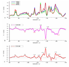
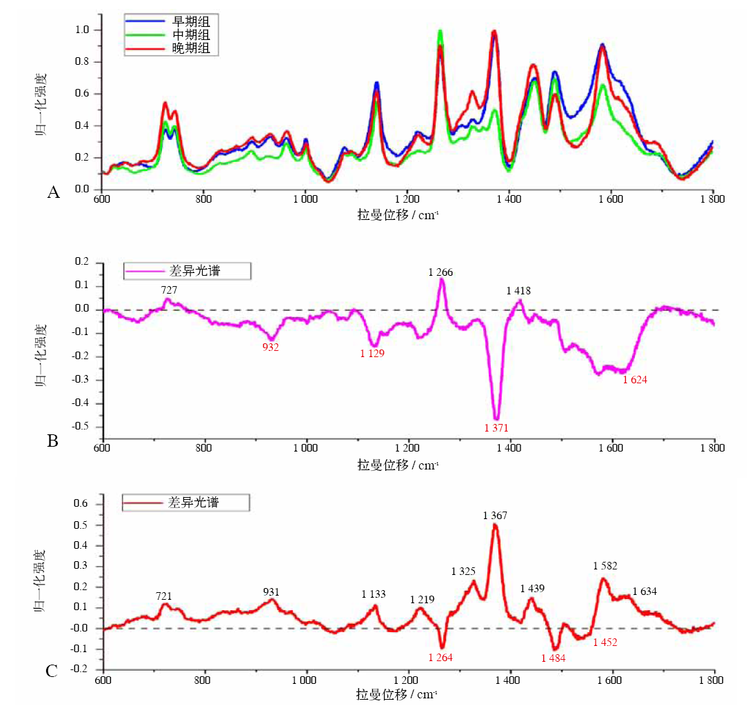
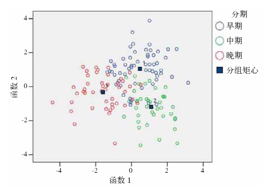
目的 应用表面增强拉曼光谱技术研究口腔鳞状细胞癌患者血清,并建立临床分期鉴别诊断模型。方法 收集144例口腔鳞状细胞癌患者血清,按照早期组、中期组及晚期组进行分组检测血清表面增强拉曼光谱,比较光谱差异,应用主成分分析法及线性判别函数法建立鉴别诊断模型,对不同分组光谱数据进行鉴别诊断。结果 共获得血清拉曼光谱144例,其中早期组59例,中期组40例及晚期组45例。血清光谱差异主要表现在721~727、931~932 、1 129~1 133、1 264~1 266、1 367~1 371 cm -1处谱峰强度差异,主要与血清中脂类、蛋白质及核酸成分和结构有关。鉴别诊断模型总体准确率达80%以上,经过交互验证后准确率为70%以上。结论 表面增强拉曼光谱检测口腔鳞状细胞癌患者血清为口腔鳞状细胞癌临床分期及预后判断提供了一种简便、准确的新方法。
中图分类号:
| [1] |
Blatt S, Krüger M, Ziebart T , et al. Biomarkers in diagnosis and therapy of oral squamous cell carcinoma: a review of the literature[J]. J Craniomaxillofac Surg, 2017,45(5):722-730.
doi: 10.1016/j.jcms.2017.01.033 pmid: 28318929 |
| [2] |
Kreppel M, Nazarli P, Grandoch A , et al. Clinical and histopathological staging in oral squamous cell carcinoma-comparison of the prognostic significance[J]. Oral Oncol, 2016,60:68-73.
doi: 10.1016/j.oraloncology.2016.07.004 pmid: 27531875 |
| [3] |
薛丽丽, 李一, 蔡巧玲 , 等. 口腔黏膜鳞状细胞癌、上皮异常增生及正常黏膜组织拉曼光谱研究[J]. 中华口腔医学杂志, 2015,50(1):18-22.
doi: 10.3760/cma.j.issn.1002-0098.2015.01.005 |
|
Xue LL, Li Y, Cai QL , et al. Raman spectral characteristics of oral squamous cell carcinoma epithelial dysplasia and normal mucosa[J]. Chin J Stomatol, 2015,50(1):18-22.
doi: 10.3760/cma.j.issn.1002-0098.2015.01.005 |
|
| [4] |
Ghantous Y, Bahouth Z , Abu El-Naaj I . Clinical and genetic signatures of local recurrence in oral squamous cell carcinoma[J]. Arch Oral Biol, 2018,95:141-148.
doi: 10.1016/j.archoralbio.2018.07.018 |
| [5] |
Xue L, Yan B, Li Y , et al. Surface-enhanced Raman spectroscopy of blood serum based on gold nanoparticles for tumor stages detection and histologic grades classification of oral squamous cell carcinoma[J]. Int J Nanomedicine, 2018,13:4977-4986.
doi: 10.2147/IJN |
| [6] | National Comprehensive Cancer Network. The NCCN clinical practice guidelines in oncology[EB/OL].. [2018-08-05] |
| [7] |
Wang J, Lin D, Lin J , et al. Label-free detection of serum proteins using surface-enhanced Raman spectroscopy for colorectal cancer screening[J]. J Biomed Opt, 2014,19(8):087003.
doi: 10.1117/1.JBO.19.8.087003 pmid: 25138208 |
| [8] |
Yan B, Li B, Wen Z , et al. Label-free blood serum detection by using surface-enhanced Raman spectroscopy and support vector machine for the preoperative diagnosis of parotid gland tumors[J]. BMC Cancer, 2015,15:650.
doi: 10.1186/s12885-015-1653-7 pmid: 4595250 |
| [9] |
Tan Y, Yan B, Xue L , et al. Surface-enhanced Raman spectroscopy of blood serum based on gold nanoparticles for the diagnosis of the oral squamous cell carcinoma[J]. Lipids Health Dis, 2017,16(1):73.
doi: 10.1186/s12944-017-0465-y pmid: 5384146 |
| [10] |
Fleischmann M, Hendra PJ , McQuillan AJ . Raman spectra of pyridine adsorbed at a silver electrode[J]. Chem Phys Lett, 1974,26(2):163-166.
doi: 10.1016/0009-2614(74)85388-1 |
| [11] |
Lin D, Pan J, Huang H , et al. Label-free blood plasma test based on surface-enhanced Raman scattering for tumor stages detection in nasopharyngeal cancer[J]. Sci Rep, 2014,4:4751.
doi: 10.1038/srep04751 pmid: 3996462 |
| [12] |
Lin JS, Sun FJ, Lin PY , et al. Clinicopathological and prognostic significance of preoperative serum epidermal growth factor levels in patients with oral squamous cell carcinoma[J]. Int J Oral Maxillofac Surg, 2018,47(10):1236-1242.
doi: 10.1016/j.ijom.2018.01.023 |
| [13] |
Gupta A, Tripathi A, Patil R , et al. Estimation of salivary and serum basic fibroblast growth factor in treated and untreated patients with oral squamous cell carcinoma[J]. J Oral Biol Craniofac Res, 2019,9(1):19-23.
doi: 10.1016/j.jobcr.2018.08.005 |
| [14] |
Rekha P, Aruna P, Bharanidharan G , et al. Near infrared Raman spectroscopic characterization of blood plasma of normal, oral premalignant and malignant conditions—a pilot study[J]. J Raman Spectrosc, 2015,46(9):735-743.
doi: 10.1002/jrs.4693 |
| [15] |
Sahu A, Nandakumar N, Sawant S , et al. Recurrence prediction in oral cancers: a serum Raman spectroscopy study[J]. Analyst, 2015,140(7):2294-2301.
doi: 10.1039/C4AN01860E |
| [1] | 周金阔,张晋弘,史晓晶,刘广顺,姜磊,刘倩峰. 长链非编码RNA小核仁RNA宿主基因22调控微小RNA-27b-3p对口腔鳞状细胞癌细胞增殖、侵袭和迁移的影响[J]. 国际口腔医学杂志, 2024, 51(1): 52-59. |
| [2] | 李立恒,王蕊,王晓明,张智轶,张璇,安峰,王芹,张凡. 环状RNA hsa_circ_0085576调控微小RNA-498/B细胞特异性莫洛尼鼠白血病病毒整合位点1轴对口腔鳞状细胞癌细胞迁移和侵袭的影响[J]. 国际口腔医学杂志, 2024, 51(1): 60-67. |
| [3] | 吴佳敏,夏斌,杨禾丰,许彪. 癌相关成纤维细胞在口腔鳞状细胞癌微环境中作用的研究进展[J]. 国际口腔医学杂志, 2023, 50(6): 711-717. |
| [4] | 刘洋,尹德强. 关于颌位调整方法的思考和改进[J]. 国际口腔医学杂志, 2023, 50(5): 499-505. |
| [5] | 柳江龙, 买买提吐逊·吐尔地. 超声造影在口腔鳞状细胞癌颈部转移性淋巴结诊断中的研究进展[J]. 国际口腔医学杂志, 2023, 50(5): 514-520. |
| [6] | 李奕君, 徐子昂, 李一. 前哨淋巴结在头颈部鳞状细胞癌检测的应用进展[J]. 国际口腔医学杂志, 2023, 50(5): 521-527. |
| [7] | 戢晓,张岚,黄定明. 牙源性与非牙源性上颌窦炎鉴别诊断及其治疗方案的研究进展[J]. 国际口腔医学杂志, 2023, 50(5): 566-572. |
| [8] | 姜玥莹,何宇添,李婷,周蓉卉. 近红外荧光探针在口腔癌诊断中应用的研究进展[J]. 国际口腔医学杂志, 2023, 50(4): 407-413. |
| [9] | 夏溦瑶,罗岩坤,贾仲林. Pierre Robin序列征的精准诊断和遗传病因学研究进展[J]. 国际口腔医学杂志, 2023, 50(3): 287-292. |
| [10] | 盛南宁,王珏,南欣荣. 性别决定基因盒9在口腔鳞状细胞癌作用机制和治疗中的研究进展[J]. 国际口腔医学杂志, 2023, 50(3): 314-320. |
| [11] | 李潭,梁新华. 盘状蛋白结构域受体1在调控恶性肿瘤进展和治疗中的作用[J]. 国际口腔医学杂志, 2023, 50(2): 230-236. |
| [12] | 秦艺纯,谭学莲,黄定明. 腺牙源性囊肿的临床研究进展[J]. 国际口腔医学杂志, 2023, 50(1): 100-107. |
| [13] | 李婷,杨学财,王俊伟. 儿童口腔颅颌面罕见畸形Williams-Beuren综合征的研究进展[J]. 国际口腔医学杂志, 2023, 50(1): 108-113. |
| [14] | 李伟光,吴亚菲,郭淑娟. 无机纳米粒子在牙周病诊疗中的研究进展[J]. 国际口腔医学杂志, 2022, 49(6): 724-730. |
| [15] | 朱星蓉,廖岚. 外胚叶发育不良综合征口腔临床诊疗的研究进展[J]. 国际口腔医学杂志, 2022, 49(6): 737-742. |
|