Int J Stomatol ›› 2023, Vol. 50 ›› Issue (5): 499-505.doi: 10.7518/gjkq.2023084
• Expert Forum • Next Articles
Liu Yang1( ),Yin Deqiang2
CLC Number:
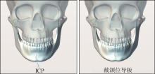
| 1 | Liu MQ, Lei JE, Han JH, et al. Metrical analysis of disc-condyle relation with different splint treatment positions in patients with TMJ disc displacement[J]. J Appl Oral Sci, 2017, 25(5): 483-489. |
| 2 | Farias-Neto A, Dias AHM, de Miranda BFS, et al. Face-bow transfer in prosthodontics: a systematic review of the literature[J]. J Oral Rehabil, 2013, 40(9): 686-692. |
| 3 | Nagy WW, Goldstein GR. Facebow use in clinical prosthodontic practice[J]. J Prosthodont, 2019, 28(7): 772-774. |
| 4 | Bernhardt O, Küppers N, Rosin M, et al. Comparative tests of arbitrary and kinematic transverse horizontal axis recordings of mandibular movements[J]. J Prosthet Dent, 2003, 89(2): 175-179. |
| 5 | Ahangari AH, Torabi K, Pour SR, et al. Evaluation of the Cadiax Compact® Ⅱ accuracy in recording preadjusted condylar inclinations on fully adjustable articulator[J]. J Contemp Dent Pract, 2012, 13(4): 504-508. |
| 6 | Wieckiewicz M, Zietek M, Nowakowska D, et al. Comparison of selected kinematic facebows applied to mandibular tracing[J]. Biomed Res Int, 2014, 2014: 818694. |
| 7 | Balch JH. Verification of the accuracy of electronic mandibular movement recording devices: an in vitro investigation[J]. Int J Exp Dent Sci, 2017(6): 84-94. |
| 8 | Fattori G, Lomax AJ, Weber DC, et al. Technical assessment of the NDI Polaris Vega optical tracking system[J]. Radiat Oncol, 2021, 16(1): 1-4. |
| 9 | Kobs G, Didziulyte A, Kirlys R, et al. Reliability of ARCUSdigma (KaVo) in diagnosing temporomandibular joint pathology[J]. Stomatologija, 2007, 9(2): 47-55. |
| 10 | Mehl A. The determination of the terminal hinge a-xis: a fundamental review and comparison of known and novel methods[J]. Int J Comput Dent, 2018, 21(3): 201-214. |
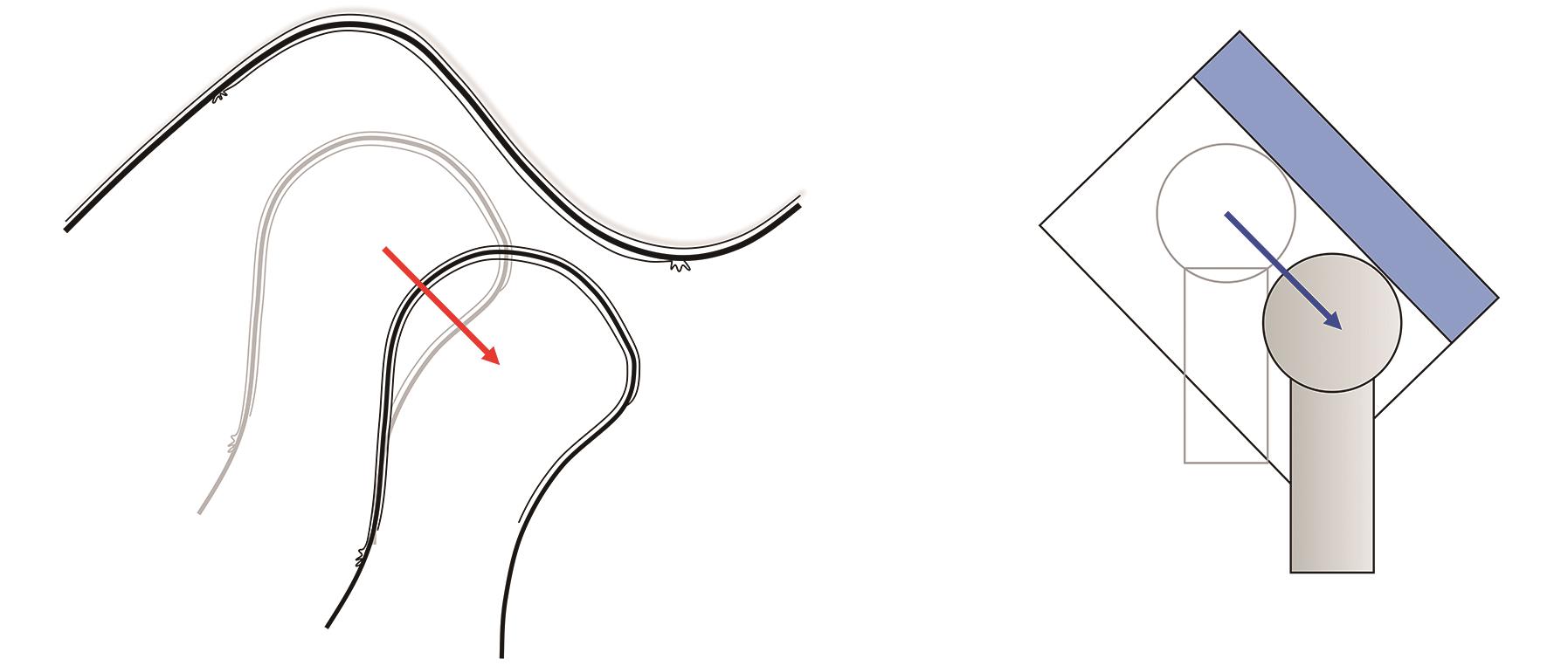
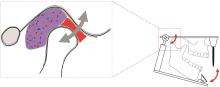
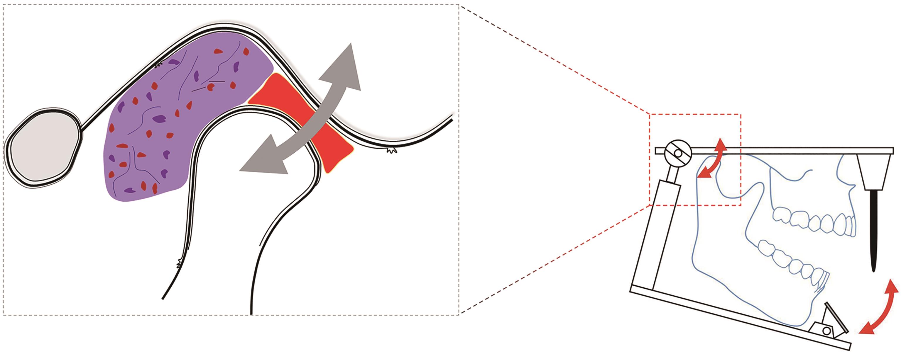
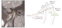
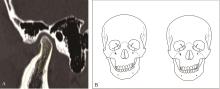
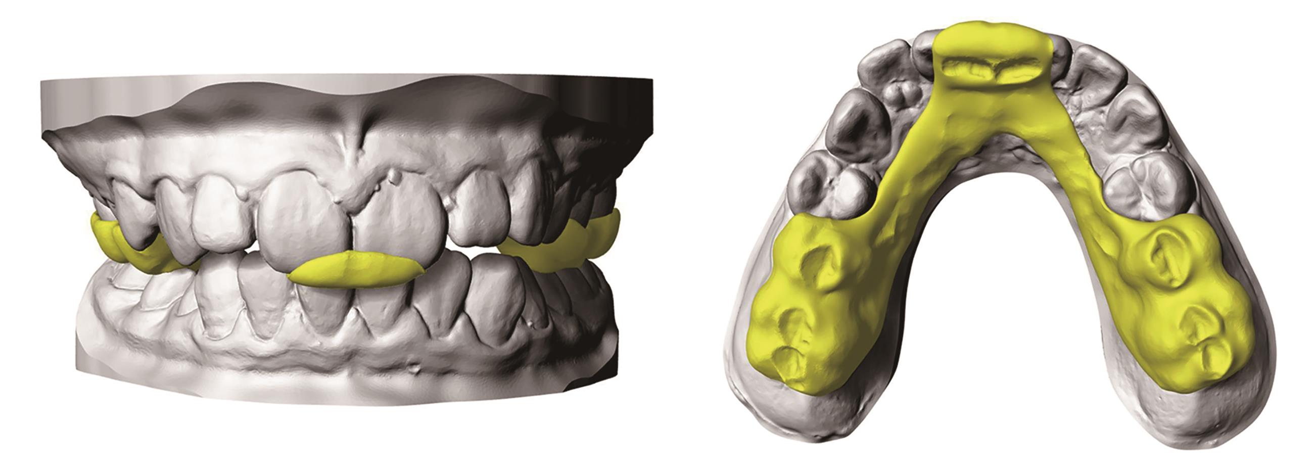
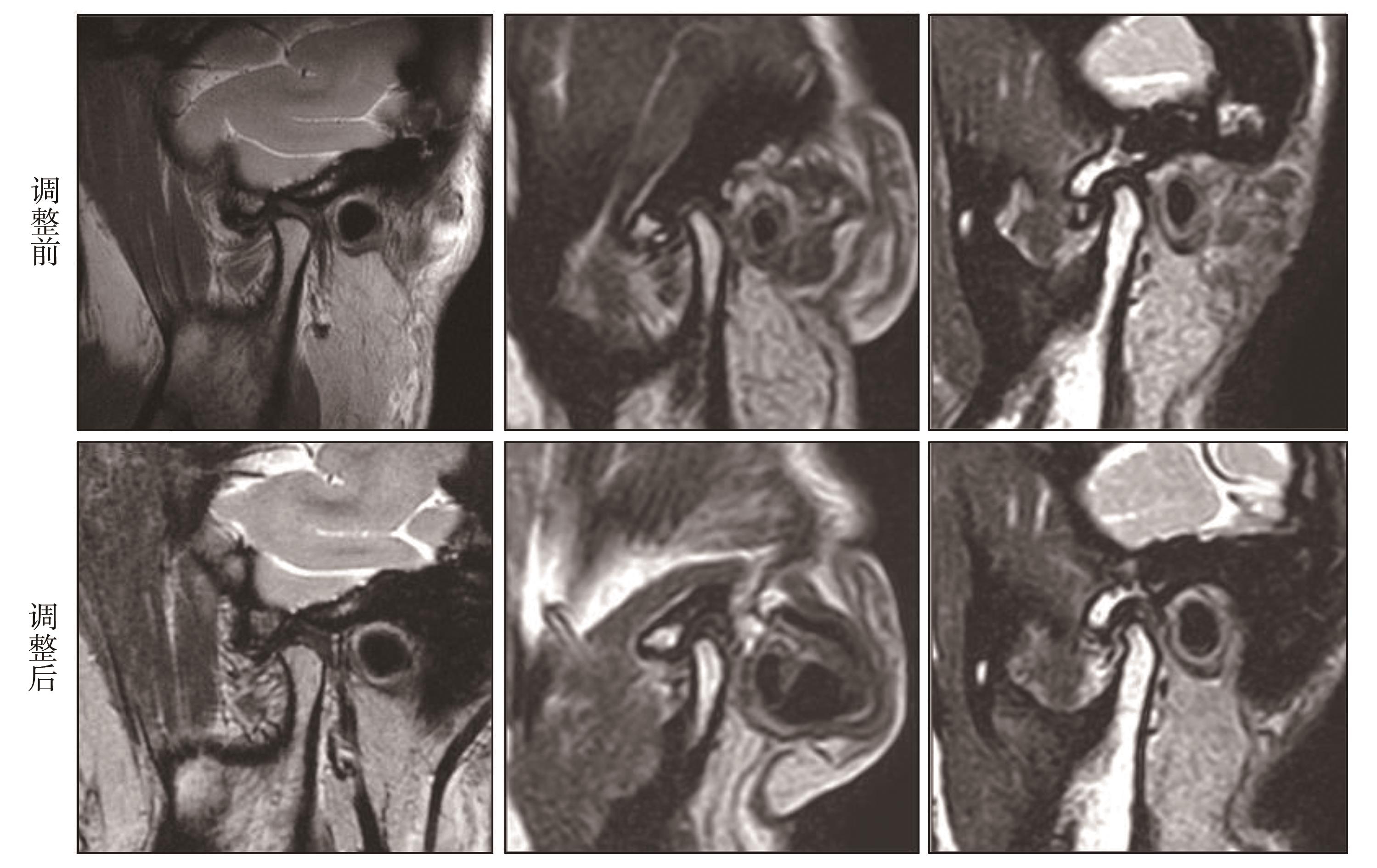
| 11 | Xiong X, Yin XL, Liu F, et al. Magnetic resonance imaging-guided disc-condyle relationship adjustment via articulation: a technical note and case series[J]. J Int Med Res, 2020, 48(8): 030006052-095105. |