Int J Stomatol ›› 2023, Vol. 50 ›› Issue (4): 423-432.doi: 10.7518/gjkq.2023063

• Original Articles • Previous Articles     Next Articles

Finite element analysis of IPS e.maxCAD and Lava Ultimate materials with different thickness in occlusal veneer

Huang Yihuan(),Li Weihang,Ma Dian,Chen Jin,Qian Jie(),Li Xudong.   

  1. Dept. of Prosthodontics, Hospital of Stomatology, Kunming Medical University, Kunming 650106, China
  • Received:2022-10-19 Revised:2023-03-08 Online:2023-07-01 Published:2023-06-21
  • Contact: Jie Qian E-mail:875673900@qq.com;Janeqian@hotmail.com
  • Supported by:
    Medical Reserve Personnel Training of Yunnan Provincial Health Committee(H-2019019);Graduate Innovation Fund of Kunming Medical University(2021S148)

Abstract:

Objective This study aimed to compare the stress distribution in the left maxillary first molar restored with an occlusal veneer made of two kinds of materials with different thickness and provide a theoretical reference for the clinical application of occlusal veneer. Methods Micro CT data of the left maxillary first molar were extracted and four finite element models of the left maxillary first molar with an occlusal veneer of different thicknesses (0.6 and 1.0 mm) and different materials [IPS e.maxCAD (LD) and Lava Ultimate (LU)] were established, respectively, LD0.6, LD1.0, LU0.6, and LU1.0. Then, the axial load F1 and lateral load F2 of the maxillary first molar were simulated. The maximum principal stress (MaxPS) and minimum principal stress (MinPS) of occlusal veneer, tooth tissue and adhesive layer were recorded under two kinds of loads. The MaxPS and MinPS were quantitatively analyzed by percentage quantification and the failure probability of the models was calculated using Weibull analysis. Results 1) Only the enamel reached the tensile strength of the material. 2) In each group, the MaxPS and the absolute value of MinPS of occlusal veneer of LD-restored teeth were significantly higher than that of LU-restored teeth, with variations of 11.4%- 34.7% and from -18.2% to -9.5%, respectively. However, the MaxPS (from -187.1% to 2.4%) and the absolute value of MinPS (from -1.4% to 16.9%) of tooth tissue and adhesive layer of LD-restored teeth were lower than those of LU-restored teeth. When the restoration thickness increased from 0.6 mm to 1.0 mm, the MaxPS and the absolute value of MinPS of occlusal veneer of LD-restored teeth significantly decreased, with variations from 20.0% to 21.0% and from -5.7% to -3.9%, respectively, whereas that of LU-restored teeth did not change. 3) The failure probability of overall models of F1 and F2 was LU0.6>LU1.0>LD0.6> LD1.0 and LU0.6>LU1.0>LD1.0>LD0.6, respectively. Moreover, the failure probability of F1 was lower than that of F2. Conclusion 1) Compared with LU, occlusal veneer made by LD takes on more stress to protect the dental tissue and adhesive layer. 2) The thickness has a significant effect on the stress distribution of LD, but not on LU.

Key words: finite element analysis, occlusal veneer, material, thickness

CLC Number: 

  • R 783.3

TrendMD: 

Fig 1

Perspective after modeling"

Tab 1

Mechanical properties of materials"

材料弹性模量/MPa泊松比拉伸强度/MPa压缩强度/MPa特征强度/MPa威布尔模量
釉质84 100[11]0.31[11]10.3[12]384.0[12]42.41[10]5.53[10]
牙本质18 600[11]0.31[11]98.7[12]297.0[12]44.45[10]3.35[10]
牙髓2[13]0.45[13]----
牙周膜0.068 9[11]0.45[11]----
皮质骨13 700[11]0.30[11]----
松质骨1 370[11]0.30[11]----
树脂粘接剂8 300[10]0.35[10]45.1[12]178.0[12]453.80[10]4.02[10]
LU12 700[10]0.45[10]160.0*516.0[14]300.64[10]10.90[10]
LD102 700[10]0.22[10]124.0[12]360.0[12]609.80[10]13.40[10]

Tab 2

Elements and nodes of models"

测量项目节点数单元数
0.6 mm试件模型1 410 785941 936
1.0 mm试件模型1 543 1871 040 054

Fig 2

Loading area"

Fig 3

The peak value of MaxPS and percentage of each component"

Fig 4

The peak value of MinPS and percentage of each component"

Tab 3

Comparison of the peak value of MaxPS percentage in each group with different restoration materials"

测量项目*??贴面釉质牙本质粘接层
F1F2F1F2F1F2F1F2
LD0.6-LU0.627.8%34.7%-183.3%-186.9%-0.5%-5.4%-38.2%0.5%
LD1.0-LU1.013.9%11.4%-187.1%-49.3%-0.6%-0.2%-18.6%2.4%

Tab 4

Comparison of the peak value of MinPS per-centage in each group with different restoration materials"

测量项目*??贴面釉质牙本质粘接层
F1F2F1F2F1F2F1F2
LD0.6-LU0.6-18.2%-14.9%16.9%2.2%7.8%7.0%16.3%15.5%
LD1.0-LU1.0-12.5%-9.5%3.3%7.2%-0.4%-1.4%13.3%6.5%

Tab 5

Comparison of the peak value of MaxPS per-centage in each group with different restoration thickness"

测量项目*??贴面釉质牙本质粘接层
F1F2F1F2F1F2F1F2
LD0.6-LD1.021.0%20.0%173.2%-31.4%1.3%-3.0%12.5%0.8%
LU0.6-LU1.07.1%-3.3%169.4%106.2%1.2%2.2%32.1%2.7%

Tab 6

Comparison of the peak value of MinPS per-centage in each group with different restoration thickness"

测量项目*??贴面釉质牙本质粘接层
F1F2F1F2F1F2F1F2
LD0.6-LD1.0-5.7%-3.9%-19.1%-1.0%-1.9%1.4%-34.4%-12.7%
LU0.6-LU1.00.0%1.5%-32.7%4.0%-10.1%-7.0%-37.4%-21.7%

Fig 5

The distribution of MaxPS of occlusal veneer"

Fig 6

The failure probability of overall models"

Fig 7

The failure probability of components"

1 Ferraris F. Posterior indirect adhesive restorations (PIAR): preparation designs and adhesthetics clinical protocol[J]. Int J Esthet Dent, 2017, 12(4): 482-502.
2 Magne P, Cheung R. Numeric simulation of occlusal interferences in molars restored with ultrathin occlusal veneers[J]. J Prosthet Dent, 2017, 117(1): 132-137.
3 Moreira A, Freitas F, Marques D, et al. Aesthetic rehabilitation of a patient with bruxism using ceramic veneers and overlays combined with four-point monolithic zirconia crowns for occlusal stabilization: a 4-year follow-up[J]. Case Rep Dent, 2019: 1640563.
4 Malament KA, Margvelashvili-Malament M, Natto ZS, et al. 10.9-year survival of pressed acid etched monolithic e.max lithium disilicate glass-ceramic partial coverage restorations: performance and outcomes as a function of tooth position, age, sex, and the type of partial coverage restoration (inlay or onlay)[J]. J Prosthet Dent, 2021, 126(4): 523-532.
5 Souza J, Fuentes MV, Baena E, et al. One-year clinical performance of lithium disilicate versus resin composite CAD/CAM onlays[J]. Odontology, 2021, 109(1): 259-270.
6 Yin RZ, Kim YK, Jang YS, et al. Comparative eva-luation of the mechanical properties of CAD/CAM dental blocks[J]. Odontology, 2019, 107(3): 360-367.
7 Al-Haj Husain N, Sonderegger S, Özcan M, et al. In vitro static and fatigue behavior of ceramic occlusal veneers using CAD/CAM[J]. Eur J Prosthodont Restor Dent, 2020, 28(3): 113-120.
8 Andrade JP, Stona D, Bittencourt HR, et al. Effect of different computer-aided design/computer-aided manufacturing (CAD/CAM) materials and thicknesses on the fracture resistance of occlusal veneers[J]. Oper Dent, 2018, 43(5): 539-548.
9 王惠芸. 我国人牙的测量和统计[J]. 中华口腔科杂志, 1959, 7(3): 149-155.
Wang HY. Measurement and statistics of human teeth in China[J]. Chin J Stomatol, 1959, 7(3): 149-155.
10 Huang XQ, Hong NR, Zou LY, et al. Estimation of stress distribution and risk of failure for maxillary premolar restored by occlusal veneer with different CAD/CAM materials and preparation designs[J]. Clin Oral Investig, 2020, 24(9): 3157-3167.
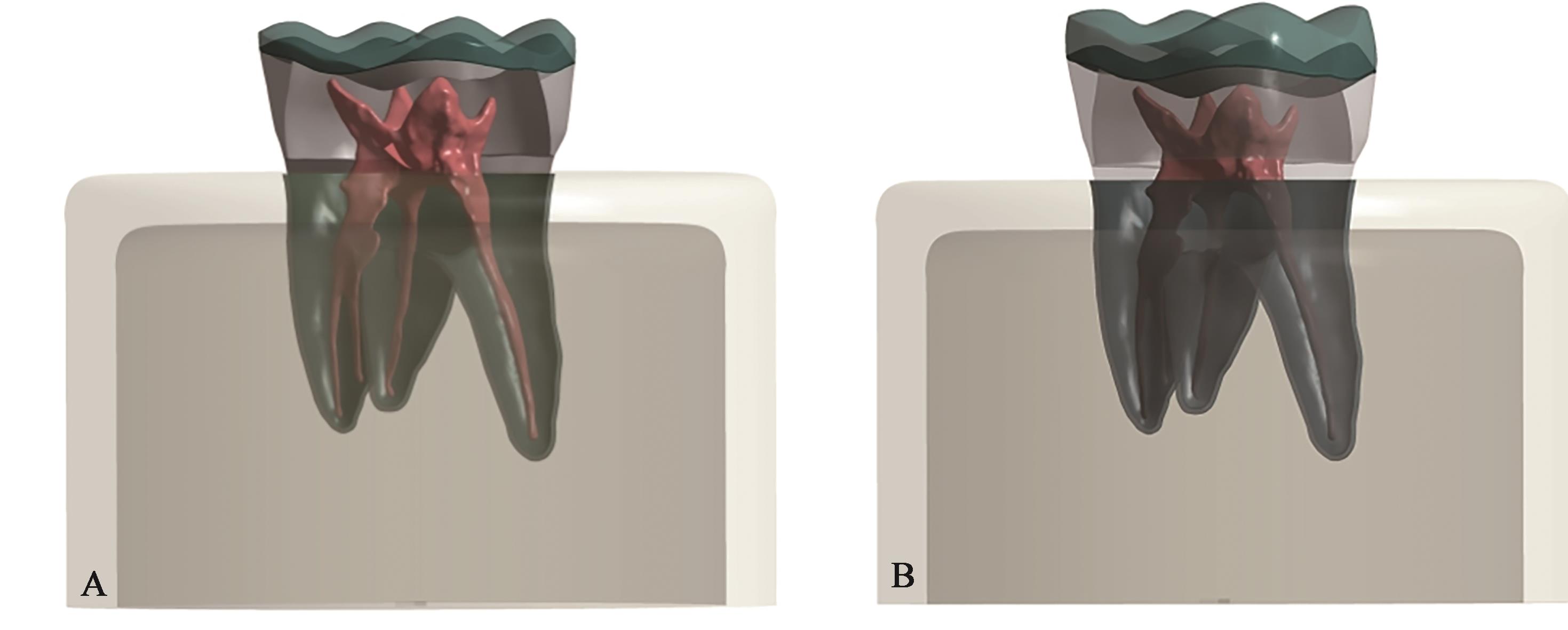
11 张孝霞. 大面积缺损的下颌第一前磨牙桩核冠与高嵌体修复的三维有限元分析[D]. 西安: 空军军医大学, 2018.
Zhang XX. Fiber post-and-core crowns or onlay: a three-dimensional finite element analysis for restoration choice of mandibular premolar with large defect[D]. Xi’an: Air Force Medical University, 2018.
12 Bragança GF, Mazão JD, Versluis A, et al. Effect of luting materials, presence of tooth preparation, and functional loading on stress distribution on ceramic laminate veneers: a finite element analysis[J]. J Prosthet Dent, 2021, 125(5): 778-787.
13 沈冬妮. 后牙𬌗贴面修复的临床应用[D]. 杭州: 浙江大学, 2020.
Shen DN. The clinical application of posterior occlusal veneer restoration[D]. Hangzhou: Zhejiang University, 2020.
14 de Kok P, Kleverlaan CJ, de Jager N, et al. Mechanical performance of implant-supported posterior crowns[J]. J Prosthet Dent, 2015, 114(1): 59-66.
15 魏振辉, 孙贺婷, 高志银, 等. 不同材料高嵌体修复大面积缺损的上颌第一前磨牙有限元分析[J]. 口腔医学研究, 2020, 36(11): 1065-1068.
Wei ZH, Sun HT, Gao ZY, et al. Finite element analysis of maxillary first premolars repaired with diffe-rent onlay materials[J]. J Oral Sci Res, 2020, 36(11): 1065-1068.
16 Ural Ç, Çağlayan E. A 3-dimensional finite element and in vitro analysis of endocrown restorations fabricated with different preparation designs and various restorative materials[J]. J Prosthet Dent, 2021, 126(4): 586.e1-586.e9.
17 Meng QZ, Zhang YJ, Chi DL, et al. Resistance fracture of minimally prepared endocrowns made by three types of restorative materials: a 3D finite element analysis[J]. J Mater Sci, 2021, 32(11): 1-9.
18 Huang XQ, Zou LY, Yao R, et al. Effect of preparation design on the fracture behavior of ceramic occlusal veneers in maxillary premolars[J]. J Dent, 2020, 97: 103346.
19 殷金萍, 王静, 孙亚丽, 等. 动态载荷下不同材料修复非龋性颈部缺损有限元分析[J]. 口腔医学研究, 2021, 37(9): 820-824.
Yin JP, Wang J, Sun YL, et al. Estimation of biomechanical behavior in non-carious cervical lesions restored by different materials: a 3D finite element analysis[J]. J Oral Sci Res, 2021, 37(9): 820-824.
20 Dejak B, Młotkowski A. A comparison of mvM stress of inlays, onlays and endocrowns made from various materials and their bonding with molars in a computer simulation of mastication-FEA[J]. Dent Mater, 2020, 36(7): 854-864.
21 Lucas PW, van Casteren A. The wear and tear of teeth[J]. Med Princ Pract, 2014, 24(): 3-13.
22 林川, 杜莉. 面载荷工况条件下三维有限元分析下颌第一磨牙的应力情况[J]. 实用口腔医学杂志, 2015, 31(3): 393-396.
Lin C, Du L. Three-dimensional finite element stress analysis of the mandible first molar under pressure loading[J]. J Pract Stomatol, 2015, 31(3): 393-396.
23 Dejak B, Młotkowski A, Langot C. Three-dimensional finite element analysis of molars with thin-walled prosthetic crowns made of various materials[J]. Dent Mater, 2012, 28(4): 433-441.
24 Zamzam H, Olivares A, Fok A. Load capacity of occlusal veneers of different restorative CAD/CAM materials under lateral static loading[J]. J Mech Behav Biomed Mater, 2021, 115: 104290.
25 de Angelis F, D’Arcangelo C, Vadini M. The effect of dentin bonding and material thickness on the fle-xural properties of a lithium-disilicate glass-ceramic[J]. J Adhes Dent, 2021, 23(4): 309-318.
26 Ruggiero MM, Soares Gomes R, Pedroso Bergamo ET, et al. Resin-matrix ceramics for occlusal veneers: effect of thickness on reliability and stress distribution[J]. Dent Mater, 2021, 37(3): e131-e139.
27 Abu-Izze FO, Ramos GF, Borges ALS, et al. Fatigue behavior of ultrafine tabletop ceramic restorations[J]. Dent Mater, 2018, 34(9): 1401-1409.
28 Albelasy E, Hamama HH, Tsoi JKH, et al. Influence of material type, thickness and storage on fracture resistance of CAD/CAM occlusal veneers[J]. J Mech Behav Biomed Mater, 2021, 119: 104485.
29 董奕彤. 新型可切削瓷材料的种类和厚度对𬌗贴面抗折性影响的实验研究[D]. 石家庄: 河北医科大学, 2019.
Dong YT. Experimental study on the influence of restoration materials and thicknesses on the fracture resistance of occlusal veneers made from chair-side CAD/CAM ceramic blocs[D]. Shijiazhuang: Hebei Medical University, 2019.
30 Ma L, Guess PC, Zhang Y. Load-bearing properties of minimal-invasive monolithic lithium disilicate and zirconia occlusal onlays: finite element and theo-retical analyses[J]. Dent Mater, 2013, 29(7): 742-751.
31 de Kok P, Kleverland CJ, Kuijs RH, et al. Influence of dentin and enamel on the fracture resistance of restorations at several thicknesses[J]. Am J Dent, 2018, 31(1): 34-38.
32 Zimmermann M, Ender A, Egli G, et al. Fracture load of CAD/CAM-fabricated and 3D-printed composite crowns as a function of material thickness[J]. Clin Oral Investig, 2019, 23(6): 2777-2784.
[1] Chang Xinnan,Liu Lei. Applications and research progress of biodegradable magnesium-based materials in craniomaxillofacial surgery [J]. Int J Stomatol, 2024, 51(1): 107-115.
[2] Zhao Yuanxi,Su Qin.. Application and development of supplementary techniques in removing the remaining filling materials of root canal retreatment [J]. Int J Stomatol, 2023, 50(5): 581-586.
[3] Li Weiguang,Wu Yafei,Guo Shujuan.. Research progress on the use of inorganic nanoparticles in the diagnosis and treatment of periodontal disease [J]. Int J Stomatol, 2022, 49(6): 724-730.
[4] Li Yanfei,Zhang Xinchun. Research progress on the dentin bone repair material [J]. Int J Stomatol, 2022, 49(2): 197-203.
[5] Meng Xiuping,Hou Jianhua,Li Yiran,Sun Mengyao. Research progress on the selection and design of base materials in deep margin elevation [J]. Int J Stomatol, 2021, 48(3): 280-286.
[6] Shen Dongni,Shi Ying,Fu Baiping. Research progress on posterior occlusal veneer [J]. Int J Stomatol, 2021, 48(3): 287-291.
[7] Chen Kenan,Guo Chuanbin. Research progress on biodegradable medical magnesium-based materials [J]. Int J Stomatol, 2021, 48(3): 322-328.
[8] Ji Mengzhen,Qi Meiyao,Du Kexin,Quan Shuqi,Zhang Yuqiang,Zheng Qinghua. Three-dimensional finite element study on the effect of pulp opening cavity on the resistance of cracked teeth after full crown restoration [J]. Int J Stomatol, 2021, 48(1): 41-49.
[9] Zhao Binbin,Zhong Weijian,Ma Guowu. Research progress on dentin as bone transplantation material [J]. Int J Stomatol, 2021, 48(1): 82-89.
[10] Zhang Xinchi,Wu Wei. Research progress on 3D printing technology and biomaterials for bone reconstruction in maxillofacial regions [J]. Int J Stomatol, 2020, 47(6): 677-685.
[11] Qian Huifen,Lin Yunhong,Wu Meiying,Dai Zichao,Li Xingxing. Effect of porcelain layer thickness and substrate on the optical properties of porcelain veneers for tetracycline stained teeth [J]. Int J Stomatol, 2020, 47(4): 413-417.
[12] Zhang Jingting,Pan Xudong,Zhang Wenyun. Effect of opaque-shade resin layer thickness on the color of polyetheretherketone-Crea.lign restoration [J]. Int J Stomatol, 2020, 47(4): 418-423.
[13] Liu Yuhao,Zhang Tao. Research progress on shape memory polymers in bone defect repair and regeneration [J]. Int J Stomatol, 2020, 47(2): 219-224.
[14] Chen Xin,Mao Bochun,Lu Yuqing,Dong Bo,Zhu Zhuoli,Yue Li,Yu Haiyang. Research on mechanical property of Co-Cr alloy and polyetheretherketone frameworks of removable partial denture: a three-dimensional finite element analysis [J]. Int J Stomatol, 2019, 46(5): 526-531.
[15] Yan Dan,Zhang Xizhong,Wang Jianguo. 3D finite element analysis for influence of microimplant thread depth on microimplant and mandible [J]. Int J Stomatol, 2019, 46(4): 387-392.
Viewed
Full text


Abstract

Cited

  Shared   
  Discussed   
[1] . [J]. Foreign Med Sci: Stomatol, 1999, 26(06): .
[2] . [J]. Foreign Med Sci: Stomatol, 1999, 26(06): .
[3] . [J]. Foreign Med Sci: Stomatol, 1999, 26(06): .
[4] . [J]. Foreign Med Sci: Stomatol, 1999, 26(06): .
[5] . [J]. Foreign Med Sci: Stomatol, 1999, 26(06): .
[6] . [J]. Foreign Med Sci: Stomatol, 1999, 26(06): .
[7] . [J]. Foreign Med Sci: Stomatol, 1999, 26(06): .
[8] . [J]. Foreign Med Sci: Stomatol, 1999, 26(06): .
[9] . [J]. Foreign Med Sci: Stomatol, 1999, 26(06): .
[10] . [J]. Foreign Med Sci: Stomatol, 1999, 26(06): .