国际口腔医学杂志 ›› 2025, Vol. 52 ›› Issue (3): 349-357.doi: 10.7518/gjkq.2025046
刘志凯1( ),刘航航2,刘士博1,李帛伦1,刘瑶1,罗恩1()
Zhikai Liu1(),Hanghang Liu2,Shibo Liu1,Bolun Li1,Yao Liu1,En Luo1()
摘要:
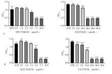
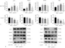
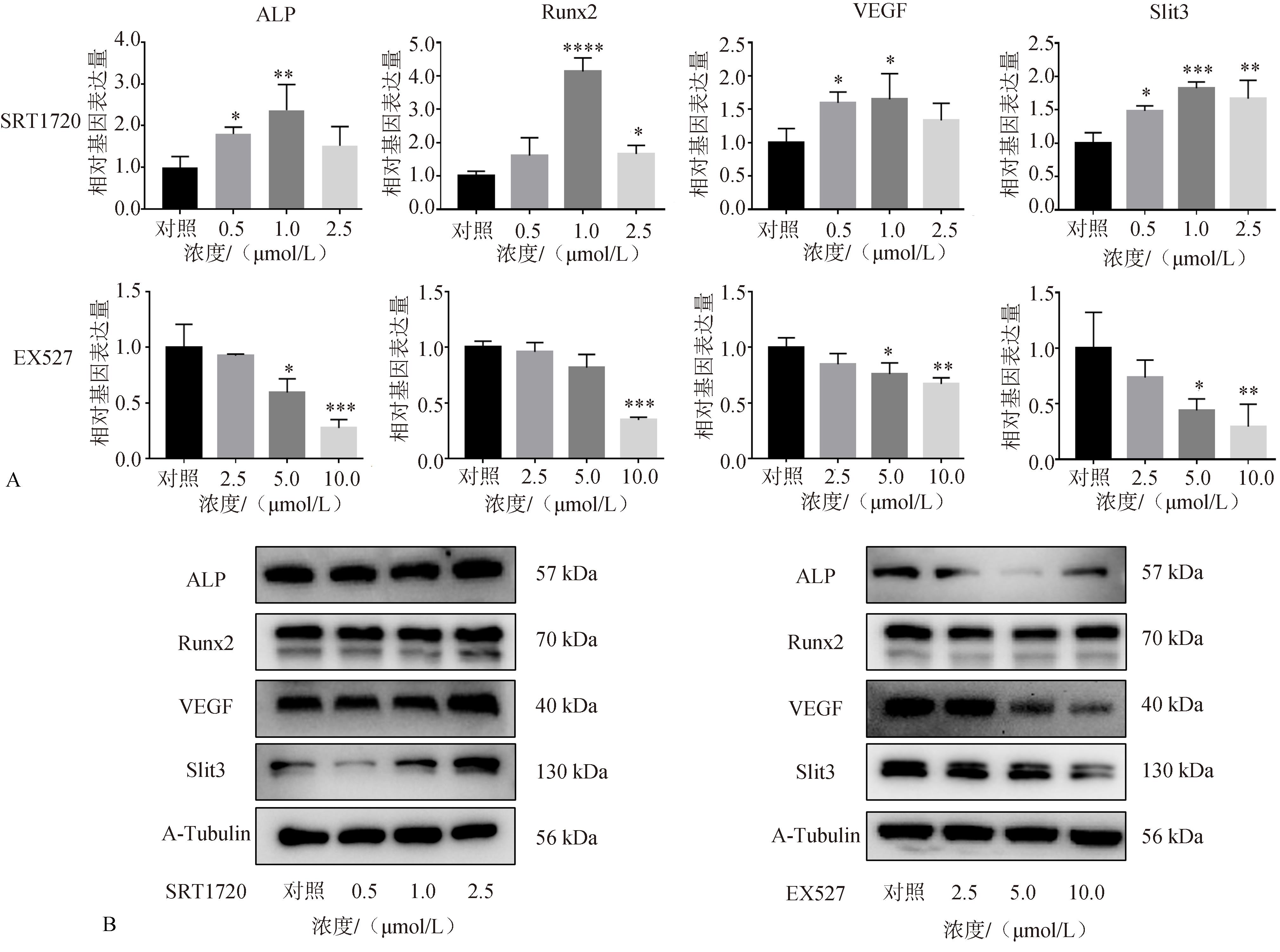
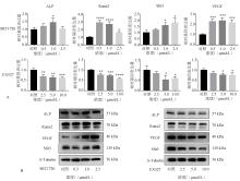
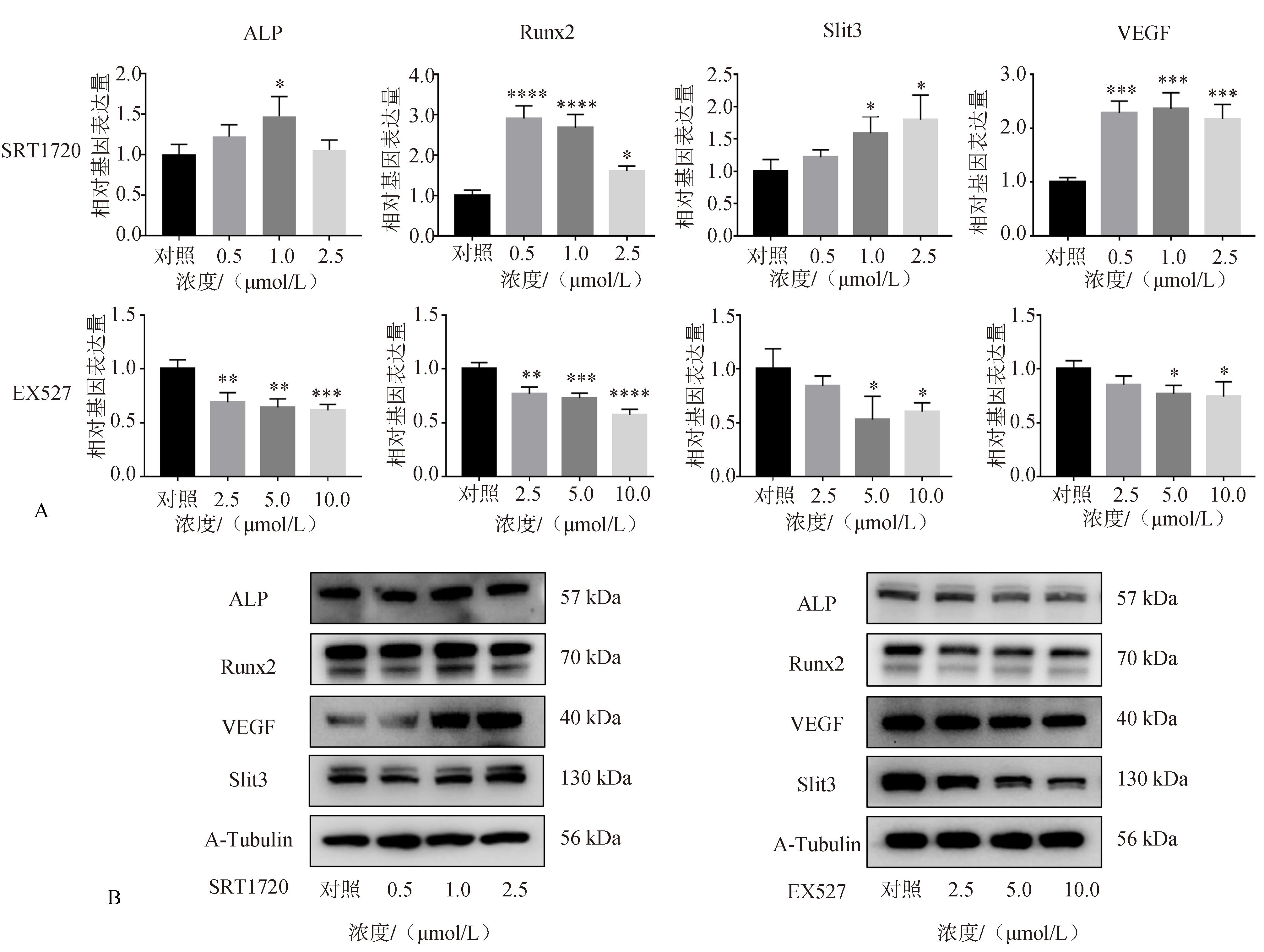
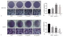
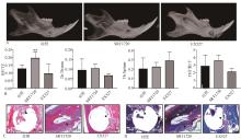
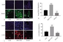
目的 探究沉默信息调节因子1(SIRT1)在体内外条件下对小鼠成骨成血管功能及颌骨缺损愈合的影响。 方法 使用SIRT1特异性激活剂及抑制剂干预小鼠胚胎前体成骨细胞(MC3T3-E1)及小鼠颌骨缺损,采用细胞计数试剂(CCK-8)、实时荧光定量聚合酶链反应、蛋白免疫印迹、碱性磷酸酶(ALP)染色、免疫荧光染色等多种方式,研究SIRT1对MC3T3-E1细胞成骨成血管因子表达、小鼠颌骨缺损愈合及颌骨缺损成骨成血管功能的影响。 结果 细胞实验中SIRT1激活时可促进MC3T3-E1细胞成骨成血管因子表达和ALP活性;动物实验中SIRT1激活可促进颌骨缺损的愈合,同时增强颌骨缺损区域成骨成血管功能;抑制SIRT1活性时则会抑制上述过程。 结论 SIRT1可通过调控小鼠颌骨成骨成血管功能促进颌骨缺损的愈合过程。
中图分类号:
| 1 | Di Maggio N, Banfi A. The osteo-angiogenic signa-ling crosstalk for bone regeneration: harmony out of complexity[J]. Curr Opin Biotechnol, 2022, 76: 102750. |
| 2 | Stegen S, van Gastel N, Carmeliet G. Bringing new life to damaged bone: the importance of angiogenesis in bone repair and regeneration[J]. Bone, 2015, 70: 19-27. |
| 3 | Stucker S, Chen JY, Watt FE, et al. Bone angiogenesis and vascular niche remodeling in stress, aging, and diseases[J]. Front Cell Dev Biol, 2020, 8: 602269. |
| 4 | Shen JJ, Sun Y, Liu XZ, et al. EGFL6 regulates angiogenesis and osteogenesis in distraction osteoge-nesis via Wnt/β-catenin signaling[J]. Stem Cell Res Ther, 2021, 12(1): 415. |
| 5 | Liu XN, Zhang PL, Gu Y, et al. Type H vessels: functions in bone development and diseases[J]. Front Cell Dev Biol, 2023, 11: 1236545. |
| 6 | Bonkowski MS, Sinclair DA. Slowing ageing by design: the rise of NAD+ and sirtuin-activating compoun-ds[J]. Nat Rev Mol Cell Biol, 2016, 17(11): 679-690. |
| 7 | 石玉, 尹贝, 李鑫, 等. 表观遗传和代谢调控间充质干细胞成骨分化的研究进展[J]. 生物医学转化, 2023, 4(2): 57-71. |
| Shi Y, Yin B, Li X, et al. Research progress on the epigenetic and metabolic regulation on osteogenesis of MSCs[J]. Biomed Transform, 2023, 4(2): 57-71. | |
| 8 | Wu QJ, Zhang TN, Chen HH, et al. The sirtuin family in health and disease[J]. Signal Transduct Target Ther, 2022, 7(1): 402. |
| 9 | Zhang WJ, Huang QB, Zeng ZH, et al. Sirt1 inhibits oxidative stress in vascular endothelial cells[J]. O-xid Med Cell Longev, 2017, 2017: 7543973. |
| 10 | Wang H, Hu ZX, Wu J, et al. Sirt1 promotes osteogenic differentiation and increases alveolar bone mass via Bmi1 activation in mice[J]. J Bone Miner Res, 2019, 34(6): 1169-1181. |
| 11 | 李明哲, 罗国厂, 张仲博, 等. 虎杖苷促进大鼠骨质疏松性骨折的作用及对SIRT1/FoxO1信号通路的影响[J]. 中国骨质疏松杂志, 2023, 29(8): 1154-1159. |
| Li MZ, Luo GC, Zhang ZB, et al. The effect of polydatin on promoting osteoporotic fracture healing in rats and its effect on SIRT1/FoxO1 signaling pathway[J]. Chin J Osteoporos, 2023, 29(8): 1154-1159. | |
| 12 | Zhang JK, Pan J, Jing W. Motivating role of type H vessels in bone regeneration[J]. Cell Prolif, 2020, 53(9): e12874. |
| 13 | Xu R, Yallowitz A, Qin A, et al. Targeting skeletal endothelium to ameliorate bone loss[J]. Nat Med, 2018, 24(6): 823-833. |
| 14 | Kim BJ, Lee YS, Lee SY, et al. Osteoclast-secreted SLIT3 coordinates bone resorption and formation[J]. J Clin Invest, 2018, 128(4): 1429-1441. |
| 15 | 张思钰, 舒晴, 贾绍辉, 等. 组蛋白乙酰化对间充质干细胞成骨分化影响机制的研究[J]. 中国骨质疏松杂志, 2023, 29(2): 232-236, 247. |
| Zhang SY, Shu Q, Jia SH, et al. Research progress on the mechanism of histone acetylation on osteogenic differentiation of mesenchymal stem cells[J]. Chin J Osteoporos, 2023, 29(2): 232-236, 247. | |
| 16 | Lemieux ME, Yang X, Jardine K, et al. The Sirt1 deacetylase modulates the insulin-like growth factor signaling pathway in mammals[J]. Mech Ageing Dev, 2005, 126(10): 1097-1105. |
| 17 | Louvet L, Leterme D, Delplace S, et al. Sirtuin 1 deficiency decreases bone mass and increases bone marrow adiposity in a mouse model of chronic energy deficiency[J]. Bone, 2020, 136: 115361. |
| 18 | Simic P, Zainabadi K, Bell E, et al. SIRT1 regulates differentiation of mesenchymal stem cells by deace-tylating β-catenin[J]. EMBO Mol Med, 2013, 5(3): 430-440. |
| 19 | Domazetovic V, Marcucci G, Falsetti I, et al. Blueberry juice antioxidants protect osteogenic activity against oxidative stress and improve long-term activation of the mineralization process in human osteoblast-like SaOS-2 cells: involvement of SIRT1[J]. Antioxidants, 2020, 9(2): 125. |
| 20 | Das A, Huang GX, Bonkowski MS, et al. Impairment of an endothelial NAD+-H2S signaling network is a reversible cause of vascular aging[J]. Cell, 2019, 176(4): 944-945. |
| 21 | Lipphardt M, Dihazi H, Müller GA, et al. Fibroge-nic secretome of sirtuin 1-deficient endothelial cells: Wnt, Notch and glycocalyx rheostat[J]. Front Phy-siol, 2018, 9: 1325. |
| 22 | Tombran-Tink J, Barnstable CJ. Osteoblasts and osteoclasts express PEDF, VEGF-A isoforms, and VEGF receptors: possible mediators of angiogenesis and matrix remodeling in the bone[J]. Biochem Biophys Res Commun, 2004, 316(2): 573-579. |
| [1] | 秦庆钊,温奥楠,高梓翔,朱玉佳,王勇,赵一姣. 口腔颌面缺损修复数字化设计方法的研究进展[J]. 国际口腔医学杂志, 2025, 52(2): 272-280. |
| [2] | 常欣楠,刘磊. 生物可降解镁基材料在颅颌面外科的应用及其研究进展[J]. 国际口腔医学杂志, 2024, 51(1): 107-115. |
| [3] | 王素杰,谭芹,韦渊,王洁,范杰,岳二丽. 口腔扁平苔藓患者血清血管生成素-2水平与叉头翼状螺旋转录因子阳性调节性T细胞及疾病活动度的相关性分析[J]. 国际口腔医学杂志, 2023, 50(6): 674-678. |
| [4] | 蒋青松,赖文莉,王艳. 骨增量技术在口腔正畸领域的研究进展[J]. 国际口腔医学杂志, 2023, 50(2): 243-250. |
| [5] | 满毅, 黄定明. 美学区种植骨增量与邻牙慢性根尖周病的联合治疗策略(下):临床诊治流程及实践病例[J]. 国际口腔医学杂志, 2022, 49(6): 621-632. |
| [6] | 满毅, 黄定明. 美学区种植骨增量与邻牙慢性根尖周病的联合治疗策略(上):应用基础及适应证[J]. 国际口腔医学杂志, 2022, 49(5): 497-505. |
| [7] | 李佩,林凌,赵玮. 乳牙牙髓干细胞在口腔组织再生修复中的研究进展[J]. 国际口腔医学杂志, 2022, 49(4): 483-488. |
| [8] | 蔡超莹,陈学鹏,胡济安. 外泌体复合支架用于口腔组织工程的研究进展[J]. 国际口腔医学杂志, 2022, 49(4): 489-496. |
| [9] | 覃思文,廖立. 牙髓再生中血管网络重建策略[J]. 国际口腔医学杂志, 2022, 49(3): 272-282. |
| [10] | 李嫣斐,张新春. 牙本质作为骨修复材料的研究进展[J]. 国际口腔医学杂志, 2022, 49(2): 197-203. |
| [11] | 刘嘉程,孟昭松,李宏捷,隋磊. 卵泡抑素在口腔颌面部发育中的作用及其治疗应用前景[J]. 国际口腔医学杂志, 2021, 48(5): 556-562. |
| [12] | 赵文俊,陈宇. 引导组织/骨再生牙周功能梯度膜的研究进展[J]. 国际口腔医学杂志, 2021, 48(4): 391-397. |
| [13] | 周丰,陈野,陈晨,张奕宁,耿瑞蔓,刘戟. 沉默信息调节因子1调控牙周炎发生发展的机制[J]. 国际口腔医学杂志, 2021, 48(3): 341-346. |
| [14] | 李佩仪,张新春. 微环境酸碱度在组织工程骨再生中作用的研究进展[J]. 国际口腔医学杂志, 2021, 48(1): 64-70. |
| [15] | 赵彬彬,仲维剑,马国武. 牙本质作为骨移植材料的研究进展[J]. 国际口腔医学杂志, 2021, 48(1): 82-89. |
|