国际口腔医学杂志 ›› 2023, Vol. 50 ›› Issue (6): 669-673.doi: 10.7518/gjkq.2023083
孙旭1( ),邓振南2,文才3,赵颖1()
Sun Xu1(),Deng Zhennan2,Wen Cai3,Zhao Ying1()
摘要:
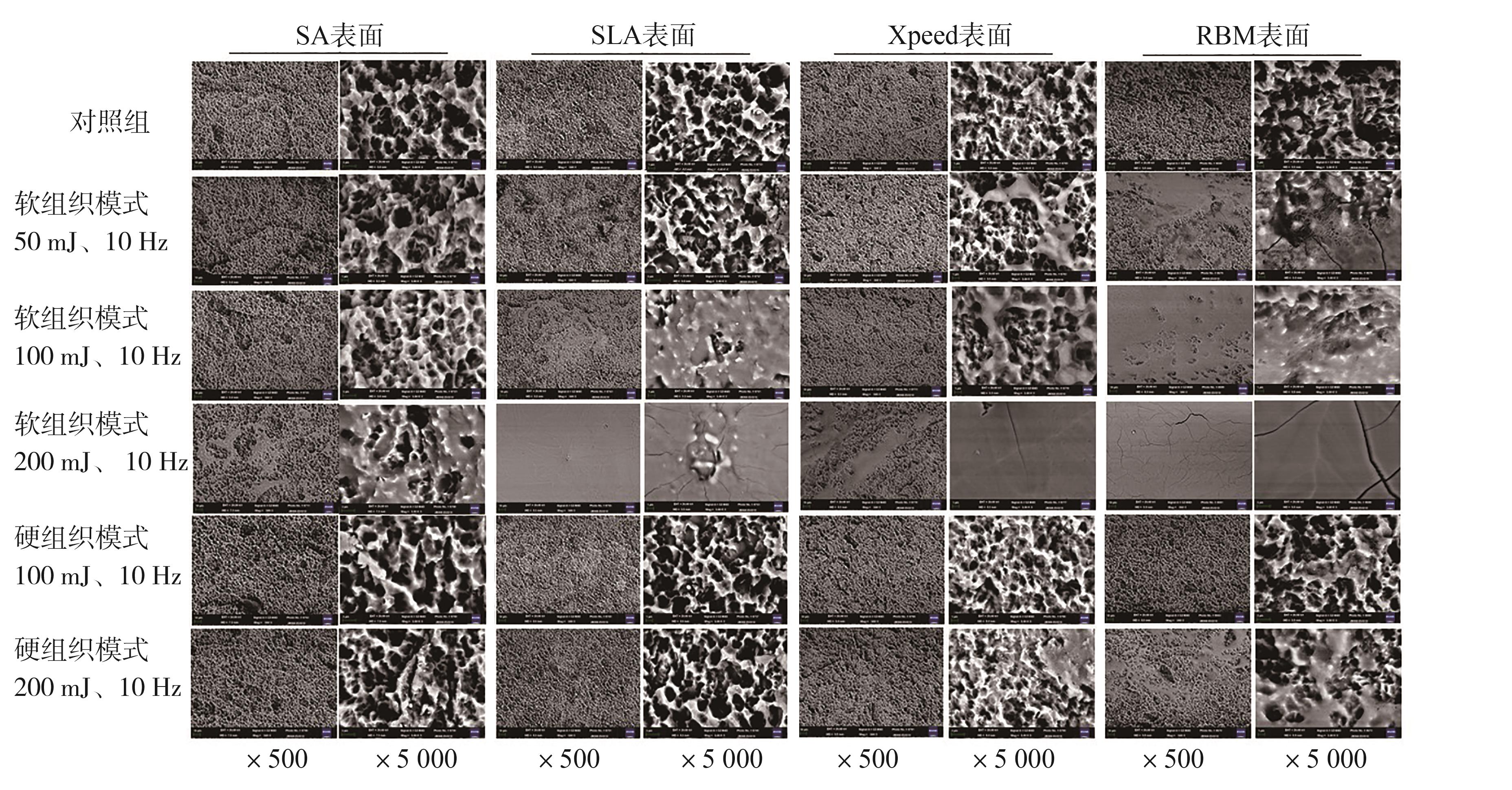
目的 探讨Er: YAG激光不同模式、不同参数照射对种植体SA、SLA、Xpeed及RBM表面微形貌的影响。 方法 选择波长为2 940 nm的Er: YAG激光并设置为软组织模式及参数50 mJ、10 Hz,100 mJ、10 Hz,200 mJ、10 Hz;设置硬组织模式及参数100 mJ、10 Hz,200 mJ、10 Hz。均在水冷却环境下,采取非接触模式,工作尖距离种植体表面1 mm,对SA、SLA、Xpeed及RBM 4种种植体表面固定一点照射5 s。用扫描电子显微镜(SEM)观察照射前后表面微形貌的变化。 结果 SEM观察可见,SA表面在软组织模式50 mJ、10 Hz及100 mJ、10 Hz照射下无变化,能量提高到200 mJ、10 Hz可见表面微形貌部分熔化;SLA表面在软组织模式50 mJ、10 Hz照射下无变化,能量提高到100 mJ、10Hz及200 mJ、10 Hz可见表面部分熔化及完全熔化;SA及SLA表面在硬组织模式100 mJ、10 Hz或200 mJ、10 Hz照射下,表面无变化。Xpeed和RBM表面在软组织模式50 mJ、10 Hz照射下分别表现为高峰塌陷和片状熔化;在能量提高到100 mJ、10 Hz及200 mJ、10 Hz均可见部分熔化及完全熔化;Xpeed及RBM表面在硬组织模式100 mJ、10 Hz照射下无变化,能量提高到200 mJ、10 Hz可见部分熔化;Xpeed表面未见涂层剥脱。 结论 本研究显示,在用Er: YAG激光处理种植体表面时,为避免对种植体表面造成损伤,SA和SLA表面用软组织模式时能量参数需设置在50 mJ、10 Hz以下,硬组织模式时参数设置200 mJ、10 Hz以下。Xpeed及RBM表面不适合用软组织模式处理,可在硬组织模式100 mJ、10 Hz以下处理。
中图分类号:
| 1 | Subramani K, Jung RE, Molenberg A, et al. Biofilm on dental implants: a review of the literature[J]. Int J Oral Maxillofac Implants, 2009, 24(4): 616-626. |
| 2 | Renvert S, Polyzois I, Maguire R. Re-osseointegration on previously contaminated surfaces: a syste-matic review[J]. Clin Oral Implants Res, 2009, 20(): 216-227. |
| 3 | Trejo PM, Bonaventura G, Weng D, et al. Effect of mechanical and antiseptic therapy on peri-implant mucositis: an experimental study in monkeys[J]. Clin Oral Implants Res, 2006, 17(3): 294-304. |
| 4 | Mizutani K, Aoki A, Coluzzi D, et al. Lasers in mi-nimally invasive periodontal and peri-implant therapy[J]. Periodontol 2000, 2016, 71(1): 185-212. |
| 5 | Taniguchi Y, Aoki A, Mizutani K, et al. Optimal Er: YAG laser irradiation parameters for debridement of microstructured fixture surfaces of titanium dental implants[J]. Lasers Med Sci, 2013, 28(4): 1057-1068. |
| 6 | Lafaurie GI, Sabogal MA, Castillo DM, et al. Microbiome and microbial biofilm profiles of peri-implantitis: a systematic review[J]. J Periodontol, 2017, 88(10): 1066-1089. |
| 7 | Sezin M, Croharé L, Ibañez JC. Microscopic study of surface microtopographic characteristics of dental implants[J]. Open Dent J, 2016, 10: 139-147. |
| 8 | Stubinger S, Etter C, Miskiewicz M, et al. Surface alterations of polished and sandblasted and acid-etched titanium implants after Er: YAG, carbon dioxide, and diode laser irradiation[J]. Int J Oral Ma-xillofac Implants, 2010, 25(1): 104-111. |
| 9 | 梁辰, 赖穗萍, 王小静, 等. Er: YAG激光照射对SLA及SLActive种植体表面结构变化的体外研究[J]. 中华老年口腔医学杂志, 2020, 18(3): 158-162. |
| Liang C, Lai SP, Wang XJ, et al. Surface alterations of sandblasted and acid-etched (SLA) and chemical modified sandblasted and acid-etched (SLActive) titanium implants after Er: YAG laser irradiation in vitro [J]. Chin J Geriatr Dent, 2020, 18(3): 158-162. | |
| 10 | 陈中仁, 陈雪, 彭琳. Er: YAG激光治疗种植体周围炎的基础研究进展[J]. 临床口腔医学杂志, 2021, 37(3): 188-191. |
| Chen ZR, Chen X, Peng L. Basic research progress of Er: YAG laser in the treatment of peri-implant in-flammation[J]. J Clin Stomatol, 2021, 37(3): 188-191. | |
| 11 | 魏雅楠, 陈筠, 李志艳. Er: YAG激光对恒牙牙本质粘接性能的影响[J]. 口腔疾病防治, 2020, 28(10): 673-676. |
| Wei YN, Chen Y, Li ZY. Effect of Er: YAG laser irradiation on the bonding strength of permanent teeth[J]. J Prev Treat Stomatol Dis, 2020, 28(10): 673-676. | |
| 12 | Nasirzade J, Kargarpour Z, Hasannia S, et al. Platelet-rich fibrin elicits an anti-inflammatory response in macrophages in vitro [J]. J Periodontol, 2020, 91(2): 244-252. |
| 13 | Nakamura M, Aizawa H, Kawabata H, et al. Platelet adhesion on commercially pure titanium plates in vitro Ⅲ: effects of calcium phosphate-blasting on titanium plate biocompatibility[J]. Int J Implant Dent, 2020, 6(1): 74. |
| 14 | Zinelis S, Silikas N, Thomas A, et al. Surface cha-racterization of SLActive dental implants[J]. Eur J Esthet Dent, 2012, 7(1): 72-92. |
| 15 | Son WW, Zhu XL, Shin HI, et al. In vivo histological response to anodized and anodized/hydrothermally treated titanium implants[J]. J Biomed Mater Res B Appl Biomater, 2003, 66(2): 520-525. |
| 16 | 穆月, 李倩, 赵继志. 激光在口腔种植中的应用[J]. 中国医学科学院学报, 2014, 36(5): 560-564. |
| Mu Y, Li Q, Zhao JZ. Applications of lasers in dental implantology[J]. Acta Acad Med Sin, 2014, 36(5): 560-564. |
| [1] | 黄元鸿,彭显,周学东. 骨碎补在治疗口腔骨相关疾病的研究进展[J]. 国际口腔医学杂志, 2023, 50(6): 679-685. |
| [2] | 龚佳明,赵瑞敏,潘宏伟,郎鑫,余占海,李健学. 动态导航与静态导航对种植体准确性的Meta分析[J]. 国际口腔医学杂志, 2023, 50(5): 538-551. |
| [3] | 陆倩,夏海斌,王敏. 种植体磨光整形术治疗种植体周围炎的研究进展[J]. 国际口腔医学杂志, 2023, 50(2): 152-158. |
| [4] | 满毅, 黄定明. 美学区种植骨增量与邻牙慢性根尖周病的联合治疗策略(上):应用基础及适应证[J]. 国际口腔医学杂志, 2022, 49(5): 497-505. |
| [5] | 曹正国. 修复治疗相关的牙周问题考量[J]. 国际口腔医学杂志, 2022, 49(1): 1-11. |
| [6] | 章善,沈树平,张舫,杨卫东. Er: YAG激光光子激活光声流技术对根管壁牙本质失水状况及牙根抗压强度的影响[J]. 国际口腔医学杂志, 2022, 49(1): 55-59. |
| [7] | 朱轩智,赵蕾. 甲状腺功能减退症与牙周炎相关性的研究进展[J]. 国际口腔医学杂志, 2021, 48(4): 380-384. |
| [8] | 黎敏,华成舸,蒋丽. 提高氧化锆陶瓷粘接性能新技术的研究进展[J]. 国际口腔医学杂志, 2021, 48(4): 485-490. |
| [9] | 路泊遥,杨大维,刘蔚晴,梁星. 超短种植体临床应用效果的影响因素[J]. 国际口腔医学杂志, 2021, 48(3): 329-328. |
| [10] | 朱俊瑾,王剑. 钛种植体表面银纳米颗粒负载方法的进展[J]. 国际口腔医学杂志, 2021, 48(3): 334-340. |
| [11] | 郑桂婷,徐燕,吴明月. 种植体周围疾病治疗的专家共识及治疗方法的进展[J]. 国际口腔医学杂志, 2020, 47(6): 725-731. |
| [12] | 童子安,姒蜜思. 种植体表面菌斑去污方式的体外研究进展[J]. 国际口腔医学杂志, 2020, 47(5): 589-594. |
| [13] | 王欢,刘洋,戚孟春,李静怡,刘梦楠,孙红. 微弧氧化技术制备钛基种植体表面涂层的研究进展[J]. 国际口腔医学杂志, 2020, 47(4): 439-444. |
| [14] | 张敏,万浩元. 种植体周围炎药物治疗与激光治疗的研究进展[J]. 国际口腔医学杂志, 2020, 47(4): 463-470. |
| [15] | 吴秋月,李治邦. 药物辅助治疗种植体周围炎的研究进展[J]. 国际口腔医学杂志, 2020, 47(4): 471-477. |
|