国际口腔医学杂志 ›› 2023, Vol. 50 ›› Issue (4): 423-432.doi: 10.7518/gjkq.2023063
 贴面修复中的有限元分析
黄依欢( ),李委航,马典,陈瑾,钱捷(),李旭东
Huang Yihuan(),Li Weihang,Ma Dian,Chen Jin,Qian Jie(),Li Xudong.
摘要:
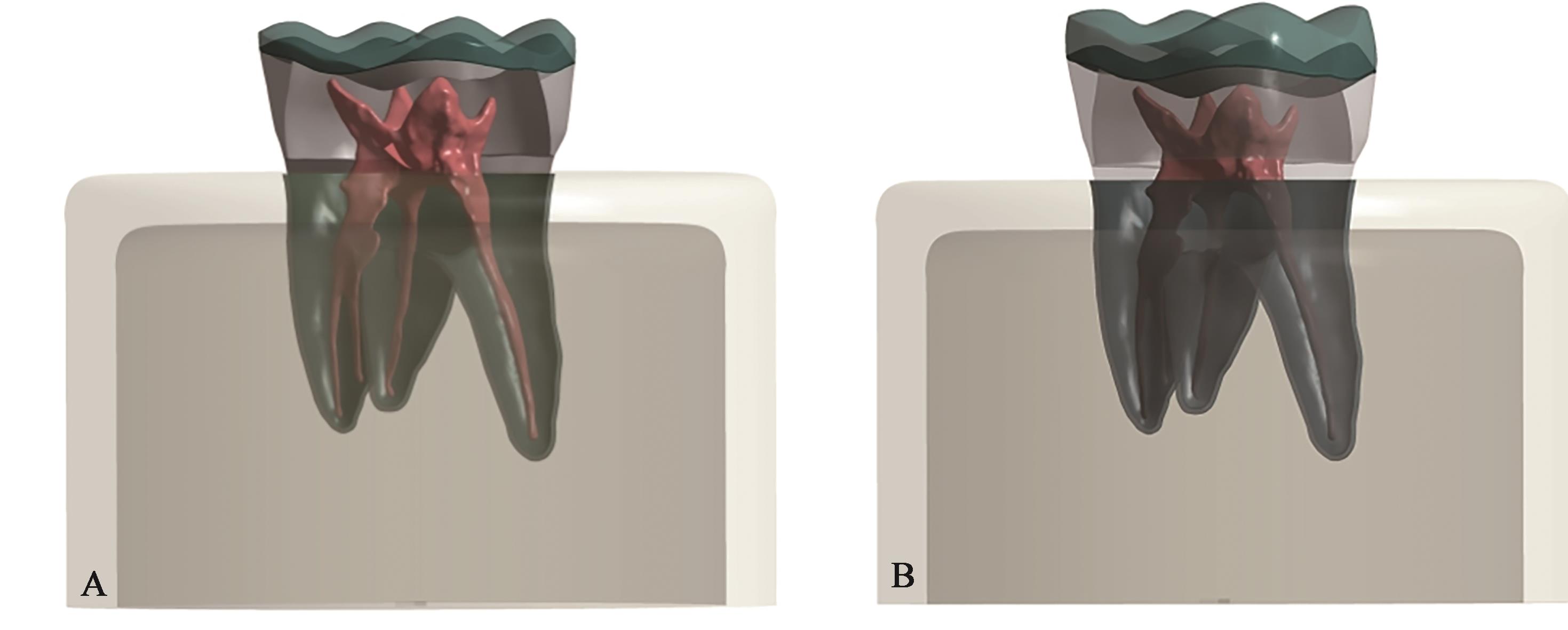
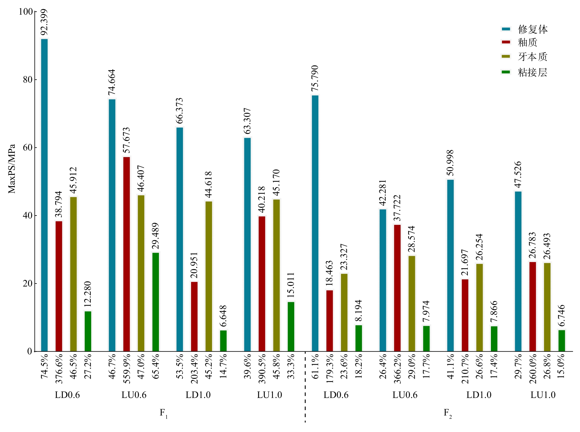
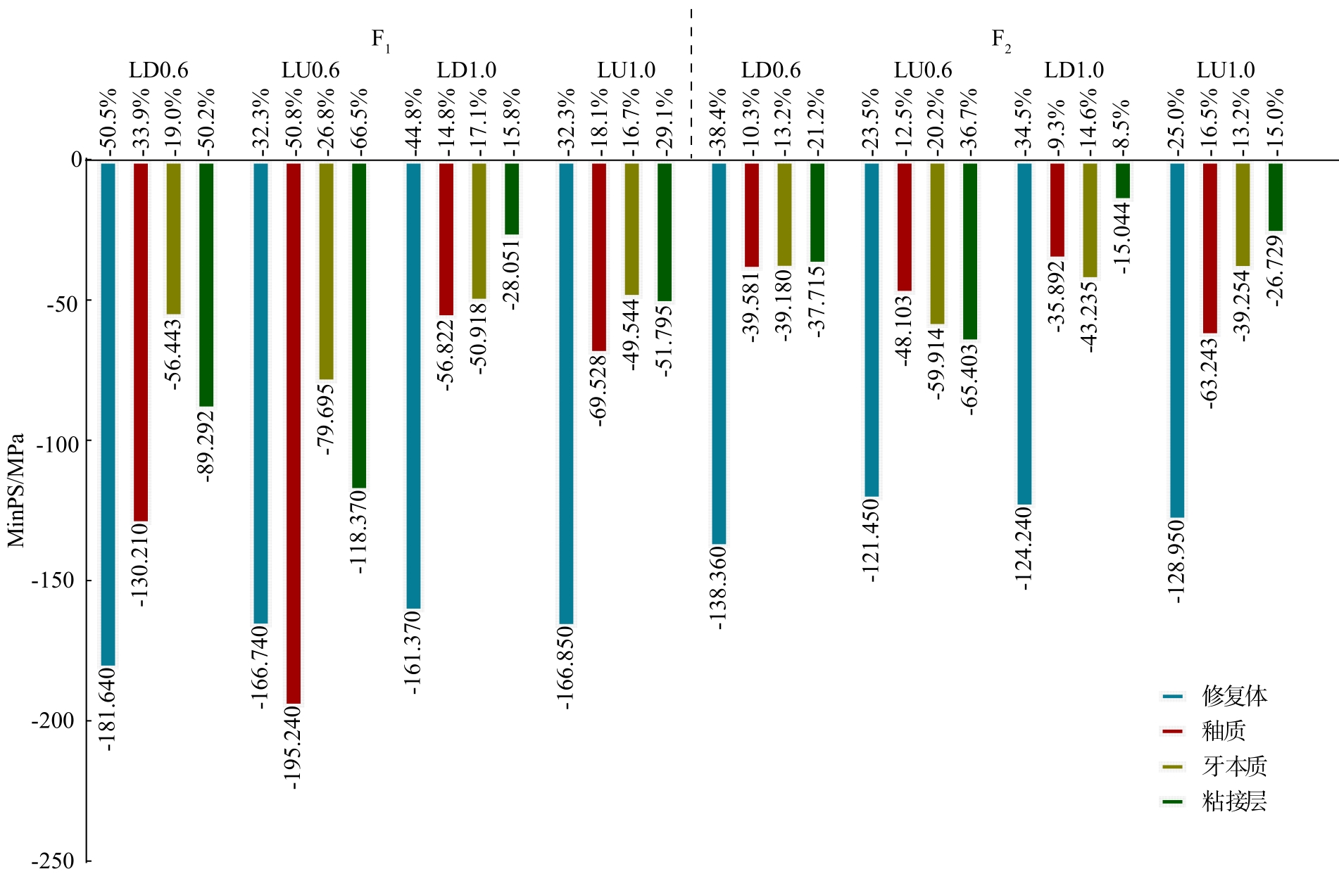
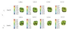
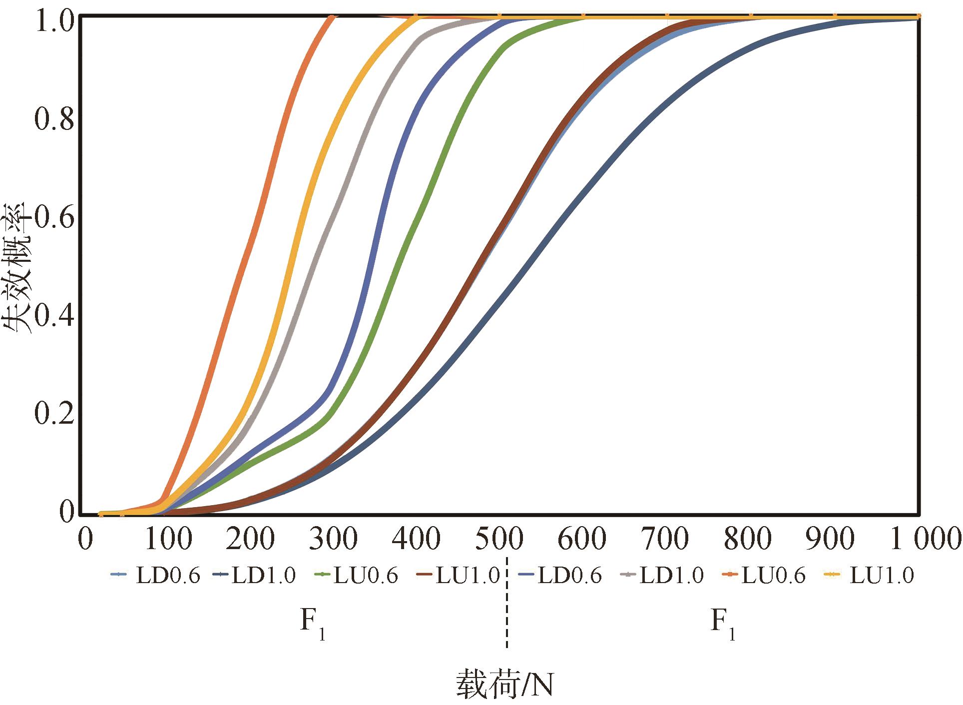
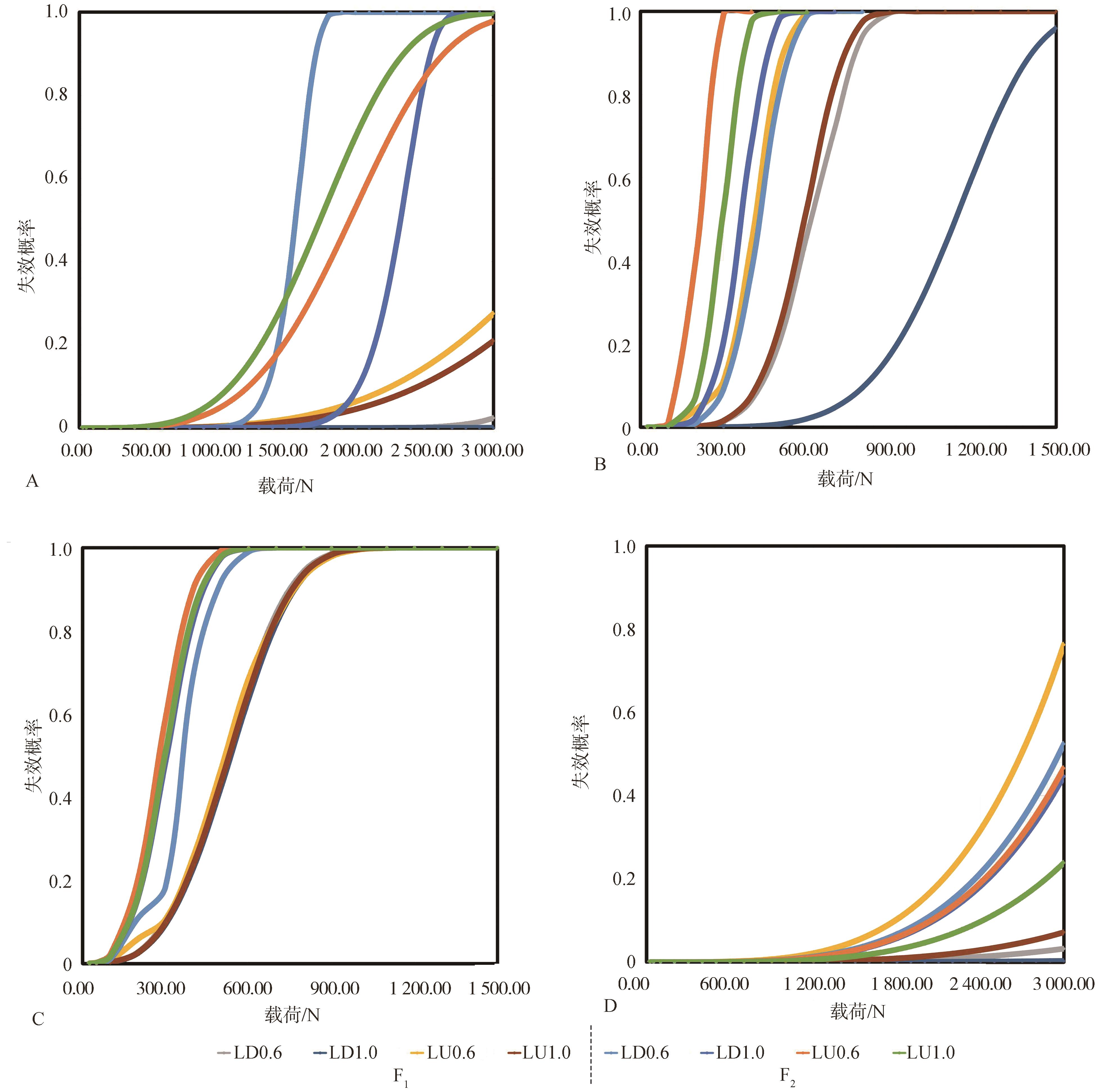
目的 对比不同厚度的2种材料制作的𬌗贴面修复左上颌第一磨牙的应力分布情况,为𬌗贴面的临床应用提供理论指导。 方法 提取左上颌第一磨牙的Micro CT数据,按不同厚度(0.6和1.0 mm)及不同材料[IPS e.maxCAD(LD)和Lava Ultimate(LU)]建立𬌗贴面修复左上颌第一磨牙的4个有限元模型,分别为LD0.6、LD1.0、LU0.6和LU1.0,模拟口内上颌第一磨牙的轴向载荷F1和侧方载荷F2,记录各载荷时𬌗贴面、牙体组织和粘接层的最大主应力(MaxPS)和最小主应力(MinPS)的大小及分布情况,对MaxPS和MinPS进行百分比量化的定量分析,并用威布尔分析计算各模型的失效概率。 结果 1)仅釉质达到该材料的拉伸强度;2)在各组中,LD组的𬌗贴面所受到MaxPS和MinPS绝对值显著大于LU组的,变化量分别为11.4%~34.7%和-18.2%~-9.5%;而LD组的牙体组织以及粘接层的MaxPS和MinPS绝对值小于LU组的,变化量分别为-187.1%~2.4%和-1.4%~16.9%。当修复体厚度由0.6增加为1.0 mm时,LD组的𬌗贴面所受到的MaxPS和MinPS绝对值显著减小,变化量分别为20.0%~21.0%和-5.7%~-3.9%;而LU组的MaxPS和MinPS绝对值变化不明显;3)F1的整体模型失效概率为LU0.6>LU1.0>LD0.6>LD1.0,轴向载荷F2的整体模型失效概率为LU0.6>LU1.0>LD1.0>LD0.6,F1的失效均低于F2。 结论 1)比起LU,LD制作的𬌗贴面承担更大的应力,从而保护其龈方的牙体组织和粘接层;2)厚度对LD的应力分布影响显著,而对LU的影响不显著。
中图分类号:
| 1 | Ferraris F. Posterior indirect adhesive restorations (PIAR): preparation designs and adhesthetics clinical protocol[J]. Int J Esthet Dent, 2017, 12(4): 482-502. |
| 2 | Magne P, Cheung R. Numeric simulation of occlusal interferences in molars restored with ultrathin occlusal veneers[J]. J Prosthet Dent, 2017, 117(1): 132-137. |
| 3 | Moreira A, Freitas F, Marques D, et al. Aesthetic rehabilitation of a patient with bruxism using ceramic veneers and overlays combined with four-point monolithic zirconia crowns for occlusal stabilization: a 4-year follow-up[J]. Case Rep Dent, 2019: 1640563. |
| 4 | Malament KA, Margvelashvili-Malament M, Natto ZS, et al. 10.9-year survival of pressed acid etched monolithic e.max lithium disilicate glass-ceramic partial coverage restorations: performance and outcomes as a function of tooth position, age, sex, and the type of partial coverage restoration (inlay or onlay)[J]. J Prosthet Dent, 2021, 126(4): 523-532. |
| 5 | Souza J, Fuentes MV, Baena E, et al. One-year clinical performance of lithium disilicate versus resin composite CAD/CAM onlays[J]. Odontology, 2021, 109(1): 259-270. |
| 6 | Yin RZ, Kim YK, Jang YS, et al. Comparative eva-luation of the mechanical properties of CAD/CAM dental blocks[J]. Odontology, 2019, 107(3): 360-367. |
| 7 | Al-Haj Husain N, Sonderegger S, Özcan M, et al. In vitro static and fatigue behavior of ceramic occlusal veneers using CAD/CAM[J]. Eur J Prosthodont Restor Dent, 2020, 28(3): 113-120. |
| 8 | Andrade JP, Stona D, Bittencourt HR, et al. Effect of different computer-aided design/computer-aided manufacturing (CAD/CAM) materials and thicknesses on the fracture resistance of occlusal veneers[J]. Oper Dent, 2018, 43(5): 539-548. |
| 9 | 王惠芸. 我国人牙的测量和统计[J]. 中华口腔科杂志, 1959, 7(3): 149-155. |
| Wang HY. Measurement and statistics of human teeth in China[J]. Chin J Stomatol, 1959, 7(3): 149-155. | |
| 10 | Huang XQ, Hong NR, Zou LY, et al. Estimation of stress distribution and risk of failure for maxillary premolar restored by occlusal veneer with different CAD/CAM materials and preparation designs[J]. Clin Oral Investig, 2020, 24(9): 3157-3167. |
| 11 | 张孝霞. 大面积缺损的下颌第一前磨牙桩核冠与高嵌体修复的三维有限元分析[D]. 西安: 空军军医大学, 2018. |
| Zhang XX. Fiber post-and-core crowns or onlay: a three-dimensional finite element analysis for restoration choice of mandibular premolar with large defect[D]. Xi’an: Air Force Medical University, 2018. | |
| 12 | Bragança GF, Mazão JD, Versluis A, et al. Effect of luting materials, presence of tooth preparation, and functional loading on stress distribution on ceramic laminate veneers: a finite element analysis[J]. J Prosthet Dent, 2021, 125(5): 778-787. |
| 13 | 沈冬妮. 后牙𬌗贴面修复的临床应用[D]. 杭州: 浙江大学, 2020. |
| Shen DN. The clinical application of posterior occlusal veneer restoration[D]. Hangzhou: Zhejiang University, 2020. | |
| 14 | de Kok P, Kleverlaan CJ, de Jager N, et al. Mechanical performance of implant-supported posterior crowns[J]. J Prosthet Dent, 2015, 114(1): 59-66. |
| 15 | 魏振辉, 孙贺婷, 高志银, 等. 不同材料高嵌体修复大面积缺损的上颌第一前磨牙有限元分析[J]. 口腔医学研究, 2020, 36(11): 1065-1068. |
| Wei ZH, Sun HT, Gao ZY, et al. Finite element analysis of maxillary first premolars repaired with diffe-rent onlay materials[J]. J Oral Sci Res, 2020, 36(11): 1065-1068. | |
| 16 | Ural Ç, Çağlayan E. A 3-dimensional finite element and in vitro analysis of endocrown restorations fabricated with different preparation designs and various restorative materials[J]. J Prosthet Dent, 2021, 126(4): 586.e1-586.e9. |
| 17 | Meng QZ, Zhang YJ, Chi DL, et al. Resistance fracture of minimally prepared endocrowns made by three types of restorative materials: a 3D finite element analysis[J]. J Mater Sci, 2021, 32(11): 1-9. |
| 18 | Huang XQ, Zou LY, Yao R, et al. Effect of preparation design on the fracture behavior of ceramic occlusal veneers in maxillary premolars[J]. J Dent, 2020, 97: 103346. |
| 19 | 殷金萍, 王静, 孙亚丽, 等. 动态载荷下不同材料修复非龋性颈部缺损有限元分析[J]. 口腔医学研究, 2021, 37(9): 820-824. |
| Yin JP, Wang J, Sun YL, et al. Estimation of biomechanical behavior in non-carious cervical lesions restored by different materials: a 3D finite element analysis[J]. J Oral Sci Res, 2021, 37(9): 820-824. | |
| 20 | Dejak B, Młotkowski A. A comparison of mvM stress of inlays, onlays and endocrowns made from various materials and their bonding with molars in a computer simulation of mastication-FEA[J]. Dent Mater, 2020, 36(7): 854-864. |
| 21 | Lucas PW, van Casteren A. The wear and tear of teeth[J]. Med Princ Pract, 2014, 24(): 3-13. |
| 22 | 林川, 杜莉. 面载荷工况条件下三维有限元分析下颌第一磨牙的应力情况[J]. 实用口腔医学杂志, 2015, 31(3): 393-396. |
| Lin C, Du L. Three-dimensional finite element stress analysis of the mandible first molar under pressure loading[J]. J Pract Stomatol, 2015, 31(3): 393-396. | |
| 23 | Dejak B, Młotkowski A, Langot C. Three-dimensional finite element analysis of molars with thin-walled prosthetic crowns made of various materials[J]. Dent Mater, 2012, 28(4): 433-441. |
| 24 | Zamzam H, Olivares A, Fok A. Load capacity of occlusal veneers of different restorative CAD/CAM materials under lateral static loading[J]. J Mech Behav Biomed Mater, 2021, 115: 104290. |
| 25 | de Angelis F, D’Arcangelo C, Vadini M. The effect of dentin bonding and material thickness on the fle-xural properties of a lithium-disilicate glass-ceramic[J]. J Adhes Dent, 2021, 23(4): 309-318. |
| 26 | Ruggiero MM, Soares Gomes R, Pedroso Bergamo ET, et al. Resin-matrix ceramics for occlusal veneers: effect of thickness on reliability and stress distribution[J]. Dent Mater, 2021, 37(3): e131-e139. |
| 27 | Abu-Izze FO, Ramos GF, Borges ALS, et al. Fatigue behavior of ultrafine tabletop ceramic restorations[J]. Dent Mater, 2018, 34(9): 1401-1409. |
| 28 | Albelasy E, Hamama HH, Tsoi JKH, et al. Influence of material type, thickness and storage on fracture resistance of CAD/CAM occlusal veneers[J]. J Mech Behav Biomed Mater, 2021, 119: 104485. |
| 29 | 董奕彤. 新型可切削瓷材料的种类和厚度对𬌗贴面抗折性影响的实验研究[D]. 石家庄: 河北医科大学, 2019. |
| Dong YT. Experimental study on the influence of restoration materials and thicknesses on the fracture resistance of occlusal veneers made from chair-side CAD/CAM ceramic blocs[D]. Shijiazhuang: Hebei Medical University, 2019. | |
| 30 | Ma L, Guess PC, Zhang Y. Load-bearing properties of minimal-invasive monolithic lithium disilicate and zirconia occlusal onlays: finite element and theo-retical analyses[J]. Dent Mater, 2013, 29(7): 742-751. |
| 31 | de Kok P, Kleverland CJ, Kuijs RH, et al. Influence of dentin and enamel on the fracture resistance of restorations at several thicknesses[J]. Am J Dent, 2018, 31(1): 34-38. |
| 32 | Zimmermann M, Ender A, Egli G, et al. Fracture load of CAD/CAM-fabricated and 3D-printed composite crowns as a function of material thickness[J]. Clin Oral Investig, 2019, 23(6): 2777-2784. |
| [1] | 常欣楠,刘磊. 生物可降解镁基材料在颅颌面外科的应用及其研究进展[J]. 国际口腔医学杂志, 2024, 51(1): 107-115. |
| [2] | 李伟光,吴亚菲,郭淑娟. 无机纳米粒子在牙周病诊疗中的研究进展[J]. 国际口腔医学杂志, 2022, 49(6): 724-730. |
| [3] | 张曦丹,孙吉宇,付馨靓,甘雪琦. 介孔硅酸钙纳米材料在牙体牙髓及颅颌面修复领域的研究进展[J]. 国际口腔医学杂志, 2022, 49(4): 476-482. |
| [4] | 李嫣斐,张新春. 牙本质作为骨修复材料的研究进展[J]. 国际口腔医学杂志, 2022, 49(2): 197-203. |
| [5] | 周易,赵玉鸣. 牙髓再生支架材料的研究进展[J]. 国际口腔医学杂志, 2022, 49(1): 19-26. |
| [6] | 孟秀萍,侯建华,李怡然,孙梦瑶. 龈壁提升术材料选择及边缘设计的研究进展[J]. 国际口腔医学杂志, 2021, 48(3): 280-286. |
| [7] | 沈冬妮,施莹,傅柏平. 后牙牙合贴面修复的研究进展[J]. 国际口腔医学杂志, 2021, 48(3): 287-291. |
| [8] | 陈克难,郭传瑸. 可降解医用镁基金属生物材料的研究进展[J]. 国际口腔医学杂志, 2021, 48(3): 322-328. |
| [9] | 赵梦珺,李杨,吴家媛. 乳牙牙髓切断术在乳牙深龋治疗中的应用[J]. 国际口腔医学杂志, 2021, 48(1): 102-109. |
| [10] | 季梦真,漆美瑶,杜珂芯,全淑琪,张煜强,郑庆华. 开髓洞型对全冠修复后隐裂牙抗力影响的三维有限元研究[J]. 国际口腔医学杂志, 2021, 48(1): 41-49. |
| [11] | 赵彬彬,仲维剑,马国武. 牙本质作为骨移植材料的研究进展[J]. 国际口腔医学杂志, 2021, 48(1): 82-89. |
| [12] | 张心驰,吴炜. 颌面骨再生领域3D打印技术及应用材料的研究进展[J]. 国际口腔医学杂志, 2020, 47(6): 677-685. |
| [13] | 钱慧芬,林云红,吴美莹,代自超,李星星. 瓷层厚度和变色基底对瓷贴面修复四环素牙光学性能的影响[J]. 国际口腔医学杂志, 2020, 47(4): 413-417. |
| [14] | 张婧婷,潘旭东,张文云. 遮色层厚度对聚醚醚酮-Crea.lign修复体颜色的影响[J]. 国际口腔医学杂志, 2020, 47(4): 418-423. |
| [15] | 刘育豪,张陶. 形状记忆高分子材料在骨缺损修复再生领域的研究进展[J]. 国际口腔医学杂志, 2020, 47(2): 219-224. |
|