国际口腔医学杂志 ›› 2019, Vol. 46 ›› Issue (6): 631-639.doi: 10.7518/gjkq.2019096
陈宏丽1,杨敬2,尹刚2,李皓缘3,乔燕4( )
)
Chen Hongli1,Yang Jing2,Yin Gang2,Li Haoyuan3,Qiao Yan4()
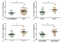
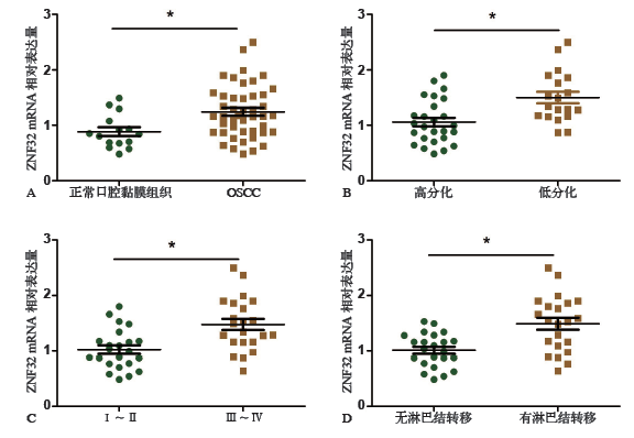
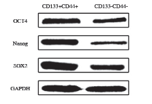
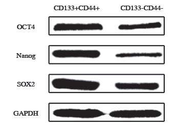
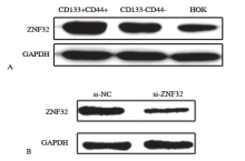
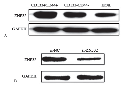
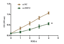
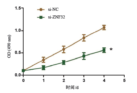
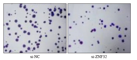
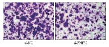
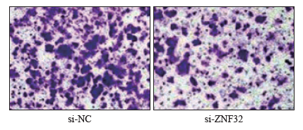
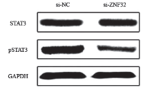
摘要: 目的 研究锌指蛋白32(ZNF32)在口腔鳞状细胞癌(OSCC)组织中的表达及与OSCC患者临床病例特征的关系,并探讨ZNF32对肿瘤干细胞(CSC)生物学特性的影响。 方法 反转录、定量即时聚合酶链反应(qRT-PCR)检测ZNF32 mRNA在45例OSCC组织和15例正常口腔黏膜组织中的表达水平,并分析OSCC组织中ZNF32的表达与临床病例特征的关系。磁珠分选OSCC细胞系Cal-27中的CSC。Western-blot检测有机阳离子/肉毒碱转运蛋白4(OCT4)、Nanog同源框(Nanog)、性别决定区Y框蛋白2(SOX2)干性标志蛋白及ZNF32在CSC中的表达;经脂质体分别转染ZNF32 siRNA(si-ZNF32)和对照(si-NC)48 h后,3-(4,5-二甲基吡啶-2-基)-5-(3-羧基甲氧基苯基)-2-(4-磺苯基)-2H-四唑(MTS)增殖实验检测各组CSC的增殖能力,平板克隆检测各组CSC克隆形成能力,Transwell实验检测各组CSC转移能力,Western-blot检测各组CSC中信号传导转录激活因子3(STAT3)和磷酸化的信号传导转录激活因子3(pSTAT3)蛋白的表达。 结果 qRT-PCR结果显示ZNF32 mRNA在OSCC组织中的表达显著高于正常口腔黏膜组织,其高表达与OSCC肿瘤低分化、TNM分期晚期及淋巴结转移显著相关(P< 0.05)。OCT4、Nanog、SOX2干性标志蛋白在CSC中表达显著增加;ZNF32在CSC中的表达均显著高于OSCC细胞Cal-27和人口腔黏膜角化(HOK)细胞(P<0.05);转染ZNF32 siRNA后,CSC细胞增殖、平板克隆形成及转移能力均下降,CSC细胞中pSTAT3蛋白的表达下降(P<0.05)。 结论 ZNF32在OSCC组织和细胞系中均高表达,且高表达与肿瘤低分化、TNM分期晚期及淋巴结转移相关,同时干扰ZNF32的表达抑制OSCC的CSC生物学特性,ZNF32可以作为治疗OSCC的潜在分子靶点。
中图分类号:
| [1] | Li YY, Xu ZM, Li J , et al. Interleukin-18 expression in oral squamous cell carcinoma: its role in tumor cell migration and invasion, and growth of tumor cell xenografts[J]. FEBS Open Bio, 2018,8(12):1953-1963. |
| [2] | Omura K . Current status of oral cancer treatment strategies: surgical treatments for oral squamous cell carcinoma[J]. Int J Clin Oncol, 2014,19(3):423-430. |
| [3] | Shimomura H, Sasahira T, Nakashima C , et al. Down-regulation of DHRS9 is associated with poor pro-gnosis in oral squamous cell carcinoma[J]. Pathology, 2018,50(6):642-647. |
| [4] | Kansy K, Mueller AA, Mücke T , et al. A worldwide comparison of the management of surgical treatment of advanced oral cancer[J]. J Craniomaxillofac Surg, 2018,46(3):511-520. |
| [5] | Brennan PA, Subramaniam S, Tsioryannis C , et al. An update on the latest evidence for managing the clinically negative neck (cN0) in oral squamous cell carcinoma[J]. Oral Dis, 2017,23(3):287-291. |
| [6] | Ortiz RC, Lopes NM, Amôr NG , et al. CD44 and ALDH1 immunoexpression as prognostic indicators of invasion and metastasis in oral squamous cell carcinoma[J]. J Oral Pathol Med, 2018,47(8):740-747. |
| [7] | Kerk SA, Finkel KA, Pearson AT , et al. 5T4-targeted therapy ablates cancer stem cells and prevents recur-rence of head and neck squamous cell carcinoma[J]. Clin Cancer Res, 2017,23(10):2516-2527. |
| [8] | Chen DM, Wu MS, Li Y , et al. Targeting BMI1 + cancer stem cells overcomes chemoresistance and inhibits metastases in squamous cell carcinoma[J]. Cell Stem Cell, 2017, 20(5): 621-634.e6. |
| [9] | Johansson AC, La Fleur L, Melissaridou S , et al. The relationship between EMT, CD44 high/EGFR low pheno-type, and treatment response in head and neck cancer cell lines [J]. J Oral Pathol Med, 2016,45(9):640-646. |
| [10] | Rhost S, Hughes É, Harrison H , et al. Sortilin inhi-bition limits secretion-induced progranulin-dependent breast cancer progression and cancer stem cell ex-pansion[J]. Breast Cancer Res, 2018,20(1):137. |
| [11] | Li YC, Xu FM, Zhang GQ , et al. Down-regulation of microRNA-21 inhibits cell proliferation and invasion of high-invasion liver cancer stem cells[J]. Eur Rev Med Pharmacol Sci, 2018,22(22):7832-7840. |
| [12] | Makena MR, Ranjan A, Thirumala V , et al. Cancer stem cells: road to therapeutic resistance and strate-gies to overcome resistance[J]. Biochim Biophys Acta Mol Basis Dis, 2018. doi: 10.1016/j.bbadis.2018.11.015. |
| [13] | Cannizzaro LA, Aronson MM, Thiesen HJ . Human zinc finger gene ZNF23 (Kox16) maps to a zinc finger gene cluster on chromosome 16q22, and ZNF32 (Kox30) to chromosome region 10q23-q24[J]. Hum Genet, 1993,91(4):383-385. |
| [14] | Li J, Ao J, Li K , et al. ZNF32 contributes to the in-duction of multidrug resistance by regulating TGF-β receptor 2 signaling in lung adenocarcinoma[J]. Cell Death Dis, 2016,7(10):e2428. |
| [15] | Li K, Gao B, Li J , et al. ZNF32 protects against oxidative stress-induced apoptosis by modulating C1QBP transcription[J]. Oncotarget, 2015,6(35):38107-38126. |
| [16] | Li YY, Gong D, Zhang L , et al. Zinc finger protein 32 promotes breast cancer stem cell-like properties through directly promoting GPER transcription[J]. Cell Death Dis, 2018,9(12):1162. |
| [17] | Gu W, Yeo E, McMillan N, et al. Silencing oncogene expression in cervical cancer stem-like cells inhibits their cell growth and self-renewal ability[J]. Cancer Gene Ther, 2011,18(12):897-905. |
| [18] | Siegel RL, Miller KD, Jemal A . Cancer statistics, 2018[J]. CA Cancer J Clin, 2018,68(1):7-30. |
| [19] | 王倩, 侯大为 . 口腔鳞状细胞癌发病及转移机制研究进展[J]. 口腔医学研究, 2018,34(11):1164-1167. |
| Wang Q, Hou DW . Research progress in pathogenesis of oral squamous cell carcinoma[J]. J Oral Sci Res, 2018,34(11):1164-1167. | |
| [20] | Haranadh S, Nandyala R, Bodagala V , et al. A pros-pective analysis of prevalence of metastasis in levels ⅡB and Ⅴ neck nodes in patients with operable oral squamous cell carcinoma[J]. Oral Oncol, 2018,83:115-119. |
| [21] | Ettinger KS, Ganry L, Fernandes RP . Oral cavity cancer[J]. Oral Maxillofac Surg Clin North Am, 2019,31(1):13-29. |
| [22] | Chen L, Li YC, Wu L , et al. TRAF6 regulates tumour metastasis through EMT and CSC phenotypes in head and neck squamous cell carcinoma[J]. J Cell Mol Med, 2018,22(2):1337-1349. |
| [23] | Rodrigues MFSD, Miguita L, De Andrade NP , et al. GLI3 knockdown decreases stemness, cell proli-feration and invasion in oral squamous cell carcinoma[J]. Int J Oncol, 2018,53(6):2458-2472. |
| [24] | Wei YY, Li K, Yao SH , et al. Loss of ZNF32 augments the regeneration of nervous lateral line system through negative regulation of SOX2 transcription[J]. Oncotarget, 2016,7(43):70420-70436. |
| [25] | Li K, Zhao G, Ao J , et al. ZNF32 induces anoikis resistance through maintaining redox homeostasis and activating Src/FAK signaling in hepatocellular carcinoma[J]. Cancer Lett, 2019,442:271-278. |
| [26] | Li YY, Zhang L, Li K , et al. ZNF32 inhibits auto-phagy through the mTOR pathway and protects MCF-7 cells from stimulus-induced cell death[J]. Sci Rep, 2015,5:9288. |
| [27] | 尹彦斌, 王建杰, 刘祎 , 等. 肿瘤干细胞分离研究进展[J]. 现代生物医学进展, 2016,16(2):382-385. |
| Yin YB, Wang JJ, Liu Y , et al. Progress in isolation of cancer stem cells[J]. Prog Mod Biomed, 2016,16(2):382-385. | |
| [28] | 俞晓毓, 吴迪, 王净 , 等. 卵巢癌细胞系ID8中肿瘤干细胞的分离及生物学特性鉴定[J]. 中国组织工程研究, 2018,22(29):4687-4691. |
| Yu XY, Wu D, Wang J , et al. Isolation and biological identification of tumor stem cells from ovarian cancer ID8 cell lines[J]. Chin J Tissue Eng Res, 2018,22(29):4687-4691. | |
| [29] | de Moraes FP, Lourenço SV, Ianez RC , et al. Expre-ssion of stem cell markers in oral cavity and oro-pharynx squamous cell carcinoma[J]. Oral Surg Oral Med Oral Pathol Oral Radiol, 2017,123(1):113-122. |
| [30] | Kaseb HO, Fohrer-Ting H, Lewis DW , et al. Identi-fication, expansion and characterization of cancer cells with stem cell properties from head and neck squamous cell carcinomas[J]. Exp Cell Res, 2016,348(1):75-86. |
| [31] | Zhang ZL, Han H, Rong YP , et al. Hypoxia poten-tiates gemcitabine-induced stemness in pancreatic cancer cells through AKT/Notch1 signaling[J]. J Exp Clin Cancer Res, 2018,37(1):291. |
| [32] | Qin T, Li B, Feng XY , et al. Abnormally elevated USP37 expression in breast cancer stem cells re-gulates stemness, epithelial-mesenchymal transition and cisplatin sensitivity[J]. J Exp Clin Cancer Res, 2018,37(1):287. |
| [33] | 赵晓晚, 范凯华, 李中悦 , 等. STAT3和EMT与肿瘤的关系[J]. 甘肃医药, 2018,37(9):779-781. |
| Zhao XW, Fan KH, Li ZY , et al. The relationship between STAT3, EMT and tumor[J]. Gansu Med J, 2018,37(9):779-781. | |
| [34] | Tong MT, Wang J, Jiang NN , et al. Correlation between p-STAT3 overexpression and prognosis in lung cancer: a systematic review and meta-analysis[J]. PLoS One, 2017,12(8):e0182282. |
| [35] | 朱正春, 姚梦群, 严芳莉 , 等. 西黄丸对肝癌HepG2细胞增殖、侵袭及STAT3信号通路的影响[J]. 安徽医科大学学报, 2018,53(12):1875-1878. |
| Zhu ZC, Yao MQ, Yan FL , et al. Effects of Xihuang pill on proliferation and invasion of HepG2 cells and STAT3 signal pathway[J]. Acta Univ Med Anhui, 2018,53(12):1875-1878. | |
| [36] | Rios-Fuller TJ, Ortiz-Soto G, Lacourt-Ventura M , et al. Ganoderma lucidum extract (GLE) impairs breast cancer stem cells by targeting the STAT3 pathway[J]. Oncotarget, 2018,9(89):35907-35921. |
| [1] | 周金阔,张晋弘,史晓晶,刘广顺,姜磊,刘倩峰. 长链非编码RNA小核仁RNA宿主基因22调控微小RNA-27b-3p对口腔鳞状细胞癌细胞增殖、侵袭和迁移的影响[J]. 国际口腔医学杂志, 2024, 51(1): 52-59. |
| [2] | 李立恒,王蕊,王晓明,张智轶,张璇,安峰,王芹,张凡. 环状RNA hsa_circ_0085576调控微小RNA-498/B细胞特异性莫洛尼鼠白血病病毒整合位点1轴对口腔鳞状细胞癌细胞迁移和侵袭的影响[J]. 国际口腔医学杂志, 2024, 51(1): 60-67. |
| [3] | 吴佳敏,夏斌,杨禾丰,许彪. 癌相关成纤维细胞在口腔鳞状细胞癌微环境中作用的研究进展[J]. 国际口腔医学杂志, 2023, 50(6): 711-717. |
| [4] | 柳江龙, 买买提吐逊·吐尔地. 超声造影在口腔鳞状细胞癌颈部转移性淋巴结诊断中的研究进展[J]. 国际口腔医学杂志, 2023, 50(5): 514-520. |
| [5] | 盛南宁,王珏,南欣荣. 性别决定基因盒9在口腔鳞状细胞癌作用机制和治疗中的研究进展[J]. 国际口腔医学杂志, 2023, 50(3): 314-320. |
| [6] | 李潭,梁新华. 盘状蛋白结构域受体1在调控恶性肿瘤进展和治疗中的作用[J]. 国际口腔医学杂志, 2023, 50(2): 230-236. |
| [7] | 赵卓平,辛鹏飞,高阳,张彩凤,张宽收,刘青梅. 光热治疗在口腔鳞状细胞癌治疗中的研究进展[J]. 国际口腔医学杂志, 2022, 49(4): 462-470. |
| [8] | 江涵,神应强,陈谦明. 毒蕈碱受体通过Yes相关蛋白信号对口腔鳞状细胞癌生物学行为的实验研究[J]. 国际口腔医学杂志, 2022, 49(2): 138-143. |
| [9] | 蒋宇磊,夏斌,饶南荃,杨禾丰,许彪. 外泌体在口腔鳞状细胞癌恶性进展及诊疗应用的研究[J]. 国际口腔医学杂志, 2021, 48(6): 711-717. |
| [10] | 甘建国,高攀,王晓毅. 循环肿瘤细胞与口腔鳞状细胞癌相关性的研究进展[J]. 国际口腔医学杂志, 2021, 48(2): 205-212. |
| [11] | 黄俊文,乔洁,梅子,陈茁,李杨,乔彬. 脂多糖结合蛋白在口腔鳞状细胞癌中的表达及其临床意义[J]. 国际口腔医学杂志, 2021, 48(1): 50-57. |
| [12] | 何宇晴,但红霞,陈谦明. 光动力疗法在口腔黏膜癌变防治中的应用[J]. 国际口腔医学杂志, 2020, 47(6): 669-676. |
| [13] | 郝福,宁毅,孙睿,郑晓旭. 口腔鳞状细胞癌中转化因子2β的表达及潜在的临床意义[J]. 国际口腔医学杂志, 2020, 47(2): 159-165. |
| [14] | 薛伶俐,李雅冬. 经首次根治性手术治疗口腔鳞状细胞癌患者的生存相关影响因素分析[J]. 国际口腔医学杂志, 2020, 47(2): 166-174. |
| [15] | 董云梅,陶艳,周瑜. 口腔黏膜癌变过程中血清生化标志物的研究进展[J]. 国际口腔医学杂志, 2020, 47(1): 43-50. |
|