国际口腔医学杂志 ›› 2025, Vol. 52 ›› Issue (6): 713-721.doi: 10.7518/gjkq.2025098
王诗雅1( ),袁国华2,邹静1()
Shiya Wang1(),Guohua Yuan2,Jing Zou1()
摘要:
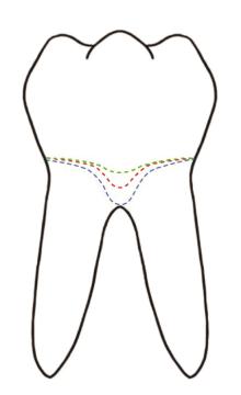
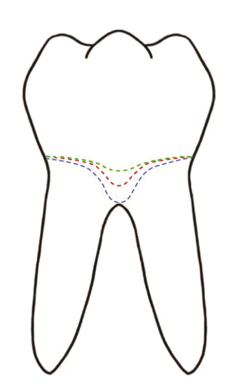
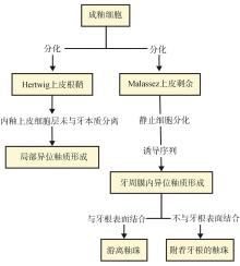
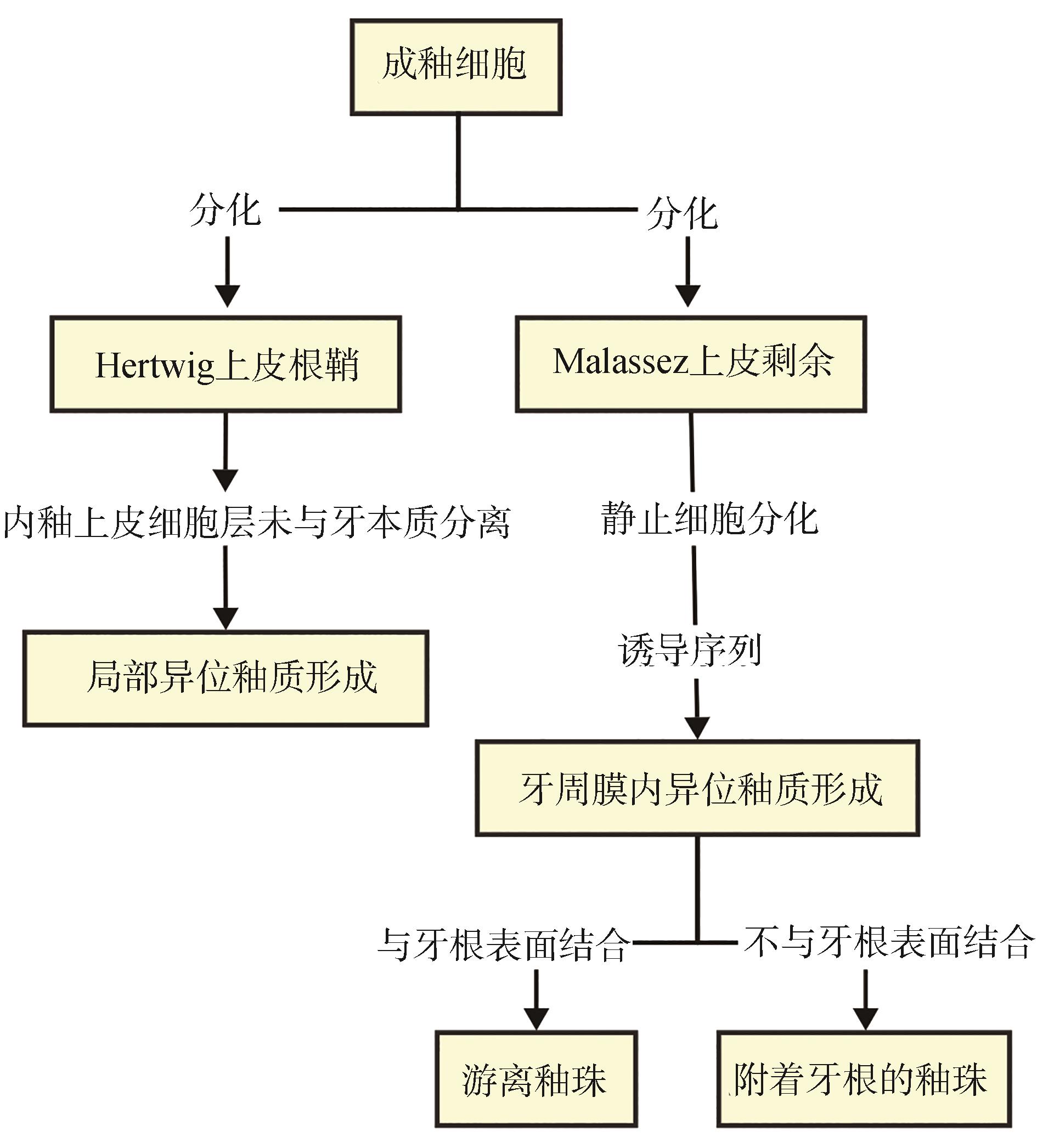
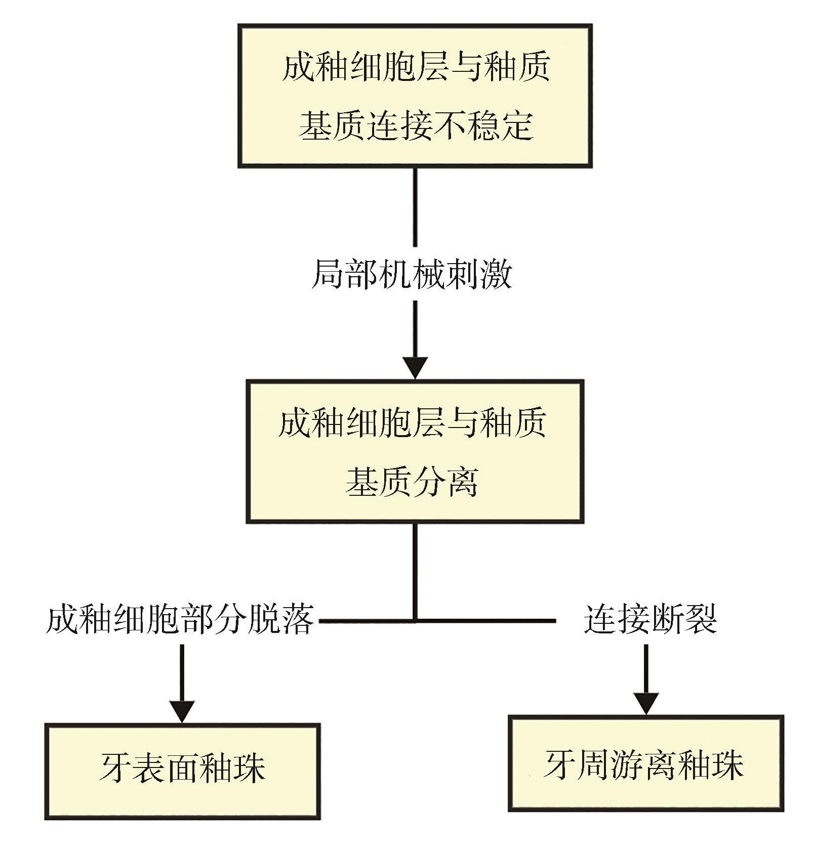
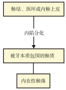
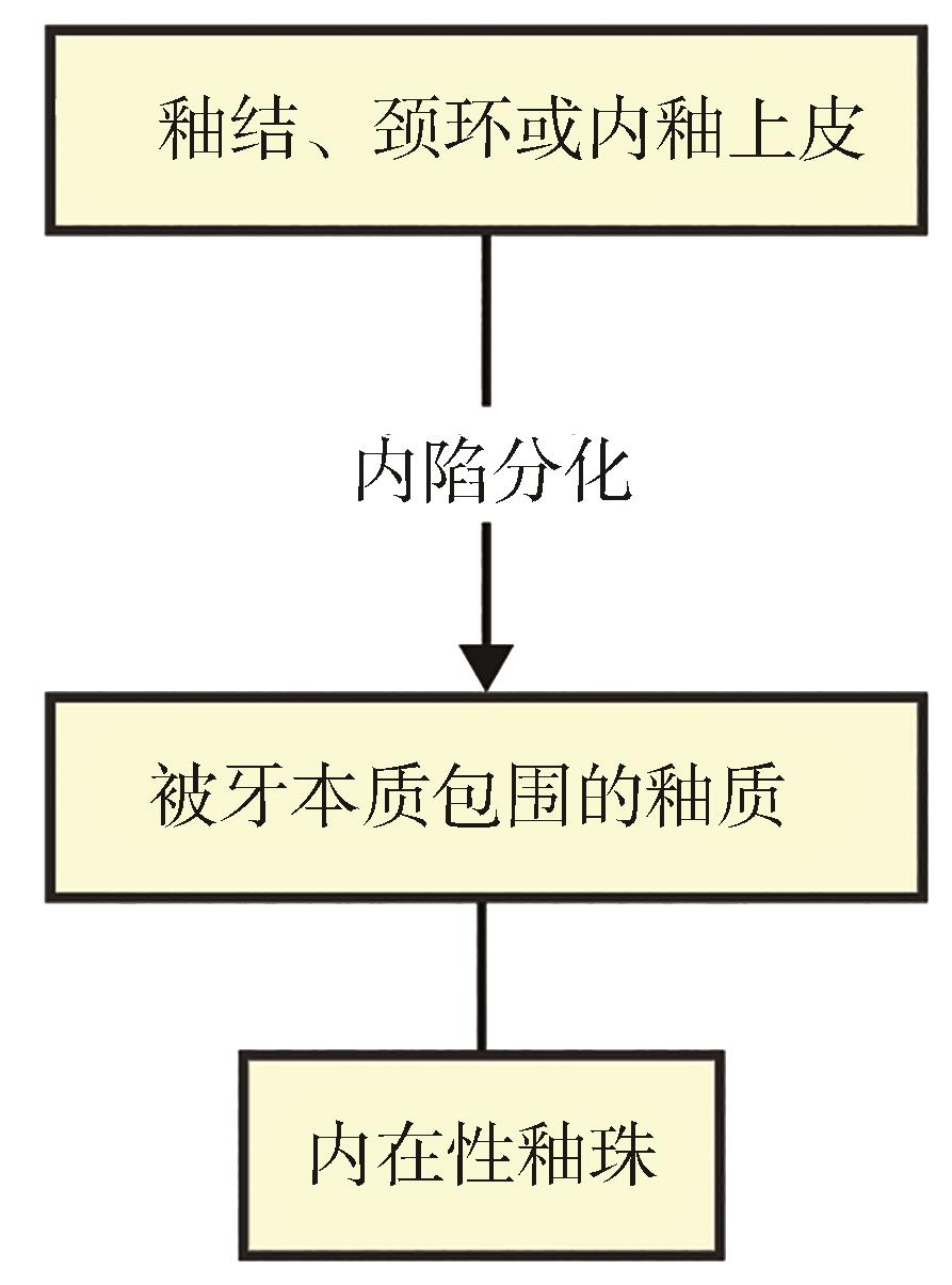
异位釉质是一种牙齿发育异常,分为釉珠和颈部釉质突起两类。异位釉质的形成可能与Hertwig上皮根鞘和Malassez 上皮剩余中的成釉细胞有关,机械因素和遗传因素也可能发挥作用。影像学检查可用于识别异位釉质、评估病情和指导临床诊疗。本文通过对异位釉质的临床特点、形成机制和诊疗策略进行综述,以期为异位釉质的临床诊疗提供参考和临床依据。
中图分类号:
| [1] | Marsico C, Grimm JR, Renteria C, et al. Characte-rizing the microstructures of mammalian enamel by synchrotron phase contrast microCT[J]. Acta Biomater, 2024, 178: 208-220. |
| [2] | Wei R, Guo S, Meng Z, et al. Mediator1 involved in functional integration of Smad3 and Notch1 promo-ting enamel mineralization[J]. Biochem Biophys Res Commun, 2023, 663: 47-53. |
| [3] | Saini T, Ogunleye A, Levering N, et al. Multiple enamel pearls in two siblings detected by volume-tric computed tomography[J]. Dentomaxillofac Radiol, 2008, 37(4): 240-244. |
| [4] | Grine FE, Holt S, Brink JS, et al. Enamel pearls: their occurrence in recent human populations and earliest manifestation in the modern human lineage[J]. Arch Oral Biol, 2019, 101: 147-155. |
| [5] | Zengin AZ, Sumer AP, Ozturk G, et al. Imaging characteristics of enamel pearls on CBCT and their co-relation with supernumerary tooth[J]. Oral Ra-diol, 2022, 38(3): 370-377. |
| [6] | Cavanha AO. Enamel pearls[J]. Oral Surg Oral Med Oral Pathol, 1965, 19: 373-382. |
| [7] | Kaugars GE. Internal enamel pearls: report of case[J]. J Am Dent Assoc, 1983, 107(6): 941-943. |
| [8] | Akgül N, Caglayan F, Durna N, et al. Evaluation of enamel pearls by cone-beam computed tomography (CBCT)[J]. Med Oral Patol Oral Cir Bucal, 2012, 17(2): e218-e222. |
| [9] | Hou GL, Tsai CC. Cervical enamel projection and intermediate bifurcational ridge correlated with molar furcation involvements[J]. J Periodontol, 1997, 68(7): 687-693. |
| [10] | Risnes S, Segura JJ, Casado A, et al. Enamel pearls and cervical enamel projections on 2 maxillary molars with localized periodontal disease: case report and histologic study[J]. Oral Surg Oral Med Oral Pathol Oral Radiol Endod, 2000, 89(4): 493-497. |
| [11] | Pedersen PO. The East Greenland Eskimo dentition, numerical variations and anatomy[M]. Copenhagen: Biance Lunos Press, 1949. |
| [12] | Moskow BS, Canut PM. Studies on root enamel (2). Enamel pearls. A review of their morphology, loca-lization, nomenclature, occurrence, classification, his-togenesis and incidence[J]. J Clin Periodontol, 1990, 17(5): 275-281. |
| [13] | Darwazeh A, Hamasha AA. Radiographic evidence of enamel pearls in jordanian dental patients[J]. Oral Surg Oral Med Oral Pathol Oral Radiol Endod, 2000, 89(2): 255-258. |
| [14] | Loh HS. A local study on enamel pearls[J]. Singapore Dent J, 1980, 5(1): 55-59. |
| [15] | Gašperšič D. Histogenetic aspects of the composition and structure of human ectopic enamel, studied by scanning electron microscopy[J]. Arch Oral Biol, 1992, 37(8): 603-611. |
| [16] | Kaminagakura E, Salmon CR, Fonseca DC, et al. Prevalence and microscopic features of enamel pearls from permanent human molars[J]. Braz J Oral Sci, 2011, 10(4): 268-271. |
| [17] | Anderson P, Elliott JC, Bose U, et al. A comparison of the mineral content of enamel and dentine in human premolars and enamel pearls measured by X-ray microtomography[J]. Arch Oral Biol, 1996, 41(3): 281-290. |
| [18] | Gašperšič D. Enamel microhardness and histological features of composite enamel pearls of different size[J]. J Oral Pathol Med, 1995, 24(4): 153-158. |
| [19] | Kim HJ, Kim SY. Cervical enamel projections from a periodontal perspective: a scoping review[J]. Clin Anat, 2024, 37(3): 353-365. |
| [20] | Marshall G, Verdelis K, Peters OA. Morphology of pulpal mineralizations: a scoping review[J]. J Dent, 2023, 139: 104745. |
| [21] | Swan RH, Hurt WC. Cervical enamel projections as an etiologic factor in furcation involvement[J]. J Am Dent Assoc, 1976, 93(2): 342-345. |
| [22] | Ko MJ, Cho CM, Jeong SN. Characteristics of the molar surface after removal of cervical enamel projections: comparison of three different rotating instruments[J]. J Periodontal Implant Sci, 2016, 46(2): 107-115. |
| [23] | Bissada NF, Abdelmalek RG. Incidence of cervical enamel projections and its relationship to furcation involvement in Egyptian skulls[J]. J Periodontol, 1973, 44(9): 583-585. |
| [24] | Blanchard SB, Derderian GM, Averitt TR, et al. Cervical enamel projections and associated pouch-like opening in mandibular furcations[J]. J Periodontol, 2012, 83(2): 198-203. |
| [25] | Kalnins V. Origin of enamel drops and cementicles in the teeth of rodents[J]. J Dent Res, 1952, 31(4): 582-590. |
| [26] | Bower RC. Furcation development of human mandibular first molar teeth. A histologic graphic reconstructional study[J]. J Periodontal Res, 1983, 18(4): 412-419. |
| [27] | Shroff NP, Xu P, Kim S, et al. Proliferation-driven mechanical compression induces signalling centre formation during mammalian organ development[J]. Nat Cell Biol, 2024, 26(4): 519-529. |
| [28] | Kim EJ, Kim HY, Li L, et al. Cuspal shape alterations by bmp4 directing cell proliferation and apoptosis[J]. J Dent Res, 2023, 102(7): 825-834. |
| [29] | Germen M, Baser U, Lacin CC, et al. Periodontitis prevalence, severity, and risk factors: a comparison of the AAP/CDC case definition and the EFP/AAP classification[J]. Int J Environ Res Public Health, 2021, 18(7): 3459. |
| [30] | Emery DC, Cerajewska TL, Seong J, et al. Comparison of blood bacterial communities in periodontal health and periodontal disease[J]. Front Cell Infect Microbiol, 2021, 10: 577485. |
| [31] | Aleksijević LH, Aleksijević M, Škrlec I, et al. Porphyromonas gingivalis virulence factors and clinical significance in periodontal disease and coronary artery diseases[J]. Pathogens, 2022, 11(10): 1173. |
| [32] | Wang BY, Cao A, Ho MH, et al. Identification of microbiological factors associated with pe-riodontal health disparities[J]. Front Cell Infect Microbiol, 2023, 13: 1137067. |
| [33] | Shahoumi LA, Saleh MHA, Meghil MM. Virulence factors of the periodontal pathogens: tools to evade the host immune response and promote carcinogenesis[J]. Microorganisms, 2023, 11(1): 115. |
| [34] | Matthews DC, Tabesh M. Detection of localized tooth-related factors that predispose to periodontal infections[J]. Periodontol 2000, 2004, 34: 136-150. |
| [35] | Atkinson SR. Changing dynamics of the growing face[J]. Am J Orthod, 1949, 35(11): 815-836. |
| [36] | Masters DH, Hoskins Jr SW. Projection of cervical enamel into molar furcations[J]. J Periodontol, 1964, 35(1): 49-53. |
| [37] | Hou GL, Tsai CC. Relationship between periodontal furcation involvement and molar cervical enamel projections[J]. J Periodontol, 1987, 58(10): 715-721. |
| [38] | Machtei EE, Wasenstein SM, Peretz B, et al. The relationship between cervical enamel projection and class Ⅱ furcation defects in humans[J]. Quintessence Int, 1997, 28(5): 315-320. |
| [39] | Chan HL, Oh TJ, Bashutski J, et al. Cervical enamel projections in unusual locations: a case report and mini-review[J]. J Periodontol, 2010, 81(5): 789-795. |
| [40] | Goldstein AR. Enamel pearls as contributing factor in periodontal breakdown[J]. J Am Dent Assoc, 1979, 99(2): 210-211. |
| [41] | Kvaratsthelia S. Prevalence of dentition, dental ar-ches and dental anomalies[J]. Georgian Med News, 2024(347): 177-180. |
| [42] | Hou L, Acharya K, Ghimire B, et al. Clinical and imaging analysis of 22 cases of supernumerary teeth in the mandibular region[J]. J Stomatol Oral Maxillofac Surg, 2024, 125(4S): 101525. |
| [43] | AlHadidi A, Lam PPY, Hassona Y. Developmental and acquired abnormalities of the teeth[J]. Dent Clin North Am, 2024, 68(2): 227-245. |
| [44] | Lisman D, Drath J, Zielińska G, et al. The evidential value of dental calculus in the identification process[J]. Sci Rep, 2023, 13(1): 21666. |
| [45] | Li Q, Luo K, Su Z, et al. Dental calculus: a repository of bioinformation indicating diseases and human evolution[J]. Front Cell Infect Microbiol, 2022, 12: 1035324. |
| [46] | Kao MC, Lin CL, Kung CY, et al. Miniature endoscopic optical coherence tomography for calculus detection[J]. Appl Opt, 2015, 54(24): 7419-7423. |
| [47] | Hsiao TY, Ho YC, Chen MR, et al. Disease activation maps for subgingival dental calculus identification based on intelligent dental optical coherence tomography[J]. Transl Biophotonics, 2021, 3: e202100-001. |
| [48] | Ishikawa M, Kanzaki H, Kodera R, et al. Early diagnosis of aortic calcification through dental X-ray examination for dental pulp stones[J]. Sci Rep, 2023, 13(1): 18576. |
| [49] | Zahran SS, Alamoudi RA. Radiographic evaluation of teeth with pulp stones and pulp canal obliteration: characteristics, and associations with dental parameters[J]. Libyan J Med, 2024, 19(1): 2306768. |
| [50] | Jolivet G, Huck O, Petit C. Evaluation of furcation involvement with diagnostic imaging methods: a systematic review[J]. Dentomaxillofac Radiol, 2022, 51(8): 20210529. |
| [51] | 邢攀峰, 孙俊毅. CBCT在根分叉病变诊疗中的准确性和实用性—文献系统性回顾: 2020年中华口腔医学会口腔颌面放射专业委员会第十八次全国口腔颌面医学影像学专题研讨会论文汇编[C]. 北京: 中华口腔医学会, 2020. |
| Xing PF, Sun JY. Accuracy and practicality of CBCT in the diagnosis and treatment of root bifurcation lesions—a systematic review of literature: Compilation of Papers from the 18th National Symposium on Oral and Maxillofacial Medical Imaging organized by the Oral and Maxillofacial Radiology Professional Committee of the Chinese Stomatological Association in 2020[C]. Beijing: Chinese Stomatological Association, 2020. | |
| [52] | Tsao YP, Neiva R, Al-Shammari K, et al. Factors influencing treatment outcomes in mandibular Class Ⅱ furcation defects[J]. J Periodontol, 2006, 77(4): 641-646. |
| [53] | Mukherjee M, Nair V, Phull T, et al. Biometric analysis of furcation area of molar teeth and its relationship with instrumentation[J]. BMC Oral Health, 2024, 24(1): 436. |
| [54] | Roussa E. Anatomic characteristics of the furcation and root surfaces of molar teeth and their significance in the clinical management of marginal pe-riodontitis[J]. Clin Anat, 1998, 11(3): 177-186. |
| [1] | 陈禹黄,梁星,李然. 牙周炎患者缺失牙修复的临床考量及预后评估[J]. 国际口腔医学杂志, 2025, 52(6): 823-831. |
| [2] | 朱然,严静,孙卫斌,吴文蕾,刘玉. 服用抗血栓药物患者牙周基础治疗期间的出血风险管理[J]. 国际口腔医学杂志, 2025, 52(5): 670-676. |
| [3] | 李欢,原韶钟. 牙周炎与非酒精性脂肪性肝病相关性的研究进展[J]. 国际口腔医学杂志, 2025, 52(5): 677-683. |
| [4] | 祝舒钰,周静,谢志刚. 辅助性T细胞17与调节性T细胞之间的制衡效应调控口腔颌面部骨损伤修复的研究进展[J]. 国际口腔医学杂志, 2025, 52(4): 514-525. |
| [5] | 于寰,康健. 牙周内镜在口腔临床诊疗中的应用进展[J]. 国际口腔医学杂志, 2025, 52(4): 544-551. |
| [6] | 李天元, 朱彤欣, 柳庆, 董迎春, 陈斌. 间充质干细胞用于牙周再生临床疗效的系统评价与Meta分析[J]. 国际口腔医学杂志, 2025, 52(3): 296-307. |
| [7] | 别梦瑶,周婕妤,吴亚菲,赵蕾. 牙龈卟啉单胞菌影响血管平滑肌细胞调节性细胞死亡及表型转换的研究进展[J]. 国际口腔医学杂志, 2025, 52(3): 308-316. |
| [8] | 范兴丽,潘乐,赵家园,项秋猛,陈启林. 竞争性内源性RNA在牙周炎中的作用及机制的研究进展[J]. 国际口腔医学杂志, 2025, 52(3): 317-322. |
| [9] | 赵美林,赵依琼,黄姣. Ⅲ期C级牙周炎正畸患者上前牙龈乳头缺陷伴牙龈退缩1例[J]. 国际口腔医学杂志, 2025, 52(3): 333-340. |
| [10] | 李晶,康健. 牙周微创手术中再生材料选择及疗效的研究进展[J]. 国际口腔医学杂志, 2025, 52(2): 161-168. |
| [11] | 张潇月,陈舒泽,周婕妤,程磊,赵蕾. 具核梭杆菌经铁死亡途径破坏体外肠道上皮屏障模型的研究[J]. 国际口腔医学杂志, 2025, 52(2): 183-194. |
| [12] | 钟良军. 数字化技术在重度牙周炎治疗中的应用[J]. 国际口腔医学杂志, 2025, 52(1): 1-10. |
| [13] | 程守正,李太文,赵蕾. 血清淀粉样蛋白A与牙周炎相关性的研究进展[J]. 国际口腔医学杂志, 2025, 52(1): 117-122. |
| [14] | 陈梦洁,徐文华,刘青青,康毓聃,刘蓉,朱丽雷. 全身免疫炎症指数与牙周炎患者分级诊断的相关性研究[J]. 国际口腔医学杂志, 2024, 51(6): 706-712. |
| [15] | 陈蕊,范桢,郝春波. 黑色素瘤缺乏因子2炎症小体在牙周炎及糖尿病中的研究进展[J]. 国际口腔医学杂志, 2024, 51(6): 763-771. |
|

 畸形远期疗效稳定性的研究进展