国际口腔医学杂志 ›› 2025, Vol. 52 ›› Issue (1): 1-10.doi: 10.7518/gjkq.2025001
• 专家笔谈 • 下一篇
钟良军( )
)
Liangjun Zhong()
摘要:
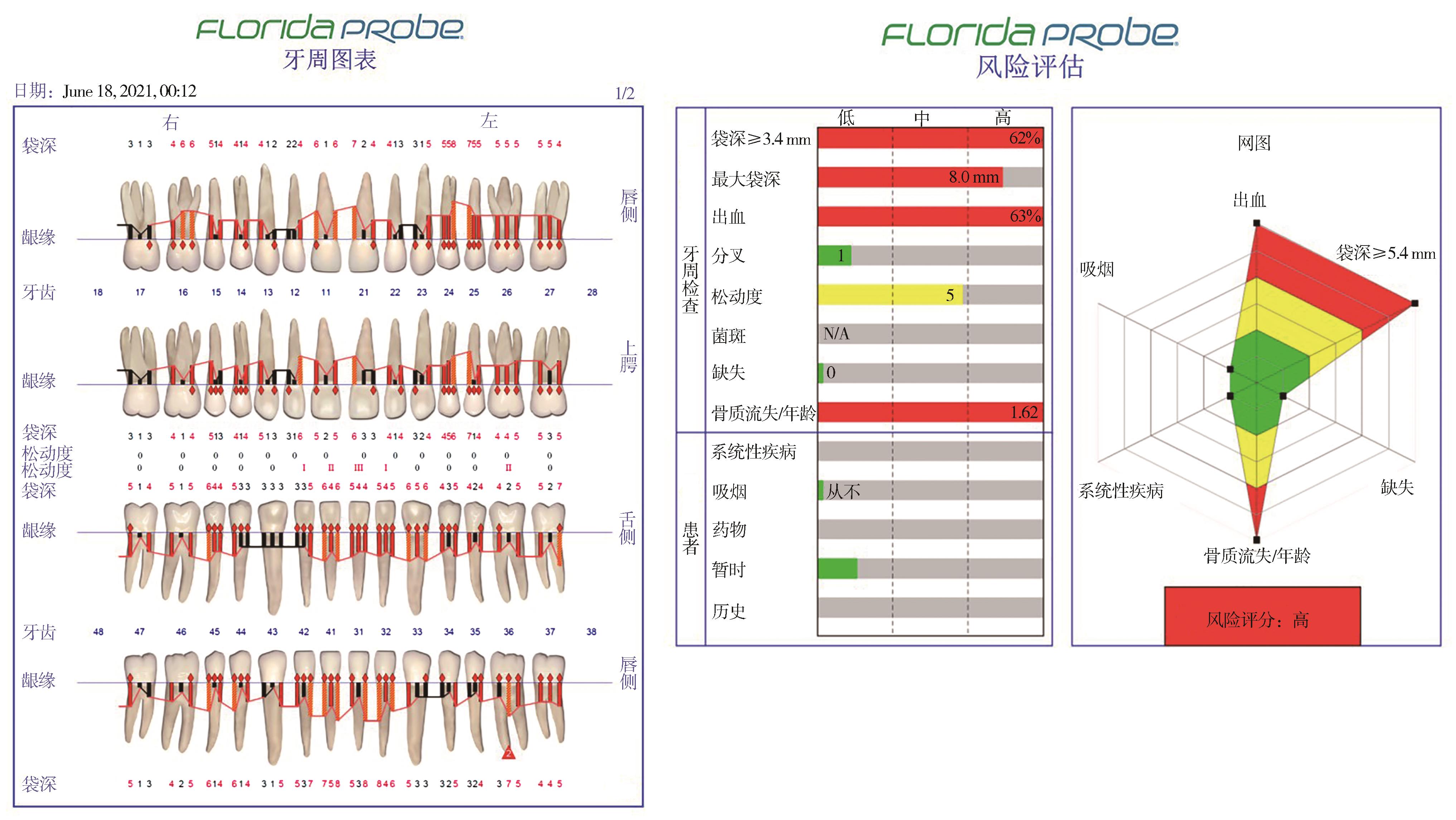
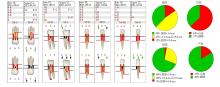
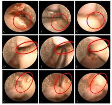
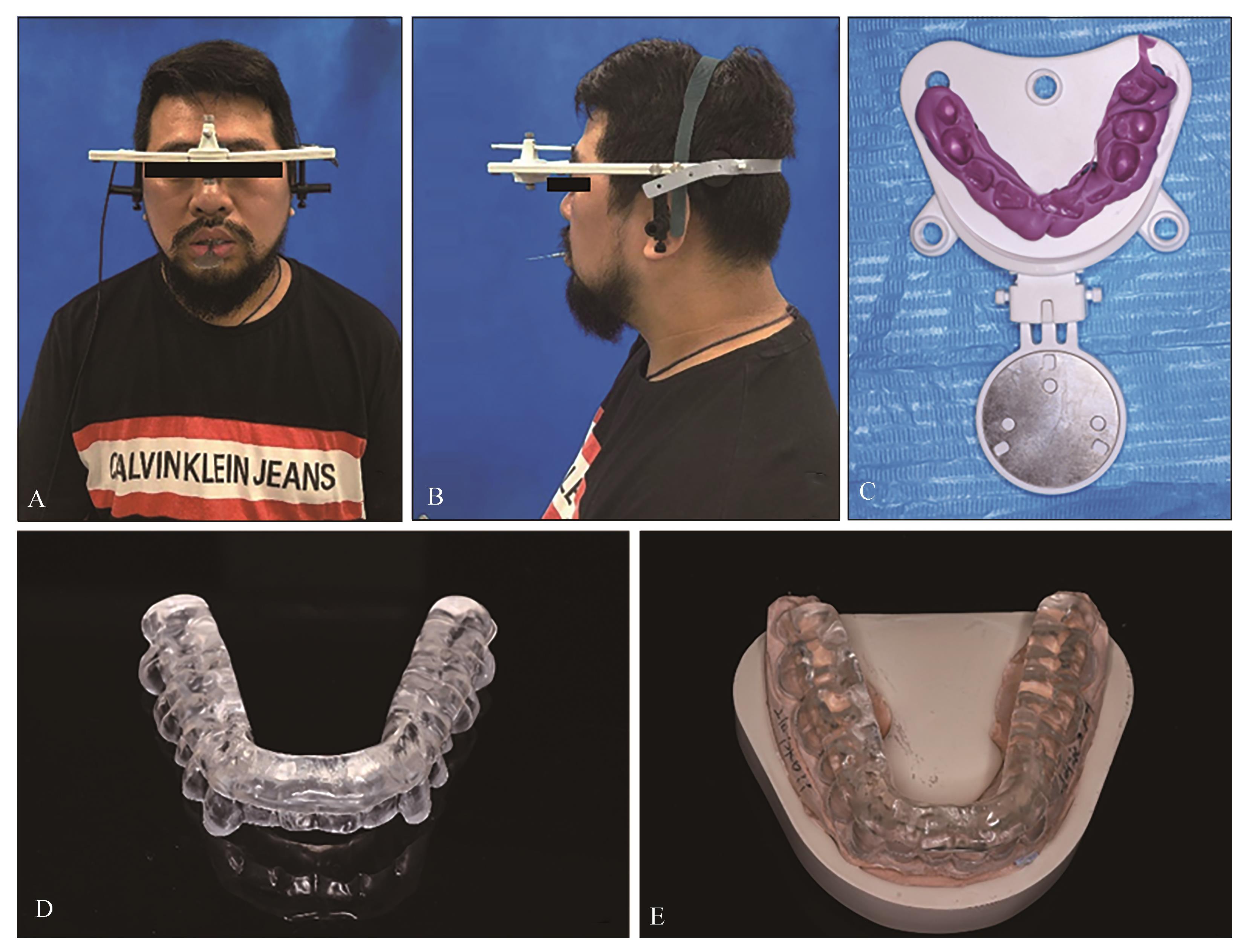
重度牙周炎损害牙齿支持组织的健康并影响咀嚼功能,甚至导致牙列缺损或缺失。在重度牙周炎的诊疗中,数字化技术的应用可以提高诊疗的精准性和高效性,为患者提供了更优的治疗效果和诊疗体验。目前口腔临床常用的数字化技术手段和器械包括数字化牙周电子探针、牙周内镜、数字化口腔扫描和3D打印、数字化电子咬合记录仪、电子面弓等。本文就上述几种数字化技术在重度牙周炎诊疗中,从临床检查、诊断、治疗规划到治疗实施等各个环节的应用进行阐述。
中图分类号:
| 1 | Khan SA, Kong EF, Meiller TF, et al. Periodontal diseases: bug induced, host promoted[J]. PLoS Pathog, 2015, 11(7): e1004952. |
| 2 | Tonetti MS, Greenwell H, Kornman KS. Staging and grading of periodontitis: framework and proposal of a new classification and case definition[J]. J Periodontol, 2018, 89(): S159-S172. |
| 3 | Pihlstrom BL, Michalowicz BS, Johnson NW. Perio-dontal diseases[J]. Lancet, 2005, 366(9499): 1809-1820. |
| 4 | Agrawal P, Nikhade P. Artificial intelligence in dentistry: past, present, and future[J]. Cureus, 2022, 14(7): e27405. |
| 5 |
Haidar ZS. Digital dentistry: past, present, and future[J]. Digit Med Healthcare Technol, 2023. doi:10.5772/dmht.17 .
doi: 10.5772/dmht.17 |
| 6 | Rekow ED. Digital dentistry: the new state of the art-is it disruptive or destructive[J]. Dent Mater, 2020, 36(1): 9-24. |
| 7 | Dhopte A, Bagde H. Smart smile: revolutionizing dentistry with artificial intelligence[J]. Cureus, 2023, 15(6): e41227. |
| 8 | Schierz O, Hirsch C, Krey KF, et al. Digital dentis-try and its impact on oral health-related quality of life[J]. J Evid Based Dent Pract, 2024, 24(1S): 101946. |
| 9 | Al Shayeb KNA, Turner W, Gillam DG. Periodontal probing: a review[J]. Prim Dent J, 2014, 3(3): 25-29. |
| 10 | Polson AM, Caton JG, Yeaple RN, et al. Histological determination of probe tip penetration into gingival sulcus of humans using an electronic pressure-sensitive probe[J]. J Clin Periodontol, 1980, 7(6): 479-488. |
| 11 | Koirala PK, Pradhan S. Gold standards in periodontics: a review[J]. J Nepal Soc Perio Oral Implantology, 2021, 5(1): 49-53. |
| 12 | Ko TJ, Byrd KM, Kim SA. The chairside periodontal diagnostic toolkit: past, present, and future[J]. Dia-gnostics (Basel), 2021, 11(6): 932. |
| 13 | Renatus A, Trentzsch L, Schönfelder A, et al. Evalua-tion of an electronic periodontal probe versus a ma-nual probe[J]. J Clin Diagn Res, 2016, 10(11): ZH03-ZH07. |
| 14 |
Salvi GE, Roccuzzo A, Imber JC, et al. Clinical perio-dontal diagnosis[J]. Periodontol 2000, 2023. doi: 10.1111/prd.12487 .
doi: 10.1111/prd.12487 |
| 15 | Elashiry M, Meghil MM, Arce RM, et al. From manual periodontal probing to digital 3-D imaging to endoscopic capillaroscopy: recent advances in periodontal disease diagnosis[J]. J Periodontal Res, 2019, 54(1): 1-9. |
| 16 | Kulkarni MR. Digitization in periodontics[M]//Jain P, Gupta M. Digitization in dentistry. Cham: Sprin-ger International Publishing, 2021: 305-333. |
| 17 |
Shakibaie F, Walsh L. Optical diagnostics to improve periodontal diagnosis and treatment[M]//Perio-dontology and dental implantology. Intech Open, 2019. doi:10.5772/intechopen.76888 .
doi: 10.5772/intechopen.76888 |
| 18 | Wright HN, Mayer ET, Lallier TE, et al. Utilization of a periodontal endoscope in nonsurgical periodontal therapy: a randomized, split-mouth clinical trial[J]. J Periodontol, 2023, 94(8): 933-943. |
| 19 | Wu J, Lin LY, Xiao JP, et al. Efficacy of scaling and root planning with periodontal endoscopy for resi-dual pockets in the treatment of chronic periodontitis: a randomized controlled clinical trial[J]. Clin Oral Investig, 2022, 26(1): 513-521. |
| 20 | Ardila CM, Vivares-Builes AM. Efficacy of perio-dontal endoscopy during subgingival debridement to treat periodontitis: a systematic review of randomized clinical trials[J]. Dent J, 2023, 11(5): 112. |
| 21 | Shi JH, Wang JM, Yang ZY, et al. A novel periodontal endoscopy-aided non-incisional periodontal regeneration technique in the treatment of intrabony defects: a retrospective cohort study[J]. BMC Oral Health, 2023, 23(1): 962. |
| 22 | Graetz C, Schorr S, Christofzik D, et al. How to train periodontal endoscopy? Results of a pilot study removing simulated hard deposits in vitro [J]. Clin Oral Investig, 2020, 24(2): 607-617. |
| 23 | Pauwels R. History of dental radiography: evolution of 2D and 3D imaging modalities[J]. Med Phys Int, 2020, 8(1): 235-77. |
| 24 | Haleem A, Javaid M. 3D scanning applications in medical field: a literature-based review[J]. Clin Epidemiol Glob Health, 2019, 7(2): 199-210. |
| 25 | Bota M. Accuracy of intraoral and extraoral scanners in fixed prosthodontics[D]. Zagreb: University of Zagreb, School of Dental Medicine, 2024. |
| 26 | Siqueira R, Galli M, Chen ZZ, et al. Intraoral scanning reduces procedure time and improves patient comfort in fixed prosthodontics and implant dentis-try: a systematic review[J]. Clin Oral Investig, 2021, 25(12): 6517-6531. |
| 27 | Zimmermann M, Ender A, Mehl A. Local accuracy of actual intraoral scanning systems for single-tooth preparations in vitro [J]. J Am Dent Assoc, 2020, 151(2): 127-135. |
| 28 | Jánosi KM, Cerghizan D, Mártha KI, et al. Evaluation of intraoral full-arch scan versus conventional preliminary impression[J]. J Clin Med, 2023, 12(17): 5508. |
| 29 | Sehrawat S, Kumar A, Grover S, et al. Study of 3D scanning technologies and scanners in orthodontics[J]. Mater Today Proc, 2022, 56: 186-193. |
| 30 | Joda T, Brägger U. Digital vs. conventional implant prosthetic workflows: a cost/time analysis[J]. Clin Oral Implants Res, 2015, 26(12): 1430-1435. |
| 31 | Hou XY, Xu XT, Zhao MH, et al. An overview of three-dimensional imaging devices in dentistry[J]. J Esthet Restor Dent, 2022, 34(8): 1179-1196. |
| 32 | Cicciù M, Fiorillo L, D’Amico C, et al. 3D digital impression systems compared with traditional techniques in dentistry: a recent data systematic review[J]. Materials, 2020, 13(8): 1982. |
| 33 | Liu YC, Bai SZ, Zhong S, et al. Digital workflow for periodontal splinting with a guided device[J]. J Esthet Restor Dent, 2023, 35(4): 621-624. |
| 34 | Zhang CR, Liu Q, Yang JW, et al. A digital technique for splinting periodontally compromised mobile teeth in the mandibular anterior region[J]. J Prosthet Dent, 2021, 125(4): 560-563. |
| 35 | Soares PB, Fernandes Neto AJ, Magalhães D, et al. Effect of bone loss simulation and periodontal splinting on bone strain: periodontal splints and bone strain[J]. Arch Oral Biol, 2011, 56(11): 1373-1381. |
| 36 | Dimitrova M, Vlahova A, Kalachev Y, et al. Recent advances in 3D printing of polymers for application in prosthodontics[J]. Polymers, 2023, 15(23): 4525. |
| 37 | Rapone B, Palmisano C, Ferrara E, et al. The accuracy of three intraoral scanners in the oral environment with and without saliva: a comparative study[J]. Appl Sci, 2020, 10(21): 7762. |
| 38 | Carey JP, Craig M, Kerstein RB, et al. Determining a relationship between applied occlusal load and articulating paper mark area[J]. Open Dent J, 2007, 1: 1-7. |
| 39 | Qadeer S, Kerstein R, Kim RJY, et al. Relationship between articulation paper mark size and percentage of force measured with computerized occlusal analysis[J]. J Adv Prosthodont, 2012, 4(1): 7-12. |
| 40 | Maness WL, Benjamin M, Podoloff R, et al. Computerized occlusal analysis: a new technology[J]. Quintessence Int, 1987, 18(4): 287-292. |
| 41 | Huang YF, Wang CM, Shieh WY, et al. The correlation between two occlusal analyzers for the measurement of bite force[J]. BMC Oral Health, 2022, 22(1): 472. |
| 42 | Gu YZ, Bai YX, Xie XJ. Bite force transducers and measurement devices[J]. Front Bioeng Biotechnol, 2021, 9: 665081. |
| 43 | Revilla-León M, Kois DE, Zeitler JM, et al. An overview of the digital occlusion technologies: intraoral scanners, jaw tracking systems, and computeri-zed occlusal analysis devices[J]. J Esthet Restor Dent, 2023, 35(5): 735-744. |
| 44 | Afrashtehfar KI, Qadeer S. Computerized occlusal analysis as an alternative occlusal indicator[J]. Cranio, 2016, 34(1): 52-57. |
| 45 | Birnbaum NS, Aaronson HB. Dental impressions using 3D digital scanners: virtual becomes reality[J]. Compend Contin Educ Dent, 2008, 29(8): 494, 496, 498-505. |
| 46 | Tomášik J, Zsoldos M, Oravcová Ľ, et al. AI and face-driven orthodontics: a scoping review of digital advances in diagnosis and treatment planning[J]. AI, 2024, 5(1): 158-176. |
| 47 | Lee JD, Nguyen O, Lin YC, et al. Facial scanners in dentistry: an overview[J]. Prosthesis, 2022, 4(4): 664-678. |
| 48 | Baxi S, Shadani K, Kesri R, et al. Recent advanced diagnostic aids in orthodontics[J]. Cureus, 2022, 14(11): e31921. |
| [1] | 杨育泽,艾璐莹,张自亮,肖康,毛雨典,吴赟,陈凌. 断冠粘接结合数字化贴面修复在上前牙复杂冠折中的应用1例[J]. 国际口腔医学杂志, 2024, 51(2): 172-175. |
| [2] | 赵喆,王富,郑秀丽,安娜,陈吉华. 功能载荷下牙移动测量方法的研究进展[J]. 国际口腔医学杂志, 2022, 49(3): 362-366. |
| [3] | 胡文杰. 牙槽嵴保存术的临床实施问题探讨[J]. 国际口腔医学杂志, 2021, 48(3): 249-259. |
| [4] | 陈斌,徐蓉蓉,张家鼎,闫福华. 重度牙周炎患牙的保存治疗[J]. 国际口腔医学杂志, 2020, 47(2): 125-130. |
| [5] | 张佳喻,罗宁,苗棣,应绚,陈悦. 意向性牙再植治疗重度牙周炎患牙的临床研究[J]. 国际口腔医学杂志, 2019, 46(4): 400-406. |
| [6] | 叶畅畅, 赵蕾, 王冬青, 王晓丽, 王海燕, 游梦, 黄萍, 吴亚菲. 妊娠期牙周疾病的防治策略[J]. 国际口腔医学杂志, 2018, 45(5): 501-508. |
| [7] | 朱姗姗, 陆支越, 王爽. 牙周基础治疗对先兆流产孕妇炎症因子和妊娠结局的影响[J]. 国际口腔医学杂志, 2018, 45(3): 319-323. |
| [8] | 刘彩云1 孙剑2 孟杨3 曾宪涛4 周静1 庞光明1. 牙周基础治疗对2 型糖尿病相关性牙周炎患者血糖控制的Meta 分析[J]. 国际口腔医学杂志, 2012, 39(2): 163-167. |
| [9] | 陈斌综述 孙卫斌审校. 抑制核因子-κB 受体活化因子及其配体通路治疗牙周炎的研究进展[J]. 国际口腔医学杂志, 2012, 39(1): 101-104. |
| [10] | 陈斌综述 吴亚菲审校. 牙周治疗对系统性疾病影响的研究进展[J]. 国际口腔医学杂志, 2009, 36(4): 472-475. |
| [11] | 龚逸明, 丁小军, 任颖. 龈下刮治和根面平整术后牙本质过敏的临床评价[J]. 国际口腔医学杂志, 2009, 36(3): 256-259. |
| [12] | 桂和明,杜丽娟,黄洁英,. 拜阿蒙骨诱导人工骨治疗重度牙周炎的临床疗效[J]. 国际口腔医学杂志, 2007, 34(02): 149-151. |
| [13] | 葛少华. 菌斑细菌及牙周治疗在牙本质敏感症中的作用[J]. 国际口腔医学杂志, 2004, 31(S1): -. |
| [14] | 邹景猗,丁一. 激光在牙周临床的应用[J]. 国际口腔医学杂志, 2002, 29(02): -. |
| [15] | 俞少杰. 伴全身性疾病患者的牙周治疗[J]. 国际口腔医学杂志, 1999, 26(04): -. |
|