国际口腔医学杂志 ›› 2025, Vol. 52 ›› Issue (6): 701-712.doi: 10.7518/gjkq.2025102
• 专家笔谈 • 下一篇
冯志宏( )
)
Zhihong Feng()
摘要:
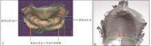
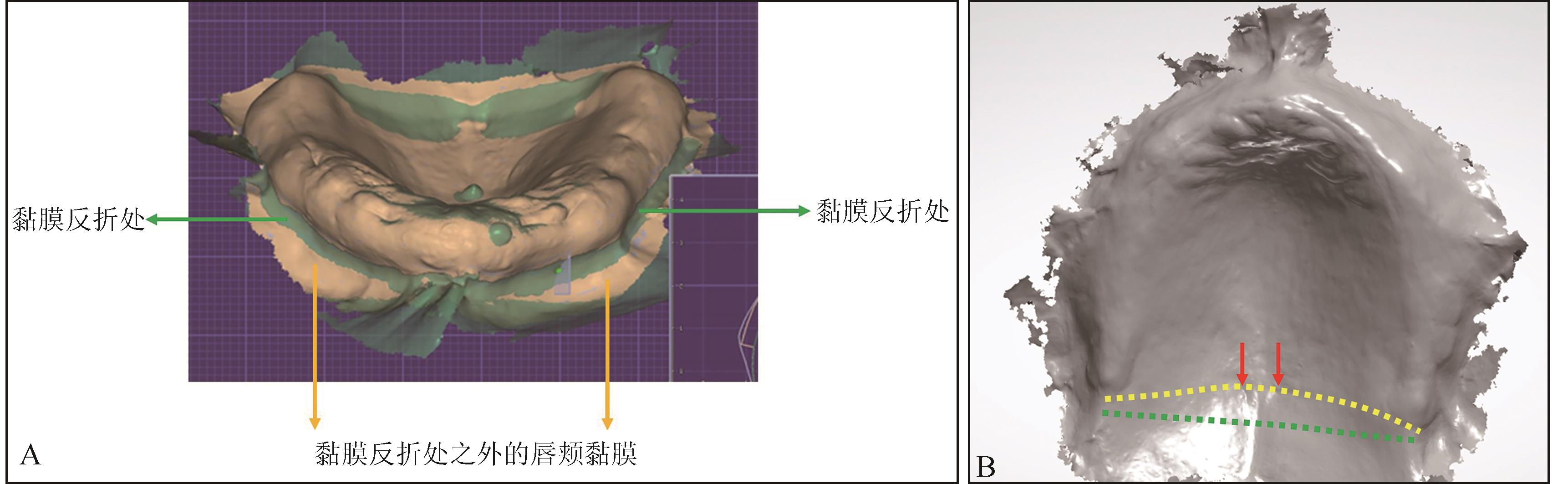
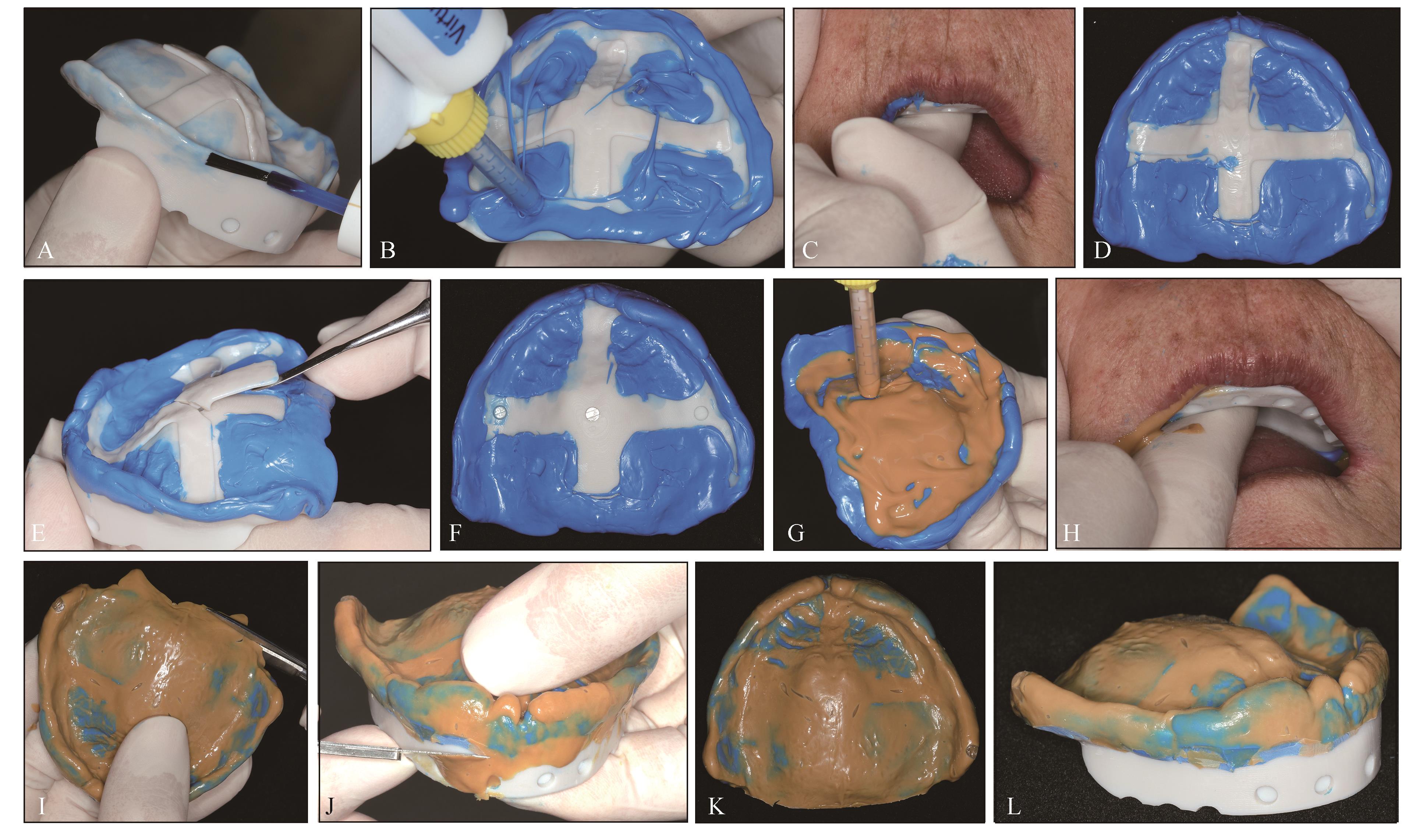
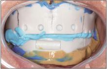
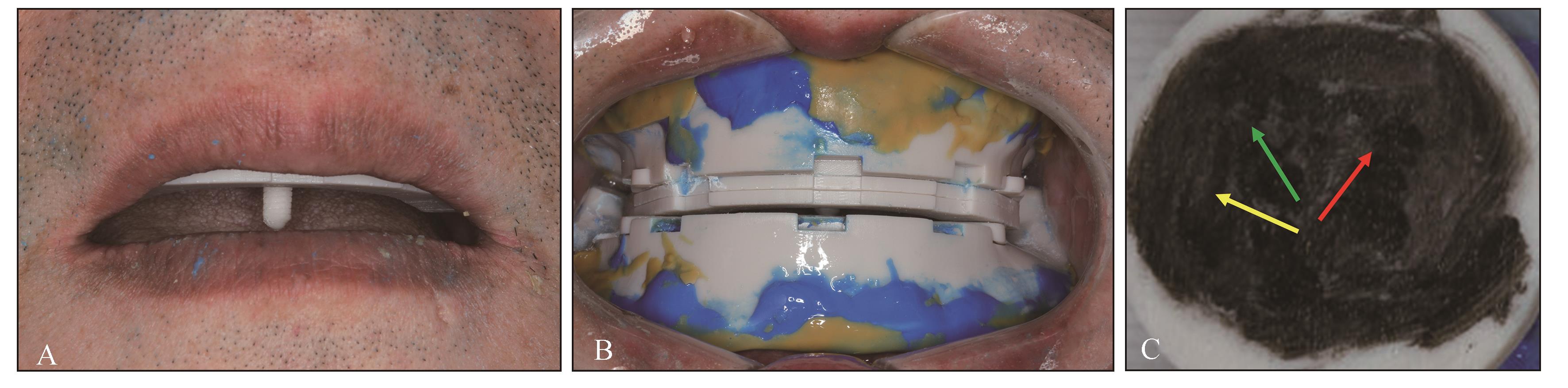
随着口腔医学数字化技术的快速发展,其已日益深入地应用于全口义齿修复的各个环节。以计算机辅助设计和计算机辅助制造(CAD/CAM)为核心,整合口内扫描、面部扫描、3D打印以及数控切削的数字化技术已经给全口义齿的修复流程带来了巨大的变化。本文就无牙颌数字化初模型、初步颌位关系记录、个别托盘复合体设计制作、无牙颌数字化终模型及最终颌位关系获取、全口义齿的数字化制作5个主要环节,探讨数字化技术在全口义齿修复中应用的注意事项及体会。相较依赖经验和手工操作的传统模式,数字化技术已经能够提高全口义齿修复的精度、可预测性和效率,同时减少就诊次数,提升患者的舒适度和就诊体验。然而,目前并未实现全口义齿修复的全程数字化,数字化技术与全口义齿修复的交叉融合、临床应用仍面临诸多亟待规范和深入研究的问题。
中图分类号:
| [1] | Chen HM, Shen K, Ji L, et al. Global and regional patterns in edentulism (1990-2021) with predictions to 2040[J]. Int Dent J, 2025, 75(2): 735-743. |
| [2] | Kutkut A, Bertoli E, Frazer R, et al. A systematic review of studies comparing conventional complete denture and implant retained overdenture[J]. J Prosthodont Res, 2018, 62(1): 1-9. |
| [3] | Hack G, Liberman L, Vach K, et al. Computerized optical impression making of edentulous jaws-an in vivo feasibility study[J]. J Prosthodont Res, 2020, 64(4): 444-453. |
| [4] | Goodacre BJ, Goodacre CJ, Baba NZ, et al. Comparison of denture base adaptation between CAD-CAM and conventional fabrication techniques[J]. J Prosthet Dent, 2016, 116(2): 249-256. |
| [5] | Steinmassl O, Dumfahrt H, Grunert I, et al. CAD/CAM produces dentures with improved fit[J]. Clin Oral Investig, 2018, 22(8): 2829-2835. |
| [6] | Hsu CY, Yang TC, Wang TM, et al. Effects of fabrication techniques on denture base adaptation: an in vitro study[J]. J Prosthet Dent, 2020, 124(6): 740-747. |
| [7] | Kattadiyil MT, AlHelal A. An update on computer-engineered complete dentures: a systematic review on clinical outcomes[J]. J Prosthet Dent, 2017, 117(4): 478-485. |
| [8] | Bidra AS, Taylor TD, Agar JR. Computer-aided technology for fabricating complete dentures: systematic review of historical background, current status, and future perspectives[J]. J Prosthet Dent, 2013, 109(6): 361-366. |
| [9] | Thu KM, Molinero-Mourelle P, Yeung AWK, et al. Which clinical and laboratory procedures should be used to fabricate digital complete dentures? A systematic review[J]. J Prosthet Dent, 2024, 132(5): 922-938. |
| [10] | Avelino MEL, Costa RTF, Vila-Nova TEL, et al. Clinical performance and patient-related outcome measures of digitally fabricated complete dentures: a systematic review and meta-analysis[J]. J Prosthet Dent, 2024, 132(4): 748.e1-748.e10. |
| [11] | Alqutaibi AY, Alghauli MA, Mahmoud II. Digital fabrication of complete dentures, compared to conventional methods, may offer a more cost-effective approach with improved patient outcomes[J]. J Evid Based Dent Pract, 2024, 24(2): 101986. |
| [12] | Russo LL, Salamini A, Troiano G, et al. Digital dentures: a protocol based on intraoral scans[J]. J Prosthet Dent, 2021, 125(4): 597-602. |
| [13] | Russo LL, Di Gioia C, Salamini A, et al. Integrating intraoral, perioral, and facial scans into the design of digital dentures[J]. J Prosthet Dent, 2020, 123(4): 584-588. |
| [14] | 赵铱民. 口腔修复学[M]. 8版. 北京: 人民卫生出版社, 2020. |
| Zhao YM. Prosthodontics[M]. 8th ed. Beijing: People’s Health Publishing House, 2020. | |
| [15] | Kattadiyil MT, Mursic Z, AlRumaih H, et al. Intraoral scanning of hard and soft tissues for partial removable dental prosthesis fabrication[J]. J Prosthet Dent, 2014, 112(3): 444-448. |
| [16] | Wu J, Li Y, Zhang YM. Use of intraoral scanning and 3-dimensional printing in the fabrication of a removable partial denture for a patient with limited mouth opening[J]. J Am Dent Assoc, 2017, 148(5): 338-341. |
| [17] | Tasaka A, Uekubo Y, Mitsui T, et al. Applying intraoral scanner to residual ridge in edentulous regions: in vitro evaluation of inter-operator validity to confirm trueness[J]. BMC Oral Health, 2019, 19(1): 264. |
| [18] | Deferm JT, Schreurs R, Baan F, et al. Validation of 3D documentation of palatal soft tissue shape, color, and irregularity with intraoral scanning[J]. Clin Oral Investig, 2018, 22(3): 1303-1309. |
| [19] | Schimmel M, Akino N, Srinivasan M, et al. Accuracy of intraoral scanning in completely and partially edentulous maxillary and mandibular jaws: an in vitro analysis[J]. Clin Oral Investig, 2021, 25(4): 1839-1847. |
| [20] | Hayama H, Fueki K, Wadachi J, et al. Trueness and precision of digital impressions obtained using an intraoral scanner with different head size in the partially edentulous mandible[J]. J Prosthodont Res, 2018, 62(3): 347-352. |
| [21] | Fang YQ, Fang JH, Jeong SM, et al. A technique for digital impression and bite registration for a single edentulous arch[J]. J Prosthodont, 2019, 28(2): e519-e523. |
| [22] | Fang JH, An XY, Jeong SM, et al. Digital intraoral scanning technique for edentulous jaws[J]. J Prosthet Dent, 2018, 119(5): 733-735. |
| [23] | Russo LL, Caradonna G, Troiano G, et al. Three-dimensional differences between intraoral scans and conventional impressions of edentulous jaws: a cli-nical study[J]. J Prosthet Dent, 2020, 123(2): 264-268. |
| [24] | 潘景光, 朱朋, 冯玥, 等. 分体式颌位关系记录托盘的设计与应用[J]. 实用口腔医学杂志, 2024, 40(3): 443-448. |
| Pan JG, Zhu P, Feng Y, et al. The design and clinical application of a split-type jaw relationship record tray[J]. J Pract Stomatol, 2024, 40(3): 443-448. | |
| [25] | Sun YC, Chen H, Li H, et al. Clinical evaluation of final impressions from three-dimensional printed custom trays[J]. Sci Rep, 2017, 7(1): 14958. |
| [26] | Deng KH, Chen H, Li R, et al. Clinical evaluation of tissue stops on 3D-printed custom trays[J]. Sci Rep, 2019, 9(1): 1807. |
| [27] | Miao XC, Luo YC, Jiang XQ, et al. Digitally prin-ted custom trays assembled with occlusion rims and Gothic arch tracing devices attached with tenon-and-mortise joints for biofunctional complete dentures: a dental technique[J]. J Prosthet Dent, 2025, 133(6): 1424-1429. |
| [28] | Qu F, Du X, Liu WC. 3D-printed custom trays with a Gothic arch for centric relation recording and definitive impression making for complete dentures: a dental technique[J]. J Prosthet Dent, 2019, 121(1): 32-36. |
| [29] | Wang X, Su JS. Evaluation of precision of custom edentulous trays fabricated with 3D printing techno-logies[J]. Int J Prosthodont, 2021, 34(1): 109-117. |
| [30] | 周永胜. 三维打印技术在口腔修复中的应用现状及发展趋势[J]. 中华口腔医学杂志, 2020, 55(10):716-721. |
| Zhou YS. Present status and outlook of prosthodontic treatments based on three-dimensional printing technologies[J]. Chin J Stomatol, 2020, 55(10): 716-721. | |
| [31] | Chen H, Yang X, Chen LT, et al. Application of FDM three-dimensional printing technology in the digital manufacture of custom edentulous mandible trays[J]. Sci Rep, 2016, 6: 19207. |
| [32] | Alhajj MN, Khalifa N, Abduo J, et al. Determination of occlusal vertical dimension for complete dentures patients: an updated review[J]. J Oral Rehabil, 2017, 44(11): 896-907. |
| [33] | 周永胜, 孙玉春, 王勇. 数字化全口义齿的临床应用和研究进展[J]. 华西口腔医学杂志, 2021, 39(1): 1-8. |
| Zhou YS, Sun YC, Wang Y. Clinical application and research progress of digital complete denture[J]. West China J Stomatol, 2021, 39(1): 1-8. | |
| [34] | de Moraes Melo Neto CL, Dos Santos DM, de Magalhães Bertoz AP, et al. Comparison of techniques for obtaining centric relation based on the reprodu-cibility of the condylar positions in centric relation-a systematic review[J]. Eur J Dent, 2022, 16(2): 251-257. |
| [35] | Revilla-León M, Zeitler JM, Kois JC. Digital maxillomandibular relationship and mandibular motion recording by using an optical jaw tracking system to acquire a dynamic virtual patient[J]. J Prosthet Dent, 2024, 132(1): 14-19. |
| [36] | Wang JR, Jin CX, Dong B, et al. Fully digital workflow for replicating treatment dentures: a technique for jaw relation transfer and dynamic occlusal adjustment[J]. J Prosthet Dent, 2023, 130(3): 288-294. |
| [37] | Koralakunte PR, Aljanakh M. The role of virtual articulator in prosthetic and restorative dentistry[J]. J Clin Diagn Res, 2014, 8(7): ZE25-ZE28. |
| [38] | Rasaie V, Abduo J. Current techniques for digital complete denture fabrication[J]. Int J Comput Dent, 2022, 25(2): 181-199. |
| [39] | Silva NRFA, Kukucka ED. Innovative subtractive production of a digital removable complete denture from start to finish: a JPD Digital video presentation[J]. J Prosthet Dent, 2022, 127(1): 1-5. |
| [1] | 钟良军. 数字化技术在重度牙周炎治疗中的应用[J]. 国际口腔医学杂志, 2025, 52(1): 1-10. |
| [2] | 孙睿哲,倪前伟,高瞻. 数字化技术在颌面部恶性肿瘤近距离照射治疗中的应用进展[J]. 国际口腔医学杂志, 2025, 52(1): 18-24. |
| [3] | 焦明阳,周煜萃,蒋正源,刘雨欣,曲柳. 数字化导板技术在牙髓治疗领域的研究进展[J]. 国际口腔医学杂志, 2024, 51(5): 550-557. |
| [4] | 杨育泽,艾璐莹,张自亮,肖康,毛雨典,吴赟,陈凌. 断冠粘接结合数字化贴面修复在上前牙复杂冠折中的应用1例[J]. 国际口腔医学杂志, 2024, 51(2): 172-175. |
| [5] | 马建斌,薛超然,王沛棋,李彬,白丁. 不同补偿间隙3D打印正颌手术𬌗板对咬合精度的影响[J]. 国际口腔医学杂志, 2022, 49(3): 296-304. |
| [6] | 赵喆,王富,郑秀丽,安娜,陈吉华. 功能载荷下牙移动测量方法的研究进展[J]. 国际口腔医学杂志, 2022, 49(3): 362-366. |
| [7] | 张心驰,吴炜. 颌面骨再生领域3D打印技术及应用材料的研究进展[J]. 国际口腔医学杂志, 2020, 47(6): 677-685. |
| [8] | 刘春煦,鲁雨晴,贾璐铭,董博,张倩倩,于海洋. 选择性激光熔融与铸造钛合金卡环的模拟摘戴固位力研究[J]. 国际口腔医学杂志, 2020, 47(2): 152-158. |
| [9] | 王勇. 全口义齿数字化技术分析[J]. 国际口腔医学杂志, 2020, 47(1): 1-9. |
| [10] | 孙淑贞,杨亚东,杨坚. 中性区技术在全口义齿修复中的研究进展[J]. 国际口腔医学杂志, 2019, 46(6): 735-739. |
| [11] | 胡常红. 回归治疗本质的无牙颌修复理念[J]. 国际口腔医学杂志, 2018, 45(6): 621-627. |
| [12] | 王珂, 项涛, 汤亚玲, 梁新华. 3D打印技术在口腔颌面外科实验教学中的应用[J]. 国际口腔医学杂志, 2018, 45(1): 119-124. |
| [13] | 王静,袁荣涛,董蒨. 计算机辅助手术系统与3D打印技术在口腔颌面部缺损修复重建中的应用[J]. 国际口腔医学杂志, 2016, 43(6): 725-728. |
| [14] | 孙学武 柳忠豪 朱详奎 杨柳. 不同颌位记录方法对全口义齿再修复患者咀嚼效率的影响[J]. 国际口腔医学杂志, 2014, 41(3): 286-288. |
| [15] | 孙学武 柳忠豪 朱详奎 杨柳. 不同颌位关系记录法对全口义齿再修复患者义齿调 量影响的研究[J]. 国际口腔医学杂志, 2014, 41(2): 146-148. |
|