国际口腔医学杂志 ›› 2024, Vol. 51 ›› Issue (5): 550-557.doi: 10.7518/gjkq.2024086
焦明阳1( ),周煜萃1,蒋正源1,刘雨欣2,曲柳2()
Mingyang Jiao1(),Yucui Zhou1,Zhengyuan Jiang1,Yuxin Liu2,Liu Qu2()
摘要:
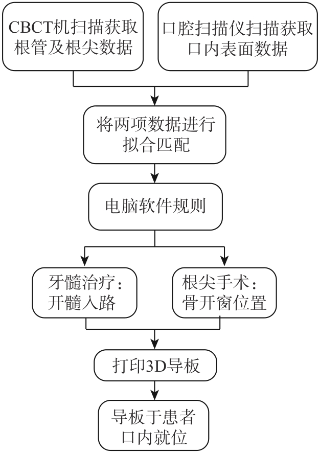
随着数字化技术和3D打印技术的飞速发展,数字化导板在口腔医学领域的应用日渐广泛。近年来,许多学者开始将这项技术应用于牙髓治疗领域,以完成更精准、更微创、更高效的牙髓治疗。本文将对数字化导板技术在牙髓治疗中的工作流程,以及该技术在非手术牙髓治疗及根尖手术中的应用现状进行阐述。
中图分类号:
| 1 | Byun C, Kim C, Cho S, et al. Endodontic treatment of an anomalous anterior tooth with the aid of a 3-dimensional printed physical tooth model[J]. J Endod, 2015, 41(6): 961-965. |
| 2 | Zehnder MS, Connert T, Weiger R, et al. Guided endodontics: accuracy of a novel method for guided access cavity preparation and root canal location[J]. Int Endod J, 2016, 49(10): 966-972. |
| 3 |
Leontiev W, Connert T, Weiger R, et al. Guided en-dodontics: three-dimensional planning and template-aided preparation of endodontic access cavities[J]. J Vis Exp, 2022(183). doi: 10.3791/63781 .
doi: 10.3791/63781 |
| 4 | 高学军, 岳林. 牙体牙髓病学[M]. 2版. 北京: 北京大学医学出版社, 2013: 356. |
| Gao XJ, Yue L. Endodontics[M]. 2nd ed. Beijing: Peking University Medical Press, 2013: 356. | |
| 5 | Tomson PL, Simon SR. Contemporary cleaning and shaping of the root canal system[J]. Prim Dent J, 2016, 5(2): 46-53. |
| 6 | McCabe PS, Dummer PM. Pulp canal obliteration: an endodontic diagnosis and treatment challenge[J]. Int Endod J, 2012, 45(2): 177-197. |
| 7 | Ballester B, Giraud T, Ahmed HMA, et al. Current strategies for conservative endodontic access cavity preparation techniques-systematic review, meta-ana-lysis, and decision-making protocol[J]. Clin Oral Investig, 2021, 25(11): 6027-6044. |
| 8 | Krastl G, Zehnder MS, Connert T, et al. Guided endo-dontics: a novel treatment approach for teeth with pulp canal calcification and apical pathology[J]. Dent Traumatol, 2016, 32(3): 240-246. |
| 9 | van der Meer WJ, Vissink A, Ng YL, et al. 3D computer aided treatment planning in endodontics[J]. J Dent, 2016, 45: 67-72. |
| 10 | Fonseca Tavares WL, Diniz Viana AC, de Carvalho Machado V, et al. Guided endodontic access of calcified anterior teeth[J]. J Endod, 2018, 44(7): 1195-1199. |
| 11 | Torres A, Shaheen E, Lambrechts P, et al. Micro-guided endodontics: a case report of a maxillary lateral incisor with pulp canal obliteration and apical periodontitis[J]. Int Endod J, 2019, 52(4): 540-549. |
| 12 | Mena‑Álvarez J, Rico-Romano C, Lobo-Galindo AB, et al. Endodontic treatment of dens evaginatus by performing a splint guided access cavity[J]. J Esthet Restor Dent, 2017, 29(6): 396-402. |
| 13 | 封琼, 王一舟, 黄雨婷, 等. 精准微创根管治疗: 3D导板指引下的钙化根管疏通术[J]. 口腔医学研究, 2017, 33(4): 427-431. |
| Feng Q, Wang YZ, Huang YT, et al. Negotiation of calcified root canal under guidance of 3D guides: precise minimally invasive root canal treatment[J]. J Oral Sci Res, 2017, 33(4): 427-431. | |
| 14 | 林捷, 林珍香, 郑志强, 等. 根管导板技术辅助冠修复后磨牙的根管治疗[J]. 实用口腔医学杂志, 2018, 34(5): 648-651. |
| Lin J, Lin ZX, Zheng ZQ, et al. Endodontic treatment of the molars following crown restoration by using endodontic guide technique[J]. J Pract Stomatol, 2018, 34(5): 648-651. | |
| 15 | 高羽轩, 汪鎏, 傅裕杰, 等. 数字化导板引导技术辅助微创治疗前牙钙化根管[J]. 华西口腔医学杂志, 2022, 40(1): 111-120. |
| Gao YX, Wang L, Fu YJ, et al. Minimally invasive treatment of calcified root canals in anterior teeth with digital guide technique[J]. West China J Stomatol, 2022, 40(1): 111-120. | |
| 16 | 陆志伟, 黄英, 邱小玲, 等. 数字化根管定位导板应用于完全钙化根管的临床报道1例[J]. 实用口腔医学杂志, 2022, 38(1): 133-135. |
| Lu ZW, Huang Y, Qiu XL, et al. Treatment of an anterior teeth with completed calcified root canal by guided endodontic access: clinical report of a case[J]. J Pract Stomatol, 2022, 38(1): 133-135. | |
| 17 | Krug R, Volland J, Reich S, et al. Guided endodontic treatment of multiple teeth with dentin dysplasia: a case report[J]. Head Face Med, 2020, 16(1): 27. |
| 18 | Maia LM, de Carvalho Machado V, da Silva NRFA, et al. Case reports in maxillary posterior teeth by guided endodontic access[J]. J Endod, 2019, 45(2): 214-218. |
| 19 | Buchgreitz J, Buchgreitz M, Mortensen D, et al. Guided access cavity preparation using cone-beam computed tomography and optical surface scans-an ex vivo study[J]. Int Endod J, 2016, 49(8): 790-795. |
| 20 | Connert T, Krug R, Eggmann F, et al. Guided endo-dontics versus conventional access cavity preparation: a comparative study on substance loss using 3-dimensional-printed teeth[J]. J Endod, 2019, 45(3): 327-331. |
| 21 | Loureiro MAZ, Elias MRA, Capeletti LR, et al. Guided endodontics: volume of dental tissue removed by guided access cavity preparation-an ex vivo study[J]. J Endod, 2020, 46(12): 1907-1912. |
| 22 | Kostunov J, Rammelsberg P, Klotz AL, et al. Minimization of tooth substance removal in normally calcified teeth using guided endodontics: an in vitro pilot study[J]. J Endod, 2021, 47(2): 286-290. |
| 23 | Buchgreitz J, Buchgreitz M, Bjørndal L. Guided root canal preparation using cone beam computed tomography and optical surface scans-an observational study of pulp space obliteration and drill path depth in 50 patients[J]. Int Endod J, 2019, 52(5): 559-568. |
| 24 | Zhang CF, Zhao X, Chen C, et al. The accuracy of using guided endodontics in access cavity preparation and the temperature changes of root surface: an in vitro study[J]. BMC Oral Health, 2022, 22(1): 504. |
| 25 | Fonseca Tavares WL, de Oliveira Murta Pedrosa N, Moreira RA, et al. Limitations and management of static-guided endodontics failure[J]. J Endod, 2022, 48(2): 273-279. |
| 26 | Ali A, Arslan H, Jethani B. Conservative management of type Ⅱ dens invaginatus with guided endo-dontic approach: a case series[J]. J Conserv Dent, 2019, 22(5): 503-508. |
| 27 | 王红, 张倩倩, 李旭光, 等. 3D打印根管定位数字化导板治疗OehlersⅡ型牙内陷1例[J]. 实用口腔医学杂志, 2021, 37(3): 428-431. |
| Wang H, Zhang QQ, Li XG, et al. Endodontic treatment of type Ⅱ dens invaginatus by using 3D splint guides for cavity access: a case report[J]. J Pract Stomatol, 2021, 37(3): 428-431. | |
| 28 | Zubizarreta Macho Á, Ferreiroa A, Rico-Romano C, et al. Diagnosis and endodontic treatment of type Ⅱ dens invaginatus by using cone-beam computed tomography and splint guides for cavity access: a case report[J]. J Am Dent Assoc, 2015, 146(4): 266-270. |
| 29 | Ali A, Arslan H. Guided endodontics: a case report of maxillary lateral incisors with multiple dens invaginatus[J]. Restor Dent Endod, 2019, 44(4): e38. |
| 30 | Sato M, Garcia-Sanchez A, Sanchez S, et al. Use of 3-dimensional-printed guide in hemisection and autotransplantation of a fusion tooth: a case report[J]. J Endod, 2021, 47(3): 526-531. |
| 31 | Buchgreitz J, Buchgreitz M, Bjørndal L. Guided endodontics modified for treating molars by using an intracoronal guide technique[J]. J Endod, 2019, 45(6): 818-823. |
| 32 | Torres A, Lerut K, Lambrechts P, et al. Guided en-dodontics: use of a sleeveless guide system on an upper premolar with pulp canal obliteration and apical periodontitis[J]. J Endod, 2021, 47(1): 133-139. |
| 33 | Connert T, Zehnder MS, Amato M, et al. Microgui-ded endodontics: a method to achieve minimally invasive access cavity preparation and root canal location in mandibular incisors using a novel computer-guided technique[J]. Int Endod J, 2018, 51(2): 247-255. |
| 34 | Connert T, Zehnder MS, Weiger R, et al. Microgui-ded endodontics: accuracy of a miniaturized technique for apically extended access cavity preparation in anterior teeth[J]. J Endod, 2017, 43(5): 787-790. |
| 35 | Schwindling FS, Tasaka A, Hilgenfeld T, et al. Three-dimensional-guided removal and preparation of dental root posts-concept and feasibility[J]. J Prosthodont Res, 2020, 64(1): 104-108. |
| 36 | Maia LM, Moreira Júnior G, Albuquerque RC, et al. Three-dimensional endodontic guide for adhesive fiber post removal: a dental technique[J]. J Prosthet Dent, 2019, 121(3): 387-390. |
| 37 | Maia LM, Bambirra Júnior W, Toubes KM, et al. Endodontic guide for the conservative removal of a fiber-reinforced composite resin post[J]. J Prosthet Dent, 2022, 128(1): 4-7. |
| 38 | Liu RL, Xie CY, Sun ML, et al. Guided removal of a fractured fiber post and immediate restoration with a digitally prefabricated titanium post-and-core and zirconia crown: a clinical report[J]. J Prosthet Dent, 2023, 129(5): 684-689. |
| 39 | Perez C, Finelle G, Couvrechel C. Optimisation of a guided endodontics protocol for removal of fibrereinforced posts[J]. Aust Endod J, 2020, 46(1): 107-114. |
| 40 | Xue Y, Zhang L, Cao Y, et al. A three-dimensional printed assembled sleeveless guide system for fiber-post removal[J]. J Prosthodont, 2023, 32(2): 178-184. |
| 41 | Mo S, Xu Y, Zhang L, et al. Accuracy of a 3D prin-ted sleeveless guide system used for fiber post remo-val: an in vitro study[J]. J Dent, 2023, 128: 104367. |
| 42 | Setzer FC, Kratchman SI. Present status and future directions: surgical endodontics[J]. Int Endod J, 2022, 55(): 1020-1058. |
| 43 | Geo TD, Saxena P, Gupta S. Static vs. dynamic na-vigation for endodontic microsurgery-A comparative review[J]. J Oral Biol Craniofac Res, 2022, 12(4): 410-412. |
| 44 | Ray JJ, Giacomino CM, Wealleans JA, et al. Targe-ted endodontic microsurgery: digital workflow options[J]. J Endod, 2020, 46(6): 863-871. |
| 45 | Nagy E, Braunitzer G, Gryschka DG, et al. Accuracy of digitally planned, guided apicoectomy with a conventional trephine and a custom-made endodontic trephine: an in vitro comparative study[J]. J Stomatol Oral Maxillofac Surg, 2022, 123(4): 388-394. |
| 46 | 王安琪, 吴丽, 赵丹, 等. 3D打印根尖切除手术导板的设计及体外模型效果评价[J]. 实用口腔医学杂志, 2021, 37(6): 739-742. |
| Wang AQ, Wu L, Zhao D, et al. Evaluation and case analysis of 3D printing apicoectomy guide plate[J]. J Pract Stomatol, 2021, 37(6): 739-742. | |
| 47 | 彭俐, 王祖华, 孙玉春, 等. 根尖切除手术导板的计算机辅助设计及三维打印[J]. 北京大学学报(医学版), 2018, 50(5): 905-910. |
| Peng L, Wang ZH, Sun YC, et al. Computer aided design and three-dimensional printing forapicoectomy guide template[J]. J Peking Univ Health Sci, 2018, 50(5): 905-910. | |
| 48 | Pinsky HM, Champleboux G, Sarment DP. Periapical surgery using CAD/CAM guidance: preclinical results[J]. J Endod, 2007, 33(2): 148-151. |
| 49 | Strbac GD, Schnappauf A, Giannis K, et al. Guided modern endodontic surgery: a novel approach for guided osteotomy and root resection[J]. J Endod, 2017, 43(3): 496-501. |
| 50 | Giacomino CM, Ray JJ, Wealleans JA. Targeted endodontic microsurgery: a novel approach to anatomically challenging scenarios using 3-dimensio-nal-printed guides and trephine burs-a report of 3 ca-ses[J]. J Endod, 2018, 44(4): 671-677. |
| 51 | 汤芝伟, 高莺. 靶向牙髓显微外科技术的应用与进展[J]. 国际口腔医学杂志, 2022, 49(6): 678-683. |
| Tang ZW, Gao Y. Application and progress on targeted endodontic microsurgery techniques[J]. Int J Stomatol, 2022, 49(6): 678-683. | |
| 52 | Hawkins TK, Wealleans JA, Pratt AM, et al. Targe-ted endodontic microsurgery and endodontic microsurgery: a surgical simulation comparison[J]. Int Endod J, 2020, 53(5): 715-722. |
| 53 | Ahn SY, Kim NH, Kim S, et al. Computer-aided design/computer-aided manufacturing-guided endodontic surgery: guided osteotomy and apex localization in a mandibular molar with a thick buccal bone plate[J]. J Endod, 2018, 44(4): 665-670. |
| 54 | Smith BG, Pratt AM, Anderson JA, et al. Targeted endodontic microsurgery: implications of the grea-ter palatine artery[J]. J Endod, 2021, 47(1): 19-27. |
| 55 | George R, Cameron A, Meer M. Streamlining and simplification of surgical stent fabrication for micro-endodontic surgery[J]. Aust Endod J, 2020, 46(3): 445-451. |
| 56 | Benjamin G, Ather A, Bueno MR, et al. Preserving the neurovascular bundle in targeted endodontic microsurgery: a case series[J]. J Endod, 2021, 47(3): 509-519. |
| 57 | Antal M, Nagy E, Braunitzer G, et al. Accuracy and clinical safety of guided root end resection with a trephine: a case series[J]. Head Face Med, 2019, 15(1): 30. |
| 58 | Popowicz W, Palatyńska-Ulatowska A, Kohli MR. Targeted endodontic microsurgery: computed tomography-based guided stent approach with platelet-rich fibrin graft: a report of 2 cases[J]. J Endod, 2019, 45(12): 1535-1542. |
| 59 | Buniag AG, Pratt AM, Ray JJ. Targeted endodontic microsurgery: a retrospective outcomes assessment of 24 cases[J]. J Endod, 2021, 47(5): 762-769. |
| 60 | 满毅, 黄定明. 美学区种植骨增量与邻牙慢性根尖周病的联合治疗策略(上): 应用基础及适应证[J]. 国际口腔医学杂志, 2022, 49(5): 497-505. |
| Man Y, Huang DM. Combined treatment strategy of oral implantology and endodontic microsurgery for bone augmentation and endodontic diseases in aesthetic area (part 1): application basis and indications[J]. Int J Stomatol, 2022, 49(5): 497-505. | |
| 61 | 满毅, 黄定明. 美学区种植骨增量与邻牙慢性根尖周病的联合治疗策略(下): 临床诊治流程及实践病例[J]. 国际口腔医学杂, 2022, 49(6): 621-632. |
| Man Y, Huang DM. Combined treatment strategy of oral implantology and endodontics microsurgery: clinical protocol and practical cases (part 2)[J]. Int J Stomatol, 2022, 49(6): 621-632. | |
| 62 | Mainkar A, Zhu Q, Safavi K. Incidence of altered sensation after mandibular premolar and molar periapical surgery[J]. J Endod, 2020, 46(1): 29-33. |
| 63 | Wang J, Luo YL, Tan XL, et al. Horizontal bone augmentation of the edentulous area with simultaneous endodontic microsurgery of the adjacent tooth: a digitally-driven multidisciplinary case report with a 1-year follow-up[J]. Int J Oral Implantol, 2021, 14(4): 435-451. |
| 64 | Gambarini G, Galli M, Morese A, et al. Precision of dynamic navigation to perform endodontic ultraconservative access cavities: a preliminary in vitro ana-lysis[J]. J Endod, 2020, 46(9): 1286-1290. |
| 65 | Dianat O, Gupta S, Price JB, et al. Guided endodontic access in a maxillary molar using a dynamic na-vigation system[J]. J Endod, 2021, 47(4): 658-662. |
| 66 | Gambarini G, Galli M, Stefanelli LV, et al. Endodontic microsurgery using dynamic navigation system: a case report[J]. J Endod, 2019, 45(11): 1397-1402.e6. |
| 67 | Ribeiro D, Reis E, Marques JA, et al. Guided en-dodontics: static vs. dynamic computer-aided techniques-a literature review[J]. J Pers Med, 2022, 12(9): 1516. |
| [1] | 杨雨楠,刘鹏,王虎,游梦. 上颌窦黏膜增厚的锥形束CT影像分析[J]. 国际口腔医学杂志, 2023, 50(3): 302-307. |
| [2] | 汤芝伟,高莺. 靶向牙髓显微外科技术的应用与进展[J]. 国际口腔医学杂志, 2022, 49(6): 678-683. |
| [3] | 蔡娉娉,卓盈颖,林捷,郑志强. 计算机辅助技术在纤维桩拆除中的应用[J]. 国际口腔医学杂志, 2022, 49(6): 731-736. |
| [4] | 吴文智,冯达兴,陈垂壮,周丽鹃. 海口地区下颌第一恒磨牙近中中央根管发生率及相关因素[J]. 国际口腔医学杂志, 2022, 49(4): 420-425. |
| [5] | 庞瑜,刘显,王了. 数字化导板在埋伏多生牙拔除中的应用[J]. 国际口腔医学杂志, 2022, 49(4): 448-452. |
| [6] | 马建斌,薛超然,王沛棋,李彬,白丁. 不同补偿间隙3D打印正颌手术𬌗板对咬合精度的影响[J]. 国际口腔医学杂志, 2022, 49(3): 296-304. |
| [7] | 叶泽林,刘璐,龙虎,游梦. 弯曲前牙的影像评价及治疗的研究进展[J]. 国际口腔医学杂志, 2022, 49(2): 173-181. |
| [8] | 田浩楠,林敏,谢丛蔓,任嫒姝. 上颌腭侧阻生尖牙与寰椎后桥相关性的锥形束CT研究[J]. 国际口腔医学杂志, 2021, 48(5): 536-540. |
| [9] | 施丹妮,杨鑫,吴建勇. 锥形束CT三维头影测量参考坐标系的研究进展[J]. 国际口腔医学杂志, 2021, 48(4): 398-404. |
| [10] | 丁张帆,郭陟永,苗诚,李春洁,宣鸣,王晓毅,张壮. 基于锥形束CT的三维可视化技术在颌骨囊性病变手术中的应用[J]. 国际口腔医学杂志, 2021, 48(2): 180-186. |
| [11] | 王奔,许喆桢,韦曦. 数字化微创技术在牙髓根尖周病学中的应用与进展[J]. 国际口腔医学杂志, 2021, 48(1): 110-118. |
| [12] | 张心驰,吴炜. 颌面骨再生领域3D打印技术及应用材料的研究进展[J]. 国际口腔医学杂志, 2020, 47(6): 677-685. |
| [13] | 唐蓓,赵文俊,王虎,郑广宁,游梦. 根管超填导致下牙槽神经损伤2例[J]. 国际口腔医学杂志, 2020, 47(3): 293-296. |
| [14] | 刘春煦,鲁雨晴,贾璐铭,董博,张倩倩,于海洋. 选择性激光熔融与铸造钛合金卡环的模拟摘戴固位力研究[J]. 国际口腔医学杂志, 2020, 47(2): 152-158. |
| [15] | 章婷婷,胡常红,彭燕,周文翘,张慧聪,刘蝶. 300例不同年龄段有牙颌人群上唇软组织侧貌的锥形束CT三维测量分析[J]. 国际口腔医学杂志, 2020, 47(2): 182-188. |
|