国际口腔医学杂志 ›› 2022, Vol. 49 ›› Issue (5): 561-568.doi: 10.7518/gjkq.2022094
曾杨林( ),谭学莲,宋东哲,黄定明()
Zeng Yanglin(),Tan Xuelian,Song Dongzhe,Huang Dingming.()
摘要:
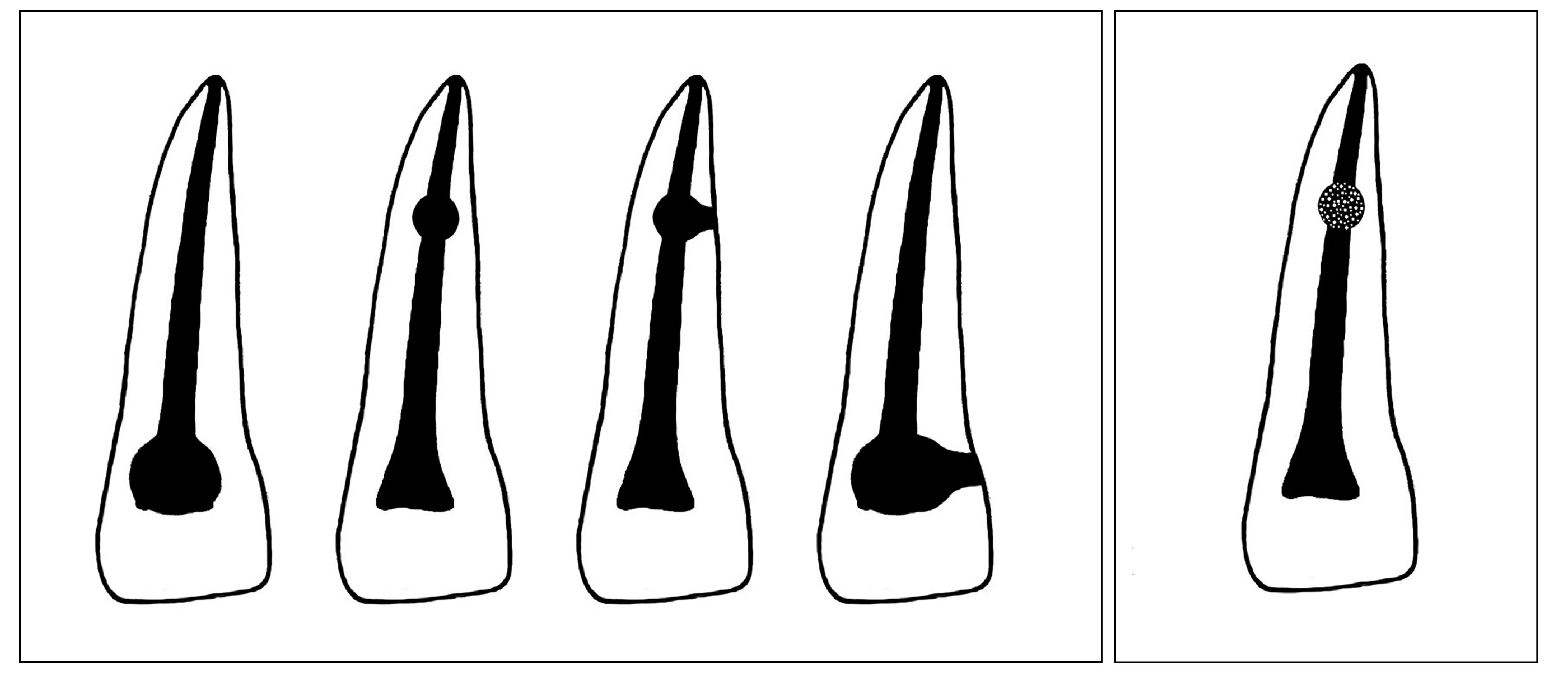
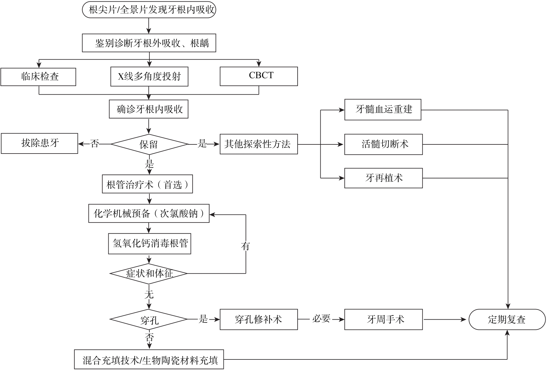
牙根内吸收是牙髓处于炎症状态时破牙本质细胞导致的髓室及根管壁牙本质丧失。其发病率低,主要与外伤和感染密切相关,具体的发病机制还不够明确。牙根内吸收一般无主观症状,常通过影像学检查发现,且易与牙根外吸收混淆。一旦确诊应尽快治疗,阻止疾病继续进展,常用的治疗方法为根管治疗术。迄今国内未见牙根内吸收的综述报道。本文就牙根内吸收的病因及发病机制、分类、诊断与鉴别诊断、治疗和预后的研究进展进行总结,为牙根内吸收的临床诊治提供参考。
中图分类号:
| 1 | Patel S, Ricucci D, Durak C, et al. Internal root resorption: a review[J]. J Endod, 2010, 36(7): 1107-1121. |
| 2 | Calişkan MK, Türkün M. Prognosis of permanent teeth with internal resorption: a clinical review[J]. Endod Dent Traumatol, 1997, 13(2): 75-81. |
| 3 | de Araujo LCG, Lins CV, de Lima GA, et al. Study of prevalence of internal resorption in periapical radiography of anteriors permanents tooth[J]. Int J Morphol, 2009, 27(1): 227-230. |
| 4 | Marinescu IR, Bănică AC, Mercuţ V, et al. Root resorption diagnostic: role of digital panoramic radio-graphy[J]. Curr Health Sci J, 2019, 45(2): 156-166. |
| 5 | Endal U, Shen Y, Knut A, et al. A high-resolution computed tomographic study of changes in root canal isthmus area by instrumentation and root filling[J]. J Endod, 2011, 37(2): 223-227. |
| 6 | Koehne T, Zustin J, Amling M, et al. Radiological and histopathological features of internal tooth resorption[J]. In Vivo, 2020, 34(4): 1875-1882. |
| 7 | Nilsson E, Bonte E, Bayet F, et al. Management of internal root resorption on permanent teeth[J]. Int J Dent, 2013, 2013: 929486. |
| 8 | Liu HM, Peng XX, Sun HC, et al. Clinical and histopathological characterization of root resorption in replanted teeth: two case reports[J]. Medicine, 2020, 99(3): e18869. |
| 9 | Darcey J, Qualtrough A. Resorption: part 1. Pathology, classification and aetiology[J]. Br Dent J, 2013, 214(9): 439-451. |
| 10 | Souza BDM, Dutra KL, Kuntze MM, et al. Incidence of root resorption after the replantation of avulsed teeth: a meta-analysis[J]. J Endod, 2018, 44(8): 1216-1227. |
| 11 | Kalender A, Oztan MD, Basmaci F, et al. CBCT evaluation of multiple idiopathic internal resorptions in permanent molars: case report[J]. BMC Oral Heal, 2014, 14: 39. |
| 12 | Urban D, Mincik J. Monozygotic twins with idiopathic internal root resorption: a case report[J]. Aust Endod J, 2010, 36(2): 79-82. |
| 13 | Levy DH, Rozenfeld S, Itzhak JB, et al. A rare case of multiple internal root resorption after the delayed treatment of a traumatic injury: a case report[J]. J Contemp Dent Pract, 2021, 22(2): 194-198. |
| 14 | Duan XH, Yang T, Zhang YL, et al. Odontoblast-like MDPC-23 cells function as odontoclasts with RANKL/M-CSF induction[J]. Arch Oral Biol, 2013, 58(3): 272-278. |
| 15 | Zheng Y, Chen M, He L, et al. Mesenchymal dental pulp cells attenuate dentin resorption in homeostasis[J]. J Dent Res, 2015, 94(6): 821-827. |
| 16 | Fernandes M, de Ataide I, Wagle R. Tooth resorption part Ⅰ-pathogenesis and case series of internal resorption[J]. J Conserv Dent, 2013, 16(1): 4-8. |
| 17 | Wedenberg C, Zetterqvist L. Internal resorption in human teeth: a histological, scanning electron microscopic, and enzyme histochemical study[J]. J Endod, 1987, 13(6): 255-259. |
| 18 | Ne RF, Witherspoon DE, Gutmann JL. Tooth resorption[J]. Quintessence Int, 1999, 30(1): 9-25. |
| 19 | Aidos H, Diogo P, Santos JM. Root resorption classifications: a narrative review and a clinical aid proposal for routine assessment[J]. Eur Endod J, 2018, 3(3): 134-145. |
| 20 | Gartner AH, Mack T, Somerlott RG, et al. Differential diagnosis of internal and external root resorption[J]. J Endod, 1976, 2(11): 329-334. |
| 21 | Cohen RG, Razzano MV. Immediate dentin sealing using an antibacterial self-etching bonding system[J]. Pract Proced Aesthet Dent, 2006, 18(9): 561-566. |
| 22 | Nascimento EHL, Gaêta-Araujo H, Galvão NS, et al. Effect of brightness and contrast variation for detectability of root resorption lesions in digital intraoral radiographs[J]. Clin Oral Investig, 2019, 23(8): 3379-3386. |
| 23 | Patel S, Saberi N. The ins and outs of root resorption[J]. Br Dent J, 2018, 224(9): 691-699. |
| 24 | Al-Momani Z, Nixon PJ. Internal and external root resorption: etiology, diagnosis and treatment options[J]. Dent Update, 2013, 40(2): 102-104, 107-108, 111-112. |
| 25 | Madani Z, Moudi E, Bijani A, et al. Diagnostic accuracy of cone-beam computed tomography and periapical radiography in internal root resorption[J]. Iran Endod J, 2016, 1(1): 51-56. |
| 26 | Lima TF, Gamba TO, Zaia A, et al. Evaluation of cone beam computed tomography and periapical radiography in the diagnosis of root resorption[J]. Aust Dent J, 2016, 61(4): 425-431. |
| 27 | Mehra N, Yadav M, Kaushik M, et al. Clinical ma-nagement of root resorption: a report of three cases[J]. Cureus, 2018, 10(8): e3215. |
| 28 | Sharma V, Tanwar R, Gupta V, et al. Impacted stapler pin in fractured maxillary central incisor with open apex: advanced endodontic management using biodentine as innovative apical matrix[J]. Indian J Dent Res, 2015, 26(6): 637-640. |
| 29 | Umashetty G, Hoshing U, Patil S, et al. Management of inflammatory internal root resorption with biodentine and thermoplasticised gutta-percha[J]. Case Rep Dent, 2015, 2015: 452609. |
| 30 | Aktemur Türker S, Uzunoğlu E, Deniz Sungur D, et al. Fracture resistance of teeth with simulated perforating internal resorption cavities repaired with different calcium silicate-based cements and backfil-ling materials[J]. J Endod, 2018, 44(5): 860-863. |
| 31 | Ulusoy Öİ, Paltun YN. Fracture resistance of roots with simulated internal resorption defects and obturated using different hybrid techniques[J]. J Dent Sci, 2017, 12(2): 121-125. |
| 32 | Aslan T, Üstün Y, Esim E. Stress distributions in internal resorption cavities restored with different materials at different root levels: a finite element analysis study[J]. Aust Endod J, 2019, 45(1): 64-71. |
| 33 | Yıldırım S, Elbay M. Multidisciplinary treatment approach for perforated internal root resorption: three-year follow-up[J]. Case Rep Dent, 2019, 2019: 5848272. |
| 34 | Torabinejad M, Parirokh M, Dummer PMH. Mine-ral trioxide aggregate and other bioactive endodontic cements: an updated overview-part Ⅱ: other clinical applications and complications[J]. Int Endod J, 2018, 51(3): 284-317. |
| 35 | Esnaashari E, Pezeshkfar A, Fazlyab M. Nonsurgical management of an extensive perforative internal root resorption with calcium-enriched mixture cement[J]. Iran Endod J, 2015, 10(1): 75-78. |
| 36 | Subay RK, Subay MO, Tuzcu SB. Endodontic ma-nagement of root perforating internal replacement resorption[J]. Eur J Dent, 2018, 12(3): 450-453. |
| 37 | Pereira da Costa R, Quaresma SA, Lopes FC, et al. Management of a perforating internal root resorption using mineral trioxide aggregate: a case report with 5-year follow-up[J]. Aust Endod J, 2020, 46(3): 452-457. |
| 38 | Küçükkaya Eren S, Aksel H, Askerbeyli Örs S, et al. Obturation quality of calcium silicate-based cements placed with different techniques in teeth with perforating internal root resorption: a micro-compu-ted tomographic study[J]. Clin Oral Investig, 2019, 23(2): 805-811. |
| 39 | Tek V, Türker SA. A micro-computed tomography evaluation of voids using calcium silicate-based materials in teeth with simulated internal root resorption[J]. Restor Dent Endod, 2020, 45(1): e5. |
| 40 | 王密, 尹世海, 王奇, 等. iRoot BP修复磨牙髓室底穿孔的研究[J]. 华西口腔医学杂志, 2013, 31(3): 257-259, 266. |
| Wang M, Yin SH, Wang Q, et al. Study of molar furcal perforation repaired with iRoot BP[J]. West China J Stomatol, 2013, 31(3): 257-259, 266. | |
| 41 | Jacobovitz M, de Lima RKP. Treatment of inflammatory internal root resorption with mineral trioxide aggregate: a case report[J]. Int Endod J, 2008, 41(10): 905-912. |
| 42 | Li FC, Hung WC. Repair of a perforating internal resorption: two case reports[J]. J Dent Sci, 2016, 11(3): 338-342. |
| 43 | Altundasar E, Demir B. Management of a perfora-ting internal resorptive defect with mineral trioxide aggregate: a case report[J]. J Endod, 2009, 35(10): 1441-1444. |
| 44 | Saoud TMA, Mistry S, Kahler B, et al. Regenerative endodontic procedures for traumatized teeth after horizontal root fracture, avulsion, and perfora-ting root resorption[J]. J Endod, 2016, 42(10): 1476-1482. |
| 45 | Arnold M. Reparative endodontic treatment of a perforating internal inflammatory root resorption: a case report[J]. J Endod, 2021, 47(1): 146-155. |
| 46 | Yang Y, Zhang B, Huang C, et al. Intentional replantation of a second premolar with internal resorption and root fracture: a case report[J]. J Contemp Dent Pract, 2021, 22(5): 562-567. |
| 47 | Krupp C, Bargholz C, Brüsehaber M, et al. Treatment outcome after repair of root perforations with mineral trioxide aggregate: a retrospective evaluation of 90 teeth[J]. J Endod, 2013, 39(11): 1364-1368. |
| 48 | Estrela C, Decurcio DDA, Rossi-Fedele G, et al. Root perforations: a review of diagnosis, prognosis and materials[J]. Braz Oral Res, 2018, 32(): e73. |
| 49 | Gorni FG, Andreano A, Ambrogi F, et al. Patient and clinical characteristics associated with primary healing of iatrogenic perforations after root canal treatment: results of a long-term Italian study[J]. J Endod, 2016, 42(2): 211-215. |
| [1] | 刘世一, 陈中, 张素欣. 程序性死亡受体/配体免疫治疗策略在人乳头瘤病毒阳性头颈部鳞状细胞癌中的研究进展[J]. 国际口腔医学杂志, 2024, 51(1): 21-27. |
| [2] | 和子慕, 李风兰. 数字化口腔定位支架在头颈部肿瘤放射治疗中的应用现状[J]. 国际口腔医学杂志, 2024, 51(1): 28-35. |
| [3] | 傅豫, 何薇, 黄兰. 铁死亡在口腔疾病中的研究进展[J]. 国际口腔医学杂志, 2024, 51(1): 36-44. |
| [4] | 韩冲,何东宁,余飞燕,吴东潮. 口腔种植术后疼痛机制及治疗的研究进展[J]. 国际口腔医学杂志, 2024, 51(1): 99-106. |
| [5] | 胡佳,王秀清,卢国英,黄晓晶. 再生性牙髓治疗在成人根尖发育不全恒牙应用的研究进展[J]. 国际口腔医学杂志, 2023, 50(6): 686-695. |
| [6] | 陆磊,王鑫,康泽标,谢富强. 计算机辅助导航手术在复杂颌面部骨折中的应用新进展[J]. 国际口腔医学杂志, 2023, 50(6): 696-703. |
| [7] | 杨静,柳登高. 内镜下激光碎石术治疗唾液腺结石病的研究进展[J]. 国际口腔医学杂志, 2023, 50(6): 704-710. |
| [8] | 杨冬叶,朱萍,吴淑仪. 舌位的影响因素及临床意义[J]. 国际口腔医学杂志, 2023, 50(6): 723-728. |
| [9] | 刘洋,尹德强. 关于颌位调整方法的思考和改进[J]. 国际口腔医学杂志, 2023, 50(5): 499-505. |
| [10] | 李奕君, 徐子昂, 李一. 前哨淋巴结在头颈部鳞状细胞癌检测的应用进展[J]. 国际口腔医学杂志, 2023, 50(5): 521-527. |
| [11] | 戢晓,张岚,黄定明. 牙源性与非牙源性上颌窦炎鉴别诊断及其治疗方案的研究进展[J]. 国际口腔医学杂志, 2023, 50(5): 566-572. |
| [12] | 赵苑汐,苏勤. 根管再治疗中根管充填物去除辅助技术的应用与发展[J]. 国际口腔医学杂志, 2023, 50(5): 581-586. |
| [13] | 宋文鹏,龚蓓文,李聃,曾剑玉,仇玲玲. 机械疗法在正畸治疗中应用的研究进展[J]. 国际口腔医学杂志, 2023, 50(5): 603-612. |
| [14] | 刘婷,武秀萍. 唐氏综合征的口腔-颅颌面表征及治疗进展[J]. 国际口腔医学杂志, 2023, 50(5): 618-622. |
| [15] | 吴思佳,舒畅,王洋,王媛,邓淑丽,王慧明. 根管内感染控制对年轻恒牙牙髓再生治疗的影响及研究进展[J]. 国际口腔医学杂志, 2023, 50(4): 388-394. |
|