国际口腔医学杂志 ›› 2022, Vol. 49 ›› Issue (3): 332-336.doi: 10.7518/gjkq.2022030
雷彬1( ),陈柯2()
Lei Bin1(),Chen Ke2()
摘要:
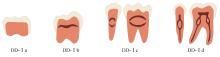
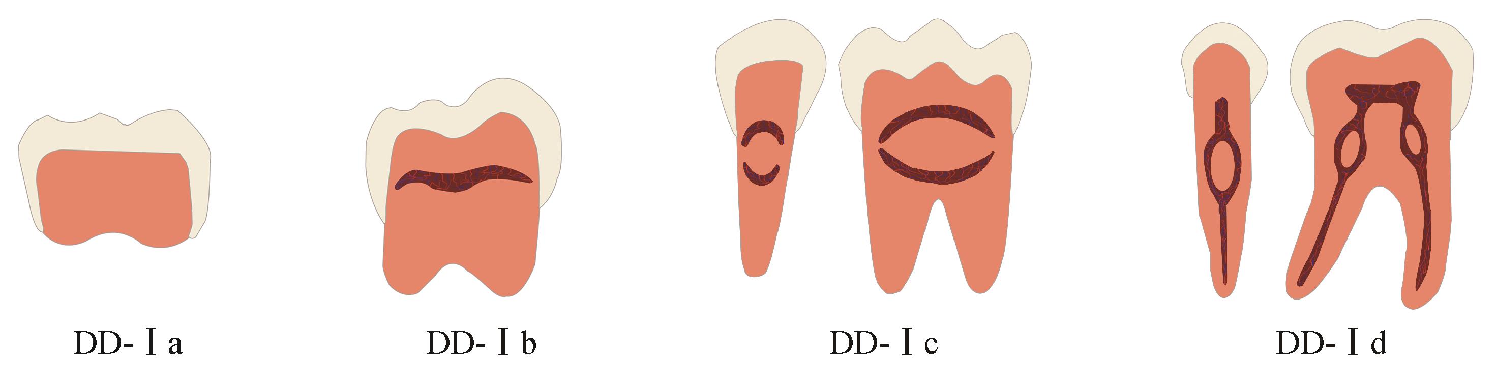
牙本质发育不良Ⅰ型(DD-Ⅰ)是一种罕见的遗传性牙本质形成障碍疾病,乳恒牙均可受累。该病在临床上表现为牙冠外形色泽正常,牙齿松动明显,可伴有自发性牙槽脓肿或囊肿等。影像学检查则可见牙髓腔消失或呈“新月形”牙髓残余,根短钝或无牙根等表现。关于DD-Ⅰ的发病机制已为大多数学者所研究,其临床治疗通常具有挑战性,本文对近年来DD-Ⅰ的临床分型及表现、致病基因、组织学特点、治疗相关研究进行综述,以期为临床诊治该病提供一定的指导。
中图分类号:
| 1 | Pitak-Arnnop P, Subbalekha K, Sirintawat N, et al. Clinical approach to rhizomicry based on a case of dentine dysplasia type 1[J]. J Stomatol Oral Maxillofac Surg, 2020, 121(2): 179-185. |
| 2 | Witkop CJ Jr, Jaspers MT. Teeth with short, thin, dilacerated roots in patients with short stature: a dominantly inherited trait[J]. Oral Surg Oral Med Oral Pathol, 1982, 54(5): 553-559. |
| 3 | O Carroll MK, Duncan WK, Perkins TM. Dentin dysplasia: review of the literature and a proposed subclassification based on radiographic findings[J]. Oral Surg Oral Med Oral Pathol, 1991, 72(1): 119-125. |
| 4 | Shields ED, Bixler D, el-Kafrawy AM. A proposed classification for heritable human dentine defects with a description of a new entity[J]. Arch Oral Biol, 1973, 18(4): 543-553. |
| 5 | Krug R, Volland J, Reich S, et al. Guided endodontic treatment of multiple teeth with dentin dysplasia: a case report[J]. Head Face Med, 2020, 16(1): 27. |
| 6 | Buchanan GD, Tredoux S, Nel C, et al. Endodontic treatment of dentin dysplasia type Ⅰ D[J]. Aust Endod J, 2021, 47(2): 343-349. |
| 7 | Kim JW, Simmer JP. Hereditary dentin defects[J]. J Dent Res, 2007, 86(5): 392-399. |
| 8 | Toomarian L, Mashhadiabbas F, Mirkarimi M, et al. Dentin dysplasia type Ⅰ: a case report and review of the literature[J]. J Med Case Rep, 2010, 4: 1. |
| 9 | de La Dure-Molla M, Philippe Fournier B, Berdal A. Isolated dentinogenesis imperfecta and dentin dysplasia: revision of the classification[J]. Eur J Hum Genet, 2015, 23(4): 445-451. |
| 10 | Filippi A. Dentin dysplasia Type 1. Root dysplasia[J]. Schweiz Monatsschr Zahnmed, 2013, 123(2): 106-107. |
| 11 | Kim JW, Nam SH, Jang KT, et al. A novel splice acceptor mutation in the DSPP gene causing dentinogenesis imperfecta type Ⅱ[J]. Hum Genet, 2004, 115(3): 248-254. |
| 12 | Zhang XQ, Chen LY, Liu JY, et al. A novel DSPP mutation is associated with type Ⅱ dentinogenesis imperfecta in a Chinese family[J]. BMC Med Ge-net, 2007, 8: 52. |
| 13 | Lee SK, Hu JC, Lee KE, et al. A dentin sialophosphoprotein mutation that partially disrupts a splice acceptor site causes type Ⅱ dentin dysplasia[J]. J Endod, 2008, 34(12): 1470-1473. |
| 14 | Hart PS, Hart TC. Disorders of human dentin[J]. Cells Tissues Organs, 2007, 186(1): 70-77. |
| 15 | 刘芬. 遗传性牙本质发育异常的临床及相关基础研究[D]. 西安: 第四军医大学, 2013. |
| Liu F. Clinical and related basic research for hereditary dentinogenesis imperfecta[D]. Xi’an: The Fourth Military Medical University, 2013. | |
| 16 | Yang Q, Chen D, Xiong F, et al. A splicing mutation in VPS4B causes dentin dysplasia Ⅰ[J]. J Med Ge-net, 2016, 53(9): 624-633. |
| 17 | 邓敏, 胡爱琴, 熊符. 遗传性牙本质发育异常的分子遗传学研究进展[J]. 国际遗传学杂志, 2017, 40(6): 361-367. |
| Deng M. Hu AQ, Xiong F. Progress in molecular genetics of inherited dentine dysplasia[J]. Int J Genet, 2017, 40(6): 361-367. | |
| 18 | Chen D, Li X, Lu F, et al. Dentin dysplasia type Ⅰ-a dental disease with genetic heterogeneity[J]. Oral Dis, 2019, 25(2): 439-446. |
| 19 | Sloan AJ, Smith AJ. Stem cells and the dental pulp: potential roles in dentine regeneration and repair[J]. Oral Dis, 2007, 13(2): 151-157. |
| 20 | Pan YH, Lu T, Peng L, et al. Vacuolar protein sor-ting 4B regulates the proliferation and odontoblastic differentiation of human dental pulp stem cells through the Wnt‑β‑catenin signalling pathway[J]. Artif Cells Nanomed Biotechnol, 2019, 47(1): 2575-2584. |
| 21 | Hu AQ, Lu T, Chen DN, et al. Vps4b heterozygous mice do not develop tooth defects that replicate human dentin dysplasia Ⅰ[J]. BMC Genet, 2019, 20(1): 7. |
| 22 | Xiong F, Ji ZS, Liu YH, et al. Mutation in SSUH2 causes autosomal-dominant dentin dysplasia type Ⅰ[J]. Hum Mutat, 2017, 38(1): 95-104. |
| 23 | Vannahme C, Gösling S, Paulsson M, et al. Characterization of SMOC-2, a modular extracellular cal-cium-binding protein[J]. Biochem J, 2003, 373(Pt 3): 805-814. |
| 24 | Bloch-Zupan A, Jamet X, Etard C, et al. Homozygosity mapping and candidate prioritization identify mutations, missed by whole-exome sequencing, in SMOC2, causing major dental developmental defects[J]. Am J Hum Genet, 2011, 89(6): 773-781. |
| 25 | Alfawaz S, Fong F, Plagnol V, et al. Recessive oligodontia linked to a homozygous loss-of-function mutation in the SMOC2 gene[J]. Arch Oral Biol, 2013, 58(5): 462-466. |
| 26 | Pintor A, Alexandria A, Marques A, et al. Histological and ultrastructure analysis of dentin dysplasia type Ⅰ in primary teeth: a case report[J]. Ultrastruct Pathol, 2015, 39(4): 281-285. |
| 27 | Melnick M, Levin LS, Brady J. Dentin dysplasia type Ⅰ: a scanning electron microscopic analysis of the primary dentition[J]. Oral Surg Oral Med Oral Pathol, 1980, 50(4): 335-340. |
| 28 | Ye X, Li KY, Liu L, et al. Dentin dysplasia type Ⅰ-novel findings in deciduous and permanent teeth[J]. BMC Oral Health, 2015, 15: 163. |
| 29 | Da Rós Gonçalves L, Oliveira CA, Holanda R, et al. Periodontal status of patients with dentin dysplasia type Ⅰ: report of three cases within a family[J]. J Periodontol, 2008, 79(7): 1304-1311. |
| 30 | Depprich RA, Ommerborn MA, Handschel JG, et al. Dentin dysplasia type Ⅰ: a challenge for treatment with dental implants[J]. Head Face Med, 2007, 3: 31. |
| 31 | Muñoz-Guerra MF, Naval-Gías L, Escorial V, et al. Dentin dysplasia type Ⅰ treated with onlay bone grafting, sinus augmentation, and osseointegrated implants[J]. Implant Dent, 2006, 15(3): 248-253. |
| 32 | Khandelwal S, Gupta D, Likhyani L. A case of dentin dysplasia with full mouth rehabilitation: a 3-year longitudinal study[J]. Int J Clin Pediatr Dent, 2014, 7(2): 119-124. |
| 33 | Bespalez-Filho R, Couto Sde A, Souza PH, et al. Orthodontic treatment of a patient with dentin dysplasia type Ⅰ[J]. Am J Orthod Dentofacial Orthop, 2013, 143(3): 421-425. |
| [1] | 刘世一, 陈中, 张素欣. 程序性死亡受体/配体免疫治疗策略在人乳头瘤病毒阳性头颈部鳞状细胞癌中的研究进展[J]. 国际口腔医学杂志, 2024, 51(1): 21-27. |
| [2] | 和子慕, 李风兰. 数字化口腔定位支架在头颈部肿瘤放射治疗中的应用现状[J]. 国际口腔医学杂志, 2024, 51(1): 28-35. |
| [3] | 傅豫, 何薇, 黄兰. 铁死亡在口腔疾病中的研究进展[J]. 国际口腔医学杂志, 2024, 51(1): 36-44. |
| [4] | 韩冲,何东宁,余飞燕,吴东潮. 口腔种植术后疼痛机制及治疗的研究进展[J]. 国际口腔医学杂志, 2024, 51(1): 99-106. |
| [5] | 夏溦瑶,贾仲林. 维生素与唇腭裂发生相关性的研究进展[J]. 国际口腔医学杂志, 2023, 50(6): 632-638. |
| [6] | 胡佳,王秀清,卢国英,黄晓晶. 再生性牙髓治疗在成人根尖发育不全恒牙应用的研究进展[J]. 国际口腔医学杂志, 2023, 50(6): 686-695. |
| [7] | 陆磊,王鑫,康泽标,谢富强. 计算机辅助导航手术在复杂颌面部骨折中的应用新进展[J]. 国际口腔医学杂志, 2023, 50(6): 696-703. |
| [8] | 杨冬叶,朱萍,吴淑仪. 舌位的影响因素及临床意义[J]. 国际口腔医学杂志, 2023, 50(6): 723-728. |
| [9] | 戢晓,张岚,黄定明. 牙源性与非牙源性上颌窦炎鉴别诊断及其治疗方案的研究进展[J]. 国际口腔医学杂志, 2023, 50(5): 566-572. |
| [10] | 赵苑汐,苏勤. 根管再治疗中根管充填物去除辅助技术的应用与发展[J]. 国际口腔医学杂志, 2023, 50(5): 581-586. |
| [11] | 宋文鹏,龚蓓文,李聃,曾剑玉,仇玲玲. 机械疗法在正畸治疗中应用的研究进展[J]. 国际口腔医学杂志, 2023, 50(5): 603-612. |
| [12] | 刘婷,武秀萍. 唐氏综合征的口腔-颅颌面表征及治疗进展[J]. 国际口腔医学杂志, 2023, 50(5): 618-622. |
| [13] | 吴思佳,舒畅,王洋,王媛,邓淑丽,王慧明. 根管内感染控制对年轻恒牙牙髓再生治疗的影响及研究进展[J]. 国际口腔医学杂志, 2023, 50(4): 388-394. |
| [14] | 高宇天,苏勤. 酸性氧化电位水在根管治疗中的研究与应用[J]. 国际口腔医学杂志, 2023, 50(4): 401-406. |
| [15] | 范琳,孙江. 微针在口腔医学中的应用[J]. 国际口腔医学杂志, 2023, 50(4): 472-478. |
|