国际口腔医学杂志 ›› 2020, Vol. 47 ›› Issue (6): 627-634.doi: 10.7518/gjkq.2020091
尹圆圆( ),马华钰,李昕怡,徐静晨,柳汀,陈嵩,何姝姝()
Yin Yuanyuan(),Ma Huayu,Li Xinyi,Xu Jingchen,Liu Ting,Chen Song,He Shushu()
摘要:
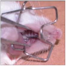
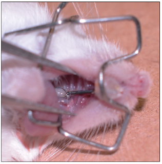
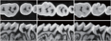
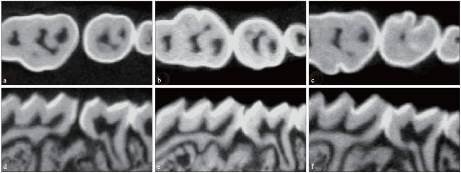
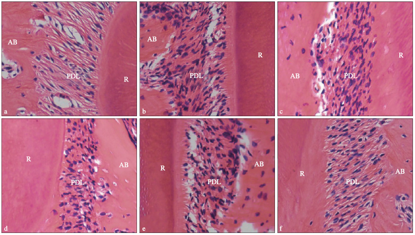
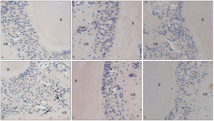
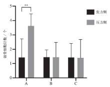
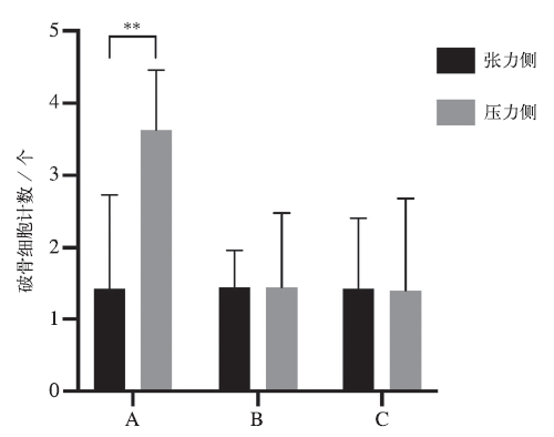
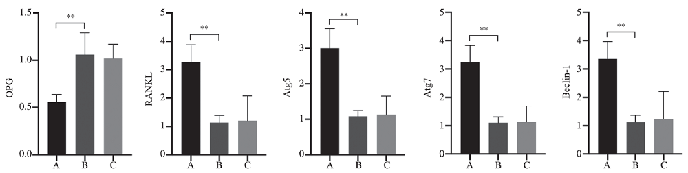
目的 研究小鼠正畸牙移动模型中移动牙牙周组织自噬相关基因的表达,探讨自噬在正畸牙移动骨塑建中的作用。方法 30只11周龄雄性C57B/6小鼠随机分为3组,每组10只。A组构建小鼠正畸牙移动模型,安放镍钛拉簧并施加矫治力;B组放置拉簧但不加矫治力;C组空白组。加力装置均安放于上颌左侧第一磨牙与左侧切牙之间,加力至第12 d处死小鼠。取左侧上颌骨标本,制作左侧上颌第一磨牙牙周组织标本切片,采用微计算机断层扫描技术测量牙移动距离,使用苏木精-伊红(HE)染色、抗酒石酸酸性磷酸酶(TRAP)染色和实时荧光定量聚合酶联式反应(RT-qPCR)观察牙移动情况,检测牙周组织中骨塑建相关基因[骨保护因子(OPG)和破骨细胞核因子κB受体活化因子配体(RANKL)]和自噬相关基因(包括Atg5、Atg7和Beclin-1)的表达。结果 A组平均牙移动距离0.09 mm,HE染色显示A组牙根张力侧与压力侧的牙周膜间隙不均等,压力侧细胞受到挤压、张力侧细胞受到拉伸。TRAP染色显示A组第一磨牙牙根近中侧见到阳性破骨细胞且A组压力侧破骨细胞数大于张力侧,差异有统计学意义(P<0.05)。RT-qPCR显示A组牙周组织OPG信使RNA(mRNA)表达显著降低,RANKL、Atg5、Atg7及Beclin-1 mRNA表达显著增加,差异有统计学意义(P<0.05)。结论 正畸力作用下牙周组织自噬相关基因表达水平升高,自噬可能通过影响牙周组织中破骨水平参与了正畸牙移动骨塑建过程。
中图分类号:
| [1] |
Masella RS, Meister M. Current concepts in the biology of orthodontic tooth movement[J]. Am J Orthod Dentofacial Orthop, 2006,129(4):458-468.
pmid: 16627170 |
| [2] |
Krishnan V, Davidovitch Z. On a path to unfolding the biological mechanisms of orthodontic tooth movement[J]. J Dent Res, 2009,88(7):597-608.
doi: 10.1177/0022034509338914 pmid: 19641146 |
| [3] |
Mizushima N. Autophagy: process and function[J]. Genes Dev, 2007,21(22):2861-2873.
pmid: 18006683 |
| [4] |
Levine B, Kroemer G. Autophagy in the pathogenesis of disease[J]. Cell, 2008,132(1):27-42.
doi: 10.1016/j.cell.2007.12.018 pmid: 18191218 |
| [5] |
Pierrefite-Carle V, Santucci-Darmanin S, Breuil V, et al. Autophagy in bone: self-eating to stay in balance[J]. Ageing Res Rev, 2015,24(Pt B):206-217.
doi: 10.1016/j.arr.2015.08.004 pmid: 26318060 |
| [6] |
Jaber FA, Khan NM, Ansari MY, et al. Autophagy plays an essential role in bone homeostasis[J]. J Cell Physiol, 2019,234(8):12105-12115.
pmid: 30820954 |
| [7] |
He D, Kou X, Luo Q, et al. Enhanced M1/M2 ma-crophage ratio promotes orthodontic root resorption[J]. J Dent Res, 2015,94(1):129-139.
pmid: 25344334 |
| [8] |
Ren YJ, Maltha JC, Kuijpers-Jagtman AM. The rat as a model for orthodontic tooth movement: a critical review and a proposed solution[J]. Eur J Orthod, 2004,26(5):483-490.
doi: 10.1093/ejo/26.5.483 pmid: 15536836 |
| [9] | Norton LA, Burstone CJ. The biology of tooth move-ment[M]. Boca Raton: CRC Press, 1989: 321-334. |
| [10] |
Ren YJ, Maltha JC, Kuijpers-Jagtman AM. Optimum force magnitude for orthodontic tooth movement: a systematic literature review[J]. Angle Orthod, 2003,73(1):86-92.
doi: 10.1043/0003-3219(2003)073<0086:OFMFOT>2.0.CO;2 pmid: 12607860 |
| [11] |
Gonzales C, Hotokezaka H, Yoshimatsu M, et al. Force magnitude and duration effects on amount of tooth movement and root resorption in the rat molar[J]. Angle Orthod, 2008,78(3):502-509.
doi: 10.2319/052007-240.1 pmid: 18416627 |
| [12] |
Yoshimatsu M, Shibata Y, Kitaura H, et al. Experimental model of tooth movement by orthodontic force in mice and its application to tumor necrosis factor receptor-deficient mice[J]. J Bone Miner Metab, 2006,24(1):20-27.
doi: 10.1007/s00774-005-0641-4 pmid: 16369894 |
| [13] |
Fujimura Y, Kitaura H, Yoshimatsu M, et al. In-fluence of bisphosphonates on orthodontic tooth movement in mice[J]. Eur J Orthod, 2009,31(6):572-577.
doi: 10.1093/ejo/cjp068 pmid: 19840975 |
| [14] |
Yoshimatsu M, Kitaura H, Fujimura Y, et al. Inhibi-tory effects of IL-12 on experimental tooth move-ment and root resorption in mice[J]. Arch Oral Biol, 2012,57(1):36-43.
doi: 10.1016/j.archoralbio.2011.07.006 pmid: 21821230 |
| [15] |
Chung CJ, Soma K, Rittling SR, et al. OPN de-ficiency suppresses appearance of odontoclastic cells and resorption of the tooth root induced by experimental force application[J]. J Cell Physiol, 2008,214(3):614-620.
doi: 10.1002/jcp.21250 pmid: 17894420 |
| [16] |
King JS, Veltman DM, Insall RH. The induction of autophagy by mechanical stress[J]. Autophagy, 2011,7(12):1490-1499.
doi: 10.4161/auto.7.12.17924 pmid: 22024750 |
| [17] |
Ma KG, Shao ZW, Yang SH, et al. Autophagy is activated in compression-induced cell degeneration and is mediated by reactive oxygen species in nucleus pulposus cells exposed to compression[J]. Osteoarthr Cartil, 2013,21(12):2030-2038.
doi: 10.1016/j.joca.2013.10.002 |
| [18] |
Baskaran R, Poornima P, Priya LB, et al. Neferine prevents autophagy induced by hypoxia through activation of Akt/mTOR pathway and Nrf2 in muscle cells[J]. Biomed Pharmacother, 2016,83:1407-1413.
doi: 10.1016/j.biopha.2016.08.063 pmid: 27583981 |
| [19] |
Memmert S, Damanaki A, Weykopf B, et al. Auto-phagy in periodontal ligament fibroblasts under biomechanical loading[J]. Cell Tissue Res, 2019,378(3):499-511.
doi: 10.1007/s00441-019-03063-1 pmid: 31352550 |
| [20] | 周云. 正畸牙齿移动过程中牙周膜成纤维细胞自噬作用的初步研究[D]. 西安: 第四军医大学, 2014. |
| Zhou Y. Study on autophagy in hPDLCs during orthodontic tooth movement[D]. Xi’an: The Fourth Military Medical University, 2014. | |
| [21] |
Glick D, Barth S, MacLeod KF. Autophagy: cellular and molecular mechanisms[J]. J Pathol, 2010,221(1):3-12.
doi: 10.1002/path.2697 pmid: 20225336 |
| [22] |
Chen LY, Mo SZ, Hua YM. Compressive force-induced autophagy in periodontal ligament cells downregulates osteoclastogenesis during tooth movement[J]. J Periodontol, 2019,90(10):1170-1181.
pmid: 31077358 |
| [23] |
Kuballa P, Nolte WM, Castoreno AB, et al. Auto-phagy and the immune system[J]. Annu Rev Immunol, 2012,30:611-646.
pmid: 22449030 |
| [24] |
Zhao Y, Chen G, Zhang W, et al. Autophagy re-gulates hypoxia-induced osteoclastogenesis through the HIF-1α/BNIP3 signaling pathway[J]. J Cell Physiol, 2012,227(2):639-648.
doi: 10.1002/jcp.22768 pmid: 21465467 |
| [25] |
Shi J, Wang L, Zhang HY, et al. Glucocorticoids: dose-related effects on osteoclast formation and function via reactive oxygen species and autophagy[J]. Bone, 2015,79:222-232.
pmid: 26115910 |
| [26] |
Wijekoon S, Bwalya EC, Fang J, et al. Chronological differential effects of pro-inflammatory cytokines on RANKL-induced osteoclast differentiation of canine bone marrow-derived macrophages[J]. J Vet Med Sci, 2017,79(12):2030-2035.
doi: 10.1292/jvms.17-0393 pmid: 29109351 |
| [27] | 吕佳岭, 徐洁, 曾锦, 等. 正畸牙压力区牙周膜细胞自噬相关蛋白Beclin-1与微管相关蛋白2轻链3的表达[J]. 华西口腔医学杂志, 2019,37(2):168-173. |
| Lü JL, Xu J, Zeng J, et al. Expression of auto-phagy-related protein Beclin-1 and microtubule-associated protein 2 light chain 3 in periodontal ligament cells in orthodontic tooth pressure areas[J]. West China J Stomatol, 2019,37(2):168-173. | |
| [28] | Alhashimi N, Frithiof L, Brudvik P, et al. Ortho-dontic tooth movement and de novo synjournal of proinflammatory cytokines[J]. Am J Orthod Dento-facial Orthop, 2001,119(3):307-312. |
| [29] |
Nollet M, Santucci-Darmanin S, Breuil V, et al. Au-tophagy in osteoblasts is involved in mineralization and bone homeostasis[J]. Autophagy, 2014,10(11):1965-1977.
doi: 10.4161/auto.36182 pmid: 25484092 |
| [30] |
Li HX, Li DH, Ma ZM, et al. Defective autophagy in osteoblasts induces endoplasmic reticulum stress and causes remarkable bone loss[J]. Autophagy, 2018,14(10):1726-1741.
doi: 10.1080/15548627.2018.1483807 pmid: 29962255 |
| [31] |
Yamaguchi M. RANK/RANKL/OPG during ortho-dontic tooth movement[J]. Orthod Craniofac Res, 2009,12(2):113-119.
doi: 10.1111/j.1601-6343.2009.01444.x pmid: 19419454 |
| [1] | 古丽其合热·阿布来提,秦旭,朱光勋. 线粒体自噬在牙周炎发生发展过程中的研究进展[J]. 国际口腔医学杂志, 2024, 51(1): 68-73. |
| [2] | 叶玉琳,江莉婷,高益鸣. 舍格伦综合征唾液腺中自噬现象的研究进展[J]. 国际口腔医学杂志, 2022, 49(5): 556-560. |
| [3] | 李归平,秦旭,朱光勋. 腺苷酸活化蛋白激酶在牙周病发生发展中的研究进展[J]. 国际口腔医学杂志, 2022, 49(3): 343-348. |
| [4] | 方苓力,谭玺,叶雨丝,黄兰,何瑶. 颞下颌关节退行性变早期髁突软骨细胞行为改变的实验研究[J]. 国际口腔医学杂志, 2021, 48(4): 417-425. |
| [5] | 周丰,陈野,陈晨,张奕宁,耿瑞蔓,刘戟. 沉默信息调节因子1调控牙周炎发生发展的机制[J]. 国际口腔医学杂志, 2021, 48(3): 341-346. |
| [6] | 赵玉洁,管晓燕,李小兰,陈琦君,王倩,刘建国. 巨噬细胞极化参与正畸牙移动的研究进展[J]. 国际口腔医学杂志, 2020, 47(4): 478-483. |
| [7] | 朱俊瑾,周佳琦,伍颖颖. 哺乳动物雷帕霉素靶蛋白复合物1介导的自噬对骨代谢的调控[J]. 国际口腔医学杂志, 2020, 47(1): 84-89. |
| [8] | 杨亚,陈鹏,戴红卫,张林. 大鼠正畸牙移动过程中转化生长因子-β/Smad信号通路相关蛋白质在Malassez上皮剩余细胞的表达变化[J]. 国际口腔医学杂志, 2019, 46(3): 270-276. |
| [9] | 张鹏, 丁一, 王琪. 炎性衰老在糖尿病牙周炎中的作用机制及研究现状[J]. 国际口腔医学杂志, 2017, 44(6): 664-668. |
| [10] | 陈冠辉 侯劲松. 低氧和自噬与肿瘤的发生发展[J]. 国际口腔医学杂志, 2016, 43(5): 584-588. |
| [11] | 任静宜1 刘歆婵1 丁烨1 于洪强1 周延民1 于维先2. 细胞自噬和炎症反应的相互调控与牙周炎[J]. 国际口腔医学杂志, 2016, 43(4): 462-467. |
| [12] | 颜子淇1 何武林2 邹淑娟1. 低强度激光促进正畸治疗牙移动的研究进展[J]. 国际口腔医学杂志, 2014, 41(2): 169-171. |
| [13] | 金淑芳 蒋灿华. 细胞自噬相关蛋白8及其连接系统与头颈部恶性肿瘤[J]. 国际口腔医学杂志, 2014, 41(2): 195-198. |
| [14] | 邢雪 卢树静 金鑫综述 陈谦明 曾昕审校. 细胞自噬及其与口腔鳞状细胞癌间的相关性[J]. 国际口腔医学杂志, 2013, 40(2): 253-256. |
| [15] | 常珍1综述 李容林1 李春阳2审校. 细胞自噬在口腔扁平苔藓恶变中作用的研究进展[J]. 国际口腔医学杂志, 2012, 39(3): 416-420. |
|