国际口腔医学杂志 ›› 2023, Vol. 50 ›› Issue (1): 37-42.doi: 10.7518/gjkq.2023009
李佩桐( ),时彬冕,许春梅,谢旭东,王骏()
Li Peitong(),Shi Binmian,Xu Chunmei,Xie Xudong,Wang Jun.()
摘要:
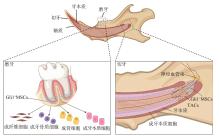
Hedgehog(Hh)信号通路在哺乳动物的组织发育和器官形成中发挥重要作用。Gli1是Hh信号通路中重要的转录因子之一,并已被证实为间充质干细胞(MSCs)可靠的体内标记物。Gli1阳性MSCs具有自我更新能力和多向分化潜能,在牙及牙周组织中可分化为多种功能细胞,包括成牙本质细胞、成牙骨质细胞、成纤维细胞和成骨细胞,参与组织的生长发育与稳态维持。本文对目前关于Gli1阳性MSCs在牙及牙周组织的分布与作用的研究进展作一综述,以期为牙及牙周组织的再生治疗提供新思路。
中图分类号:
| 1 | Dominici M, le Blanc K, Mueller I, et al. Minimal criteria for defining multipotent mesenchymal stromal cells. The International Society for Cellular Therapy position statement[J]. Cytotherapy, 2006, 8(4): 315-317. |
| 2 | Mabuchi Y, Okawara C, Méndez-Ferrer S, et al. Cellular heterogeneity of mesenchymal stem/stromal ce-lls in the bone marrow[J]. Front Cell Dev Biol, 2021, 9: 689366. |
| 3 | Wilson A, Webster A, Genever P. Nomenclature and heterogeneity: consequences for the use of mesenchymal stem cells in regenerative medicine[J]. Regen Med, 2019, 14(6): 595-611. |
| 4 | Hosoya A, Shalehin N, Takebe H, et al. Sonic hedgehog signaling and tooth development[J]. Int J Mol Sci, 2020, 21(5): E1587. |
| 5 | Men Y, Wang YH, Yi YT, et al. Gli1+ periodontium stem cells are regulated by osteocytes and occlusal force[J]. Dev Cell, 2020, 54(5): 639-654.e6. |
| 6 | Kan C, Chen LJ, Hu YY, et al. Gli1-labeled adult mesenchymal stem/progenitor cells and hedgehog signaling contribute to endochondral heterotopic ossification[J]. Bone, 2018, 109: 71-79. |
| 7 | Franchi F, Peterson KM, Quandt K, et al. Impaired hedgehog-Gli1 pathway activity underlies the vascular phenotype of polycystic kidney disease[J]. Hypertension, 2020, 76(6): 1889-1897. |
| 8 | He J, Zuo QZ, Hu B, et al. A novel, liver-specific long noncoding RNA LINC01093 suppresses HCC progression by interaction with IGF2BP1 to facilitate decay of GLI1 mRNA[J]. Cancer Lett, 2019, 450: 98-109. |
| 9 | Cassandras M, Wang CQ, Kathiriya J, et al. Gli1+ mesenchymal stromal cells form a pathological niche to promote airway progenitor metaplasia in the fib-rotic lung[J]. Nat Cell Biol, 2020, 22(11): 1295-1306. |
| 10 | Guan WW, Zhang J, Chen J. Connection of GLI1 variants to congenital heart disease susceptibility: a case-control study[J]. Medicine (Baltimore), 2020, 99(27): e19868. |
| 11 | Kramann R, Goettsch C, Wongboonsin J, et al. Adventitial MSC-like cells are progenitors of vascular smooth muscle cells and drive vascular calcification in chronic kidney disease[J]. Cell Stem Cell, 2016, 19(5): 628-642. |
| 12 | Nüsslein-Volhard C, Wieschaus E. Mutations affecting segment number and polarity in Drosophila[J]. Nature, 1980, 287(5785): 795-801. |
| 13 | Rimkus TK, Carpenter RL, Qasem S, et al. Targeting the sonic hedgehog signaling pathway: review of smoothened and GLI inhibitors[J]. Cancers (Basel), 2016, 8(2): E22. |
| 14 | Pak E, Segal RA. Hedgehog signal transduction: key players, oncogenic drivers, and cancer therapy[J]. Dev Cell, 2016, 38(4): 333-344. |
| 15 | Pietrobono S, Gagliardi S, Stecca B. Non-canonical hedgehog signaling pathway in cancer: activation of GLI transcription factors beyond smoothened[J]. Front Genet, 2019, 10: 556. |
| 16 | Jing D, Li CY, Yao K, et al. The vital role of Gli1+ mesenchymal stem cells in tissue development and homeostasis[J]. J Cell Physiol, 2021, 236(9): 6077-6089. |
| 17 | Taipale J, Cooper MK, Maiti T, et al. Patched acts catalytically to suppress the activity of smoothened[J]. Nature, 2002, 418(6900): 892-897. |
| 18 | Sabol M, Trnski D, Musani V, et al. Role of GLI transcription factors in pathogenesis and their potential as new therapeutic targets[J]. Int J Mol Sci, 2018, 19(9): E2562. |
| 19 | Janečková E, Feng JF, Li JY, et al. Dynamic activation of Wnt, Fgf, and Hh signaling during soft pa-late development[J]. PLoS One, 2019, 14(10): e0223879. |
| 20 | Skoda AM, Simovic D, Karin V, et al. The role of the Hedgehog signaling pathway in cancer: a comprehensive review[J]. Bosn J Basic Med Sci, 2018, 18(1): 8-20. |
| 21 | 王韵, 谢旭东, 许春梅, 等. Gli1阳性细胞在牙周组织发育中的时空分布特点及功能研究[J]. 华西口腔医学杂志, 2020, 38(2): 128-132. |
| Wang Y, Xie XD, Xu CM, et al. Temporal and spatial distribution of Gli1+ cells and their function during periodontal development[J]. West China J Stomatol, 2020, 38(2): 128-132. | |
| 22 | Warshawsky H, Smith CE. Morphological classification of rat incisor ameloblasts[J]. Anat Rec, 1974, 179(4): 423-446. |
| 23 | Seidel K, Marangoni P, Tang C, et al. Resolving stem and progenitor cells in the adult mouse incisor through gene co-expression analysis[J]. Elife, 2017, 6: e24712. |
| 24 | Seidel K, Ahn CP, Lyons D, et al. Hedgehog signaling regulates the generation of ameloblast progenitors in the continuously growing mouse incisor[J]. Development, 2010, 137(22): 3753-3761. |
| 25 | Gerlach JC, Over P, Turner ME, et al. Perivascular mesenchymal progenitors in human fetal and adult liver[J]. Stem Cells Dev, 2012, 21(18): 3258-3269. |
| 26 | Zhao H, Feng JF, Seidel K, et al. Secretion of shh by a neurovascular bundle niche supports mesenchymal stem cell homeostasis in the adult mouse incisor[J]. Cell Stem Cell, 2018, 23(1): 147. |
| 27 | Bitgood MJ, McMahon AP. Hedgehog and Bmp genes are coexpressed at many diverse sites of cell-cell interaction in the mouse embryo[J]. Dev Biol, 1995, 172(1): 126-138. |
| 28 | Cobourne MT, Miletich I, Sharpe PT. Restriction of sonic hedgehog signalling during early tooth development[J]. Development, 2004, 131(12): 2875-2885. |
| 29 | Dassule HR, Lewis P, Bei M, et al. Sonic hedgehog regulates growth and morphogenesis of the tooth[J]. Development, 2000, 127(22): 4775-4785. |
| 30 | Chen S, Jing JJ, Yuan Y, et al. Runx2+ niche cells maintain incisor mesenchymal tissue homeostasis th-rough IGF signaling[J]. Cell Rep, 2020, 32(6): 108007. |
| 31 | Shi C, Yuan Y, Guo Y, et al. BMP signaling in regulating mesenchymal stem cells in incisor homeostasis[J]. J Dent Res, 2019, 98(8): 904-911. |
| 32 | Imhof T, Balic A, Heilig J, et al. Pivotal role of tenascin-W (-N) in postnatal incisor growth and periodontal ligament remodeling[J]. Front Immunol, 2020, 11: 608223. |
| 33 | Sharpe PT. Dental mesenchymal stem cells[J]. Development, 2016, 143(13): 2273-2280. |
| 34 | Pang YW, Feng JF, Daltoe F, et al. Perivascular stem cells at the tip of mouse incisors regulate tissue regeneration[J]. J Bone Miner Res, 2016, 31(3): 514-523. |
| 35 | Jernvall J, Thesleff I. Tooth shape formation and tooth renewal: evolving with the same signals[J]. Development, 2012, 139(19): 3487-3497. |
| 36 | Ishikawa Y, Nakatomi M, Ida-Yonemochi H, et al. Quiescent adult stem cells in murine teeth are regulated by Shh signaling[J]. Cell Tissue Res, 2017, 369(3): 497-512. |
| 37 | Li C, Jing Y, Wang K, et al. Dentinal mineralization is not limited in the mineralization front but occurs along with the entire odontoblast process[J]. Int J Biol Sci, 2018, 14(7): 693-704. |
| 38 | Liu Y, Feng JF, Li JY, et al. An Nfic-hedgehog signaling cascade regulates tooth root development[J]. Development, 2015, 142(19): 3374-3382. |
| 39 | Hardcastle Z, Mo R, Hui CC, et al. The Shh signalling pathway in tooth development: defects in Gli2 and Gli3 mutants[J]. Development, 1998, 125(15): 2803-2811. |
| 40 | Feng JF, Jing JJ, Li JY, et al. BMP signaling orchestrates a transcriptional network to control the fate of mesenchymal stem cells in mice[J]. Development, 2017, 144(14): 2560-2569. |
| 41 | Xie X, Xu C, Zhao H, et al. A biphasic feature of Gli1+-mesenchymal progenitors during cementogenesis that is positively controlled by wnt/β-catenin signaling[J]. J Dent Res, 2021, 100(11): 1289-1298. |
| 42 | Liu AQ, Zhang LS, Chen J, et al. Mechanosensing by Gli1+ cells contributes to the orthodontic force-induced bone remodelling[J]. Cell Prolif, 2020, 53(5): e12810. |
| 43 | Panciera T, Azzolin L, Cordenonsi M, et al. Mechanobiology of YAP and TAZ in physiology and disease[J]. Nat Rev Mol Cell Biol, 2017, 18(12): 758-770. |
| 44 | Maurer M, Lammerding J. The driving force: nuclear mechanotransduction in cellular function, fate, and di-sease[J]. Annu Rev Biomed Eng, 2019, 21: 443-468. |
| 45 | Qi J, Zhou YL, Jiao ZY, et al. Exosomes derived from human bone marrow mesenchymal stem cells promote tumor growth through hedgehog signaling pathway[J]. Cell Physiol Biochem, 2017, 42(6): 2242-2254. |
| 46 | Kan C, Ding N, Yang JZ, et al. BMP-dependent, injury-induced stem cell niche as a mechanism of heterotopic ossification[J]. Stem Cell Res Ther, 2019, 10(1): 14. |
| 47 | Ji QJ, Hou JW, Yong XQ, et al. Targeted dual small interfering ribonucleic acid delivery via non-viral polymeric vectors for pulmonary fibrosis therapy[J]. Adv Mater, 2021, 33(12): e2007798. |
| [1] | 徐书奎,张珊,谢新宇,马文盛. 上颌前方牵引矫治骨性Ⅲ类错 畸形远期疗效稳定性的研究进展[J]. 国际口腔医学杂志, 2023, 50(6): 646-652. 畸形远期疗效稳定性的研究进展[J]. 国际口腔医学杂志, 2023, 50(6): 646-652. |
| [2] | 石佳鑫,王淳艺,李精韬. Pierre Robin序列征患者腭裂临床治疗的研究进展[J]. 国际口腔医学杂志, 2023, 50(2): 237-242. |
| [3] | 张宇宁,曾妮,张焙,石冰,郑谦. 咽后壁瓣咽成形术对腭裂术后患者颌面部生长影响的初步研究[J]. 国际口腔医学杂志, 2023, 50(1): 66-71. |
| [4] | 张珊,葛晓磊,李杰,谢新宇,常维维,马文盛. 上颌前方牵引矫治对颌骨生长发育长期影响的Meta分析[J]. 国际口腔医学杂志, 2022, 49(5): 548-555. |
| [5] | 黎静文,周力. 颈椎成熟法评估下颌骨骨龄的研究进展[J]. 国际口腔医学杂志, 2022, 49(3): 337-342. |
| [6] | 施培磊,于晨浩,谢旭东,吴亚菲,王骏. 牙源性间充质干细胞应用于牙周组织缺损修复的研究进展[J]. 国际口腔医学杂志, 2021, 48(6): 690-695. |
| [7] | 刘嘉程,孟昭松,李宏捷,隋磊. 卵泡抑素在口腔颌面部发育中的作用及其治疗应用前景[J]. 国际口腔医学杂志, 2021, 48(5): 556-562. |
| [8] | 叶冠琛,余晓雯,赵飞亚,俞梦飞,王柏翔,王慧明. 上颌窦提升术前上颌窦病变评估和处理的研究进展[J]. 国际口腔医学杂志, 2021, 48(4): 468-474. |
| [9] | 邓诗勇,宫苹,谭震. 脑和肌肉芳香烃受体核转运样蛋白1基因调控口腔及全身骨代谢的作用[J]. 国际口腔医学杂志, 2021, 48(2): 198-204. |
| [10] | 陈野, 周丰, 邬琼辉, 车会凌, 李佳璇, 申佳琪, 罗恩. 脂联素对骨髓间充质干细胞的作用及其调控机制[J]. 国际口腔医学杂志, 2021, 48(1): 58-63. |
| [11] | 金作林. 颅面部生长发育与早期生长改良[J]. 国际口腔医学杂志, 2021, 48(1): 7-11. |
| [12] | 吕辉,王华,孙雯. 辅助性T细胞17与牙周炎骨免疫[J]. 国际口腔医学杂志, 2020, 47(6): 661-668. |
| [13] | 杨叶青,陈明,吴补领. 环状非编码RNA在间充质干细胞成骨向分化中作用的研究进展[J]. 国际口腔医学杂志, 2020, 47(3): 257-262. |
| [14] | 刘俊圻,陈艺尹,杨文宾. RNA腺嘌呤6-甲基化修饰调控骨髓间充质干细胞成骨向分化的研究进展[J]. 国际口腔医学杂志, 2020, 47(3): 263-269. |
| [15] | 朱明静,张清彬. 生长因子诱导间充质干细胞三维体外软骨形成的研究进展[J]. 国际口腔医学杂志, 2020, 47(3): 270-277. |
|