国际口腔医学杂志 ›› 2020, Vol. 47 ›› Issue (6): 686-692.doi: 10.7518/gjkq.2020087
吴洁林1,2( ),高莺2()
Wu Jielin1,2(),Gao Ying2()
摘要:
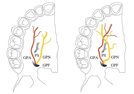
软组织增量手术是目前改善天然牙与种植体周围软组织宽度、厚度欠佳的理想术式,硬腭黏膜是该手术的常用供区。术前精准定位腭大神经血管束,术中重视所获取移植物的厚度和成分,术后保证游离移植物稳固及贴合,是提高硬腭来源游离软组织移植物成活率、确保有效实施软组织增量手术的重要基础。本文就硬腭黏膜获取游离软组织移植物的相关解剖基础、游离软组织移植物存活要素及其临床应用等方面进行总结,以期为手术提供依据。
中图分类号:
| [1] |
Cagimni P, Govsa F, Ozer MA, et al. Computerized analysis of the greater palatine foramen to gain the palatine neurovascular bundle during palatal surgery[J]. Surg Radiol Anat, 2017,39(2):177-184.
doi: 10.1007/s00276-016-1691-0 pmid: 27177906 |
| [2] |
Fu JH, Hasso DG, Yeh CY, et al. The accuracy of identifying the greater palatine neurovascular bundle: a cadaver study[J]. J Periodontol, 2011,82(7):1000-1006.
doi: 10.1902/jop.2011.100619 pmid: 21284546 |
| [3] | Wang TM, Kuo KJ, Shih C, et al. Assessment of the relative locations of the greater palatine foramen in adult Chinese skulls[J]. Acta Anat (Basel), 1988,132(3):182-186. |
| [4] | 薛绯, 段晋瑜, 张瑞. 汉族人群腭大孔解剖位置及其与腭穹隆形态关系的CBCT研究[J]. 实用口腔医学杂志, 2018,34(3):364-367. |
| Xue F, Duan JY, Zhang R. Anatomical variation of the greater palatine foremen and its relationship with palatal vault morphology in Han population studied by CBCT[J]. J Pract Stomat, 2018,34(3):364-367. | |
| [5] |
Reiser GM, Bruno JF, Mahan PE, et al. The sube-pithelial connective tissue graft palatal donor site: anatomic considerations for surgeons[J]. Int J Periodontics Restorative Dent, 1996,16(2):130-137.
pmid: 9084301 |
| [6] |
Benninger B, Andrews K, Carter W. Clinical mea-surements of hard palate and implications for subepi-thelial connective tissue grafts with suggestions for palatal nomenclature[J]. J Oral Maxillofac Surg, 2012,70(1):149-153.
pmid: 21802817 |
| [7] |
Monnet-Corti V, Santini A, Glise JM, et al. Connective tissue graft for gingival recession treatment: assess-ment of the maximum graft dimensions at the palatal vault as a donor site[J]. J Periodontol, 2006,77(5):899-902.
doi: 10.1902/jop.2006.050047 pmid: 16671884 |
| [8] |
Monsour P, Huang T. Morphology of the greater palatine grooves of the hard palate: a cone beam computed tomography study[J]. Aust Dent J, 2016,61(3):329-332.
pmid: 26435329 |
| [9] |
Yu SK, Lee MH, Park BS, et al. Topographical rela-tionship of the greater palatine artery and the palatal spine. Significance for periodontal surgery[J]. J Clin Periodontol, 2014,41(9):908-913.
doi: 10.1111/jcpe.12288 pmid: 25041323 |
| [10] |
Klosek SK, Rungruang T. Anatomical study of the greater palatine artery and related structures of the palatal vault: considerations for palate as the subepi-thelial connective tissue graft donor site[J]. Surg Radiol Anat, 2009,31(4):245-250.
pmid: 19015806 |
| [11] | 刘玲玲, 刘树泰. 上颌腭侧软组织厚度的测量方法及影响因素[J]. 国际口腔医学杂志, 2019,46(2):234-237. |
| Liu LL, Liu ST. Measurement methods and relevant factors of the soft tissue thickness in the palatal mas-ticatory mucosa of maxillary[J]. J Int Stomatol, 2019,46(2):234-237. | |
| [12] | Zuhr O, Bäumer D, Hürzeler M. The addition of soft tissue replacement grafts in plastic periodontal and implant surgery: critical elements in design and exe-cution[J]. J Clin Periodontol, 2014,41(Suppl 15):S123-S142. |
| [13] | Sanz M, Simion M, Working Group of the European Workshop on Periodontology. Surgical techniques on periodontal plastic surgery and soft tissue regene-ration: consensus report of Group 3 of the 10th Euro-pean Workshop on Periodontology[J]. J Clin Perio-dontol, 2014,41(Suppl 15):S92-S97. |
| [14] | 万双全, 邓飞龙. 上皮下结缔组织瓣在种植软组织缺陷中的应用[J]. 国际口腔医学杂志, 2018,45(1):68-73. |
| Wan SQ, Deng FL. Clinical applications of subepi-thelial connective tissue grafting in soft tissue de-ficiencies of dental implantation[J]. Int J Stomatol, 2018,45(1):68-73. | |
| [15] |
Mörmann W, Schaer F, Firestone AR. The relation-ship between success of free gingival grafts and transplant thickness. Revascularization and shrin-kage: a one year clinical study[J]. J Periodontol, 1981,52(2):74-80.
doi: 10.1902/jop.1981.52.2.74 pmid: 6164778 |
| [16] | Müller HP. Periodontology: the essentials[M]. New York: Thieme Medical Publication, 2005: 65-119. |
| [17] |
Vellis J, Kutkut A, Al-Sabbagh M. Comparison of xenogeneic collagen matrix vs. free gingival grafts to increase the zone of keratinized mucosa around functioning implants[J]. Implant Dent, 2019,28(1):20-27.
doi: 10.1097/ID.0000000000000842 pmid: 30461439 |
| [18] | 张停停, 宗娟娟. 自体结缔组织移植术的研究现状[J]. 国际口腔医学杂志, 2019,46(1):89-93. |
| Zhang TT, Zong JJ. Research progress on connective tissue graft[J]. Int J Stomatol, 2019,46(1):89-93. | |
| [19] | Zucchelli G. 膜龈美学手术精要[M]. 束蓉, 沈阳: 辽宁科学技术出版社, 2016: 425-436. |
| Zucchelli G. Mucogingival esthetic surgery[M]. Shu R, trans. Shenyang: Liaoning Science and Technology Press, 2016: 425-436. | |
| [20] |
Yu SK, Lee MH, Kim CS, et al. Thickness of the palatal masticatory mucosa with reference to auto-genous grafting: a cadaveric and histologic study[J]. Int J Periodontics Restorative Dent, 2014,34(1):115-121.
doi: 10.11607/prd.1530 pmid: 24396846 |
| [21] |
Bertl K, Pifl M, Hirtler L, et al. Relative composition of fibrous connective and fatty/glandular tissue in connective tissue grafts depends on the harvesting technique but not the donor site of the hard palate[J]. J Periodontol, 2015,86(12):1331-1339.
pmid: 26291293 |
| [22] |
da Silva Neves FL, Silveira CA, Dias SB, et al. Com-parison of two power densities on the healing of palatal wounds after connective tissue graft removal: randomized clinical trial[J]. Lasers Med Sci, 2016,31(7):1371-1378.
pmid: 27344670 |
| [23] |
Bhatavadekar NB, Gharpure AS. Controlled palatal harvest technique for harvesting a palatal subepi-thelial connective tissue graft[J]. Compend Contin Educ Dent, 2018,39(2):e9-e12.
pmid: 29388789 |
| [24] |
Madi M, Kassem A. Topical simvastatin gel as a novel therapeutic modality for palatal donor site wound healing following free gingival graft procedure[J]. Acta Odontol Scand, 2018,76(3):212-219.
pmid: 29145771 |
| [25] | Lee YJ, Kwon YH, Park JB, et al. Epithelial thick-ness of the palatal mucosa: a histomorphometric study in Koreans[J]. Anat Rec (Hoboken), 2010,293(11):1966-1970. |
| [26] |
Zucchelli G, Mele M, Stefanini M, et al. Patient morbidity and root coverage outcome after sube-pithelial connective tissue and de-epithelialized grafts: a comparative randomized-controlled clinical trial[J]. J Clin Periodontol, 2010,37(8):728-738.
pmid: 20590963 |
| [27] |
Agudio G, Nieri M, Rotundo R, et al. Free gingival grafts to increase keratinized tissue: a retrospective long-term evaluation (10 to 25 years) of outcomes[J]. J Periodontol, 2008,79(4):587-594.
doi: 10.1902/jop.2008.070414 pmid: 18380550 |
| [28] |
de Resende DRB, Greghi SLA, Siqueira AF, et al. Acellular dermal matrix allograft versus free gingival graft: a histological evaluation and split-mouth randomized clinical trial[J]. Clin Oral Investig, 2019,23(2):539-550.
pmid: 29713889 |
| [29] |
Tatakis DN, Chambrone L, Allen EP, et al. Perio-dontal soft tissue root coverage procedures: a consensus report from the AAP Regeneration Workshop[J]. J Periodontol, 2015,86(2 Suppl):S52-S55.
doi: 10.1902/jop.2015.140376 pmid: 25315018 |
| [30] |
Kaushik A, Pk P, Jhamb K, et al. Clinical evaluation of papilla reconstruction using subepithelial conne-ctive tissue graft[J]. J Clin Diagn Res, 2014, 8(9):ZC77-ZC81.
pmid: 25386529 |
| [31] |
Lee CT, Chang PC, Touchan N, et al. Root coverage with a modified laterally positioned flap combined with a subepithelial connective tissue graft in advanced recession[J]. J Periodontal Implant Sci, 2014,44(6):300-306.
pmid: 25568811 |
| [32] |
Basegmez C, Karabuda ZC, Demirel K, et al. The comparison of acellular dermal matrix allografts with free gingival grafts in the augmentation of peri-implant attached mucosa: a randomised controlled trial[J]. Eur J Oral Implantol, 2013,6(2):145-152.
pmid: 23926586 |
| [33] |
Zucchelli G, Mazzotti C, Mounssif I, et al. A novel surgical-prosthetic approach for soft tissue dehiscence coverage around single implant[J]. Clin Oral Implants Res, 2013,24(9):957-962.
pmid: 22924841 |
| [34] |
Kan JY, Rungcharassaeng K, Lozada JL, et al. Facial gingival tissue stability following immediate place-ment and provisionalization of maxillary anterior single implants: a 2- to 8-year follow-up[J]. Int J Oral Maxillofac Implants, 2011,26(1):179-187.
pmid: 21365054 |
| [35] | 富晓娇, 张宇. 种植体周围软组织退缩外科处理技术的研究进展[J]. 中华口腔医学杂志, 2019,54(4):267-272. |
| Fu XJ, Zhang Y. Surgical techniques of peri-implant soft tissue recession[J]. Chin J Stomatol, 2019,54(4):267-272. | |
| [36] |
Siegel RJ. Palatal grafts for eyelid reconstruction[J]. Plast Reconstr Surg, 1985,76(3):411-414.
pmid: 4034758 |
| [37] |
Emesz M, Krall EM, Rasp M, et al. Transplants from the hard palate: method for mucosal graft reconstruction of the upper eyelid[J]. Ophthalmologe, 2014,111(9):853-861.
doi: 10.1007/s00347-013-3002-z pmid: 24549685 |
| [1] | 傅豫, 何薇, 黄兰. 铁死亡在口腔疾病中的研究进展[J]. 国际口腔医学杂志, 2024, 51(1): 36-44. |
| [2] | 古丽其合热·阿布来提,秦旭,朱光勋. 线粒体自噬在牙周炎发生发展过程中的研究进展[J]. 国际口腔医学杂志, 2024, 51(1): 68-73. |
| [3] | 罗晓洁,王德续,陈晓涛. 基于生物信息学分析铁死亡调控基因与牙周炎的关系[J]. 国际口腔医学杂志, 2023, 50(6): 661-668. |
| [4] | 黄元鸿,彭显,周学东. 骨碎补在治疗口腔骨相关疾病的研究进展[J]. 国际口腔医学杂志, 2023, 50(6): 679-685. |
| [5] | 廖洪林,方仲瀚,张艳艳,刘飞,沈颉飞. 牙种植术后三叉神经创伤性神经病理性疼痛的诊断与防治[J]. 国际口腔医学杂志, 2023, 50(6): 729-738. |
| [6] | 余岳霖,孔卫东. 甲状旁腺激素受体1基因相关与原发性牙齿萌出障碍的研究进展[J]. 国际口腔医学杂志, 2023, 50(5): 573-580. |
| [7] | 龚美灵,程兴群,吴红崑. 牙周炎与帕金森病相关性的研究进展[J]. 国际口腔医学杂志, 2023, 50(5): 587-593. |
| [8] | 赵玲帆, 周杨, 叶鑫鑫, 张强. 肾移植术后腮腺低分化黏液表皮样癌1例[J]. 国际口腔医学杂志, 2023, 50(4): 419-422. |
| [9] | 范琳,孙江. 微针在口腔医学中的应用[J]. 国际口腔医学杂志, 2023, 50(4): 472-478. |
| [10] | 刘云通,刘畅,高丽钞,罗瑜雪,曹钰彬,华成舸. 术后下牙槽神经功能障碍的研究进展[J]. 国际口腔医学杂志, 2023, 50(4): 479-484. |
| [11] | 孙佳,韩烨,侯建霞. 白细胞介素-6-铁调素信号轴调控牙周炎相关性贫血致病机制的研究进展[J]. 国际口腔医学杂志, 2023, 50(3): 329-334. |
| [12] | 陆倩,夏海斌,王敏. 种植体磨光整形术治疗种植体周围炎的研究进展[J]. 国际口腔医学杂志, 2023, 50(2): 152-158. |
| [13] | 蒋青松,赖文莉,王艳. 骨增量技术在口腔正畸领域的研究进展[J]. 国际口腔医学杂志, 2023, 50(2): 243-250. |
| [14] | 杨梦瑶,高现灵,邓淑丽. 静电纺丝纳米纤维在牙周再生中的应用[J]. 国际口腔医学杂志, 2023, 50(1): 10-18. |
| [15] | 刘体倩,梁星,刘蔚晴,李晓虹,朱睿. 咬合创伤在牙周炎发生发展中的作用及机制的研究进展[J]. 国际口腔医学杂志, 2023, 50(1): 19-24. |
|