Int J Stomatol ›› 2026, Vol. 53 ›› Issue (4): 476-486.doi: 10.7518/gjkq.2026226
• Interpretation of Consensus • Previous Articles Next Articles
Hanping Xia1( ),Juan Liu1,Wenlan Xiao2,Yujie Yao1,Xin Tong2,Fuhua Yan1,Bin Chen1()
CLC Number:
| [1] | Kim WJ, Cho YD, Ku Y, et al. The worldwide pa-tent landscape of dental implant technology[J]. Biomater Res, 2022, 26(1): 59. |
| [2] | Herrera D, Sanz M, Kebschull M, et al. Treatment of stage Ⅳ periodontitis: the EFP S3 level clinical practice guideline[J]. J Clin Periodontol, 2022, 49(): 4-71. |
| [3] | West N, Chapple I, Culshaw S, et al. BSP Implementation of prevention and treatment of peri-implant diseases—the EFP S3 level clinical practice guideline[J]. J Dent, 2024, 149: 104980. |
| [4] | Wang HL, Avila-Ortiz G, Monje A, et al. AO/AAP consensus on prevention and management of peri-implant diseases and conditions: summary report[J]. J Periodontol, 2025, 96(6): 519-541. |
| [5] | Derks J, Tomasi C. Peri-implant health and disease. A systematic review of current epidemiology[J]. J Clin Periodontol, 2015, 42(): S158-S171. |
| [6] | Lee CT, Huang YW, Zhu L, et al. Prevalences of peri-implantitis and peri-implant mucositis: systema-tic review and meta-analysis[J]. J Dent, 2017, 62: 1-12. |
| [7] | Romandini M, Lima C, Pedrinaci I, et al. Prevalence and risk/protective indicators of peri-implant disea-ses: a university-representative cross-sectional study[J]. Clin Oral Implants Res, 2021, 32(1): 112-122. |
| [8] | Apaza-Bedoya K, Galarraga-Vinueza ME, Correa BB, et al. Prevalence, risk indicators, and clinical characteristics of peri-implant mucositis and peri-implantitis for an internal conical connection implant system: a multicenter cross-sectional study[J]. J Periodontol, 2024, 95(6): 582-593. |
| [9] | Obreja K, Ramanauskaite A, Begic A, et al. The prevalence of peri-implant diseases around subcres-tally placed implants: a cross-sectional study[J]. Clin Oral Implants Res, 2021, 32(6): 702-710. |
| [10] | Shi JY, Jie N, Zhuang LF, et al. Peri-implant conditions and marginal bone loss around cemented and screw-retained single implant crowns in posterior regions: a retrospective cohort study with up to 4 years follow-up[J]. PLoS One, 2018, 13(2): e0191717. |
| [11] | Tavelli L, Barootchi S. Prevalence, incidence, risk, and protective factors for soft tissue dehiscences at implant sites in the absence of disease: an AO/AAP systematic review and meta-regression analysis[J]. J Periodontol, 2025, 96(6): 562-586. |
| [12] | Gamborena I, Avila-Ortiz G. Peri-implant marginal mucosa defects: classification and clinical management[J]. J Periodontol, 2021, 92(7): 947-957. |
| [13] | Romandini M, Pedrinaci I, Lima C, et al. Prevalence and risk/protective indicators of buccal soft tissue dehiscence around dental implants[J]. J Clin Periodontol, 2021, 48(3): 455-463. |
| [14] | Renvert S, Polyzois I. Treatment of pathologic peri-implant pockets[J]. Periodontol 2000, 2018, 76(1): 180-190. |
| [15] | Heitz-Mayfield LJ, Mombelli A. The therapy of peri-implantitis: a systematic review[J]. Int J Oral Maxillofac Implants, 2014, 29(): 325-345. |
| [16] | Sanz M, Solonko M, Luengo F. Key factors in prevention of peri-implant diseases[J]. Compend Contin Educ Dent, 2017, 38(8 ): 6-13. |
| [17] | Carra MC, Blanc-Sylvestre N, Courtet A, et al. Primordial and primary prevention of peri-implant di-seases: a systematic review and meta-analysis[J]. J Clin Periodontol, 2023, 50(): 77-112. |
| [18] | Lee JCY, Mattheos N, Nixon KC, et al. Residual periodontal pockets are a risk indicator for peri-implantitis in patients treated for periodontitis[J]. Clin Oral Implants Res, 2012, 23(3): 325-333. |
| [19] | Roccuzzo A, Imber JC, Marruganti C, et al. Clinical outcomes of dental implants in patients with and without history of periodontitis: a 20-year prospective study[J]. J Clin Periodontol, 2022, 49(12): 1346-1356. |
| [20] | Alhakeem M, Kanounisabet N, Nowzari H, et al. Risk indicators of long-term outcome of implant therapy in patients with a history of severe periodontitis or no history of periodontitis: a retrospective cohort study[J]. Int J Dent Hyg, 2023, 21(1): 227-237. |
| [21] | Renvert S, Polyzois I. Risk indicators for peri-implant mucositis: a systematic literature review[J]. J Clin Periodontol, 2015, 42(): S172-S186. |
| [22] | Renvert S, Quirynen M. Risk indicators for peri-implantitis. A narrative review[J]. Clin Oral Implants Res, 2015, 26(): 15-44. |
| [23] | Costa FO, Lages EJP, Cortelli SC, et al. Association between cumulative smoking exposure, span since smoking cessation, and peri-implantitis: a cross-sectional study[J]. Clin Oral Investig, 2022, 26(7): 4835-4846. |
| [24] | Galindo-Moreno P, Fauri M, Avila-Ortiz G, et al. Influence of alcohol and tobacco habits on peri-implant marginal bone loss: a prospective study[J]. Clin Oral Implants Res, 2005, 16(5): 579-586. |
| [25] | Carr BR, Boggess WJ, Coburn JF, et al. Does alcohol consumption protect against late dental implant failures[J]. Oral Surg Oral Med Oral Pathol Oral Radiol, 2021, 131(6): 631-637. |
| [26] | Monje A, Aranda L, Diaz KT, et al. Impact of maintenance therapy for the prevention of peri-implant diseases: a systematic review and meta-analysis[J]. J Dent Res, 2016, 95(4): 372-379. |
| [27] | Chan D, Pelekos G, Ho D, et al. The depth of the implant mucosal tunnel modifies the development and resolution of experimental peri-implant mucositis: a case-control study[J]. J Clin Periodontol, 2019, 46(2): 248-255. |
| [28] | Monje A, Kan JY, Borgnakke W. Impact of local predisposing/precipitating factors and systemic dri-vers on peri-implant diseases[J]. Clin Implant Dent Relat Res, 2023, 25(4): 640-660. |
| [29] | Galindo-Moreno P, León-Cano A, Ortega-Oller I, et al. Prosthetic abutment height is a key factor in peri-implant marginal bone loss[J]. J Dent Res, 2014, 93(7 ): 80S-85S. |
| [30] | Blanco J, Pico A, Caneiro L, et al. Effect of abutment height on interproximal implant bone level in the early healing: a randomized clinical trial[J]. Clin Oral Implants Res, 2018, 29(1): 108-117. |
| [31] | Katafuchi M, Weinstein BF, Leroux BG, et al. Restoration contour is a risk indicator for peri-implantitis: a cross-sectional radiographic analysis[J]. J Clin Periodontol, 2018, 45(2): 225-232. |
| [32] | Strauss FJ, Siegenthaler M, Hämmerle CHF, et al. Restorative angle of zirconia restorations cemented on non-original titanium bases influences the initial marginal bone loss: 5-year results of a prospective cohort study[J]. Clin Oral Implants Res, 2022, 33(7): 745-756. |
| [33] | Mattheos N, Janda M, Acharya A, et al. Impact of design elements of the implant supracrestal complex (ISC) on the risk of peri-implant mucositis and peri-implantitis: a critical review[J]. Clin Oral Implants Res, 2021, 32(): 181-202. |
| [34] | Pjetursson BE, Thoma D, Jung R, et al. A systema-tic review of the survival and complication rates of implant-supported fixed dental prostheses (FDPs) after a mean observation period of at least 5 years[J]. Clin Oral Implants Res, 2012, 23(): 22-38. |
| [35] | Zucchelli G, Tavelli L, Stefanini M, et al. Classification of facial peri-implant soft tissue dehiscence/deficiencies at single implant sites in the esthetic zone[J]. J Periodontol, 2019, 90(10): 1116-1124. |
| [36] | Kumar PS, Kan J, Galarraga-Vinueza ME, et al. Risk for peri-implant diseases and defects: report of workgroup 1 of the joint AO/AAP consensus confe-rence on prevention and management of peri-implant diseases[J]. Clin Adv Periodontics, 2025, 15(2): 93-102. |
| [37] | Fiorellini JP, Mojaver S, Sarmiento H, et al. Clinical translation of the 2024 AO/AAP consensus on prevention and management of peri-implant diseases and conditions[J]. Int J Periodontics Restorative Dent, 2025, 45(4): 1-23. |
| [38] | Galarraga-Vinueza ME, Pagni S, Finkelman M, et al. Prevalence, incidence, systemic, behavioral, and patient-related risk factors and indicators for peri-implant diseases: an AO/AAP systematic review and meta-analysis[J]. J Periodontol, 2025, 96(6): 587-633. |
| [39] | Monje A, Barootchi S, Rosen PS, et al. Surgical- and implant-related factors and onset/progression of peri-implant diseases: an AO/AAP systematic review[J]. J Periodontol, 2025, 96(6): 542-561. |
| [40] | Lin GH, Lee E, Barootchi S, et al. The influence of prosthetic designs on peri-implant bone loss: an AO/AAP systematic review and meta-analysis[J]. J Pe-riodontol, 2025, 96(6): 634-651. |
| [41] | Berglundh T, Armitage G, Araujo MG, et al. Peri-implant diseases and conditions: consensus report of workgroup 4 of the 2017 World Workshop on the Classification of Periodontal and Peri-Implant Di-seases and Conditions[J]. J Clin Periodontol, 2018, 45(): S286-S291. |
| [42] | Tavelli L, Barootchi S, Majzoub J, et al. Volumetric changes at implant sites: a systematic appraisal of traditional methods and optical scanning-based digital technologies[J]. J Clin Periodontol, 2021, 48(2): 315-334. |
| [43] | Tavelli L, Barootchi S, Majzoub J, et al. Prevalence and risk indicators of midfacial peri-implant soft tissue dehiscence at single site in the esthetic zone: a cross-sectional clinical and ultrasonographic study[J]. J Periodontol, 2022, 93(6): 857-866. |
| [44] | Sanz-Martín I, Regidor E, Navarro J, et al. Factors associated with the presence of peri-implant buccal soft tissue dehiscences: a case-control study[J]. J Periodontol, 2020, 91(8): 1003-1010. |
| [45] | Lin GH, Chambrone L, Rajendran Y, et al. Treatment of peri-implant mucositis: an AAP/AO syste-matic review and meta-analysis[J]. Int J Oral Maxillofac Implants, 2025(4): 49-72. |
| [46] | Ravidà A, Dias DR, Lemke R, et al. Efficacy of decontamination methods for biofilm removal from dental implant surfaces and reosseointegration: an AAP/AO systematic review on peri-implant disea-ses and conditions[J]. Int J Oral Maxillofac Implants, 2025(4): 91-160. |
| [47] | Saleh MHA, Misch C, Alrmali A, et al. Efficacy of nonreconstructive surgical treatment of peri-implantitis: an AAP/AO systematic review and meta-analysis of access flap versus osseous surgery procedures[J]. Int J Oral Maxillofac Implants, 2025(4): 73-90. |
| [48] | Barootchi S, Monje A, Sabri H, et al. Surgical reconstructive therapy for the management of peri-implantitis: an AAP/AO systematic review and network meta-analysis[J]. Int J Oral Maxillofac Implants, 2025(4): 1-48. |
| [49] | Figuero E, Graziani F, Sanz I, et al. Management of peri-implant mucositis and peri-implantitis[J]. Perio-dontol 2000, 2014, 66(1): 255-273. |
| [50] | Jepsen S, Berglundh T, Genco R, et al. Primary prevention of peri-implantitis: managing peri-implant mucositis[J]. J Clin Periodontol, 2015, 42(): S152-S157. |
| [51] | Rakic M, Tatic Z, Radovanovic S, et al. Resolution of peri-implant mucositis following standard treatment: a prospective split-mouth study[J]. J Perio-dontol, 2024, 95(9): 842-852. |
| [52] | Iorio-Siciliano V, Blasi A, Isola G, et al. Resolution of peri-implant mucositis at tissue- and bone-level implants: a 6-month prospective controlled clinical trial[J]. Clin Oral Implants Res, 2023, 34(5): 450- 462. |
| [53] | Herrera D, Berglundh T, Schwarz F, et al. Prevention and treatment of peri-implant diseases—the EFP S3 level clinical practice guideline[J]. J Clin Periodontol, 2023, 50(): 4-76. |
| [54] | Roccuzzo M, Layton DM, Roccuzzo A, et al. Clinical outcomes of peri-implantitis treatment and supportive care: a systematic review[J]. Clin Oral Implants Res, 2018, 29(): 331-350. |
| [55] | Butera A, Maiorani C, Gallo S, et al. Evaluation of adjuvant systems in non-surgical peri-implant treatment: a literature review[J]. Healthcare, 2022, 10(5): 886. |
| [56] | Blanco C, Pico A, Dopico J, et al. Adjunctive benefits of systemic metronidazole on non-surgical treatment of peri-implantitis. A randomized placebo-controlled clinical trial[J]. J Clin Periodontol, 2022, 49(1): 15-27. |
| [57] | Ramanauskaite A, Schwarz F. Current concepts for the treatment of peri-implant disease[J]. Int J Pros-thodont, 2024, 37(2): 124-134. |
| [58] | Schwarz F, John G, Mainusch S, et al. Combined surgical therapy of peri-implantitis evaluating two methods of surface debridement and decontamination. A two-year clinical follow up report[J]. J Clin Periodontol, 2012, 39(8): 789-797. |
| [59] | Monje A, Pons R, Peña P. Electrolytic surface decontamination in the reconstructive therapy of peri-implantitis: single-center outcomes[J]. Int J Perio-dontics Restorative Dent, 2025, 45(2): 185-198. |
| [60] | Carcuac O, Derks J, Abrahamsson I, et al. Surgical treatment of peri-implantitis: 3-year results from a randomized controlled clinical trial[J]. J Clin Perio-dontol, 2017, 44(12): 1294-1303. |
| [61] | Karlsson K, Derks J, Håkansson J, et al. Interventions for peri-implantitis and their effects on further bone loss: a retrospective analysis of a registry-based cohort[J]. J Clin Periodontol, 2019, 46(8): 872-879. |
| [62] | Donos N, Calciolari E, Ghuman M, et al. The efficacy of bone reconstructive therapies in the management of peri-implantitis. A systematic review and meta-analysis[J]. J Clin Periodontol, 2023, 50(): 285-316. |
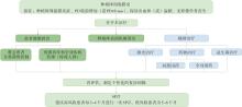
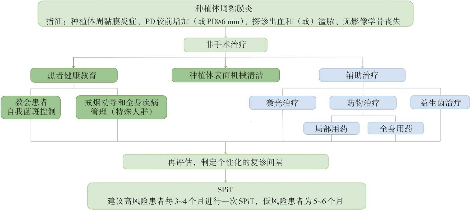
|