Int J Stomatol ›› 2026, Vol. 53 ›› Issue (4): 496-502.doi: 10.7518/gjkq.2026126
• Implantology • Previous Articles Next Articles
Weiyao Xia1( ),Jing Wang2,Yili Qu1()
CLC Number:
| [1] | Quirynen M, Vogels R, Alsaadi G, et al. Predisposing conditions for retrograde peri-implantitis, and treatment suggestions[J]. Clin Oral Implants Res, 2005, 16(5): 599-608. |
| [2] | Chan HL, Wang HL, Bashutski JD, et al. Retrograde peri-implantitis: a case report introducing an approach to its management[J]. J Periodontol, 2011, 82(7): 1080-1088. |
| [3] | Di Murro B, Papi P, Di Murro C, et al. Correlation between endodontic pulpal/periapical disease and re-trograde peri-implantitis: a case series[J]. Aust Endod J, 2021, 47(2): 358-364. |
| [4] | 曾文奕, 杜宇. 逆行性植体周炎的诊断、病因与防治[J]. 中华口腔医学杂志, 2022, 57(3): 302-306. |
| Zeng WY, Du Y. Diagnosis, etiology, prevention and treatment in retrograde peri-implantitis[J]. Chin J Stomatol, 2022, 57(3): 302-306. | |
| [5] | Quirynen M, Gijbels F, Jacobs R. An infected jawbone site compromising successful osseointegration[J]. Periodontol 2000, 2000, 33: 129-144. |
| [6] | Sarmast ND, Wang HH, Sajadi AS, et al. Classification and clinical management of retrograde peri-implantitis associated with apical periodontitis: a proposed classification system and case report[J]. J Endod, 2017, 43(11): 1921-1924. |
| [7] | Al-Ahmad A, Ameen H, Pelz K, et al. Antibiotic resistance and capacity for biofilm formation of diffe-rent bacteria isolated from endodontic infections associated with root-filled teeth[J]. J Endod, 2014, 40(2): 223-230. |
| [8] | Nelson S, Thomas G. Bacterial persistence in den-toalveolar bone following extraction: a microbiological study and implications for dental implant treatment[J]. Clin Implant Dent Relat Res, 2010, 12(4): 306-314. |
| [9] | Ayangco L, Sheridan PJ. Development and treatment of retrograde peri-implantitis involving a site with a history of failed endodontic and apicoectomy procedures: a series of reports[J]. Int J Oral Maxillofac Implants, 2001, 16(3): 412-417. |
| [10] | Alssum LR, Alghofaily MM, Aleyiydi AS, et al. The incidence of retrograde peri-implantitis in a single university dental hospital training center: a retrospective analysis[J]. Medicina, 2023, 59(3): 560. |
| [11] | Peñarrocha-Diago MA, Blaya-Tárraga JA, Menén-dez-Nieto I, et al. Implant survival after surgical treatment of early apical peri-implantitis: an ambispective cohort study covering a 20-year period[J]. Int J Oral Implantol, 2020, 13(2): 161-170. |
| [12] | Peñarrocha-Oltra D, Blaya-Tárraga JA, Menéndez-Nieto I, et al. Factors associated with early apical peri-implantitis: a retrospective study covering a 20-year period[J]. Int J Oral Implantol, 2020, 13(1): 65-73. |
| [13] | Burdurlu MÇ, Dagasan VÇ, Tunç O, et al. Retrograde peri-implantitis: evaluation and treatment protocols of a rare lesion[J]. Quintessence Int, 2021, 52(2): 112-121. |
| [14] | Lefever D, Van Assche N, Temmerman A, et al. Aetiology, microbiology and therapy of periapical lesions around oral implants: a retrospective analysis[J]. J Clin Periodontol, 2013, 40(3): 296-302. |
| [15] | Muñoz-Cámara D, Gilbel-Del Águila O, Pardo-Za-mora G, et al. Immediate post-extraction implants placed in acute periapical infected sites with immediate prosthetic provisionalization: a 1-year prospective cohort study[J]. Med Oral Patol Oral Cir Bucal, 2020, 25(6): e720-e727. |
| [16] | Di Murro B, Canullo L, Pompa G, et al. Prevalence and treatment of retrograde peri-implantitis: a retrospective cohort study covering a 20-year period[J]. Clin Oral Investig, 2021, 25(7): 4553-4561. |
| [17] | McAllister BS, Masters D, Meffert RM. Treatment of implants demonstrating periapical radiolucencies[J]. Pract Periodontics Aesthet Dent, 1992, 4(9): 37-41. |
| [18] | Pistilli R, Canullo L, Menini M, et al. Retrograde peri-implantitis associated with residual cysts: 3 ca-se reports[J]. J Am Dent Assoc, 2020, 151(12): 956-961. |
| [19] | Solomonov M, Via S, Dinur N, et al. Retrograde peri-implantitis: incidence and possible co-existing factors: a retrospective analysis[J]. Aust Dent J, 2022, 67(4): 340-343. |
| [20] | Daubert D, Black RM, Chrepa V, et al. Endodontic peri-implant defects: a new disease entity[J]. J Endod, 2020, 46(3): 444-448. |
| [21] | Zhou W, Han C, Li DH, et al. Endodontic treatment of teeth induces retrograde peri-implantitis[J]. Clin Oral Implants Res, 2009, 20(12): 1326-1332. |
| [22] | 丁锋, 王蕾, 王悦, 等. 逆行性种植体周围炎动物建模方式初探[J]. 实用口腔医学杂志, 2019, 35(2): 201-204. |
| Ding F, Wang L, Wang Y, et al. Preliminary study on animal modeling of retrograde peri-implantitis[J]. J Pract Stomatol, 2019, 35(2): 201-204. | |
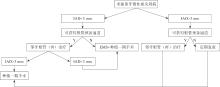
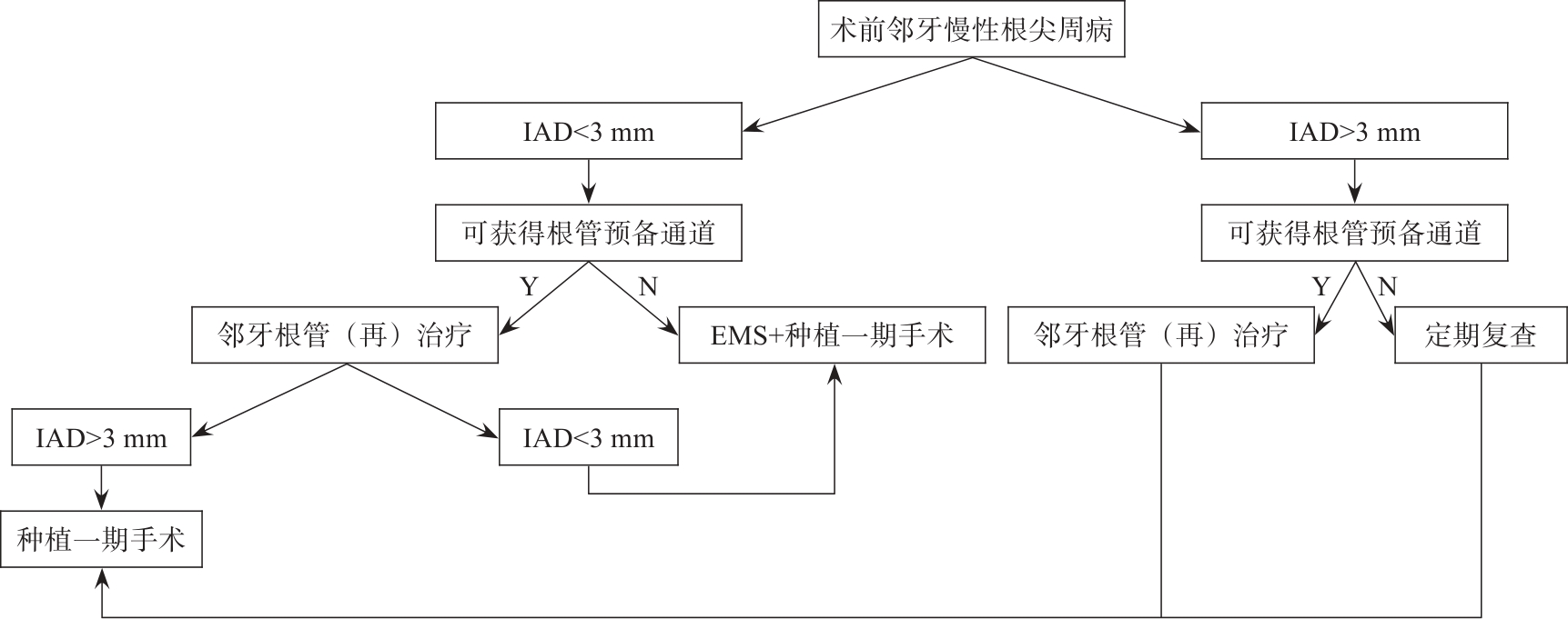
| [23] | 满毅, 黄定明. 美学区种植骨增量与邻牙慢性根尖周病的联合治疗策略(上): 应用基础及适应证[J]. 国际口腔医学杂志, 2022, 49(5): 497-505. |
| Man Y, Huang DM. Combined treatment strategy of oral implantology and endodontic microsurgery for bone augmentation and endodontic diseases in aesthetic area (part 1): application basis and indications[J]. Int J Stomatol, 2022, 49(5): 497-505. | |
| [24] | Mei F, Xie MR, Huang XF, et al. Porphyromonas gingivalis and its systemic impact: current status[J]. Pathogens, 2020, 9(11): 944. |
| [25] | Ran SJ, Huang J, Liu B, et al. Enterococcus faecalis activates NLRP3 inflammasomes leading to increa-sed interleukin-1 beta secretion and pyroptosis of THP-1 macrophages[J]. Microb Pathog, 2021, 154: 104761. |
| [26] | Salvi GE, Bosshardt DD, Lang NP, et al. Temporal sequence of hard and soft tissue healing around titanium dental implants[J]. Periodontol 2000, 2015, 68(1): 135-152. |
| [27] | Biguetti CC, Cavalla F, Silveira EM, et al. Oral implant osseointegration model in C57Bl/6 mice: microtomographic, histological, histomorphometric and molecular characterization[J]. J Appl Oral Sci, 2018, 26: e20170601. |
| [28] | Lavelle C, Wedgwood D. Effect of internal irrigation on frictional heat generated from bone drilling[J]. J Oral Surg, 1980, 38(7): 499-503. |
| [29] | Roos-Jansåker AM, Renvert S, Egelberg J. Treatment of peri-implant infections: a literature review[J]. J Clin Periodontol, 2003, 30(6): 467-485. |
| [30] | Nedir R, Bischof M, Pujol O, et al. Starch-induced implant periapical lesion: a case report[J]. Int J Oral Maxillofac Implants, 2007, 22(6): 1001-1006. |
| [31] | Langer L, Langer B, Salem D. Unintentional root fragment retention in proximity to dental implants: a series of six human case reports[J]. Int J Periodontics Restorative Dent, 2015, 35(3): 305-313. |
| [32] | Wassmann T, Kreis S, Behr M, et al. The influence of surface texture and wettability on initial bacterial adhesion on titanium and zirconium oxide dental implants[J]. Int J Implant Dent, 2017, 3(1): 32. |
| [33] | Renvert S, Polyzois I, Claffey N. How do implant surface characteristics influence peri-implant disease[J]. J Clin Periodontol, 2011, 38(): 214-222. |
| [34] | Amiel C, Ostertag A, Slama L, et al. BMD is reduced in HIV-infected men irrespective of treatment[J]. J Bone Miner Res, 2004, 19(3): 402-409. |
| [35] | Erbe M, Rickerts V, Bauersachs RM, et al. Acquired protein C and protein S deficiency in HIV-infected patients[J]. Clin Appl Thromb Hemost, 2003, 9(4): 325-331. |
| [36] | Shivakumar B, Mohamed J, Sudarsan S, et al. Retrograde peri-implantitis[J]. J Indian Soc Periodontol, 2010, 14(1): 57. |
| [37] | Sussman HI. Periapical implant pathology[J]. J Oral Implantol, 1998, 24(3): 133-138. |
| [38] | Waasdorp J, Reynolds M. Nonsurgical treatment of retrograde peri-implantitis: a case report[J]. Int J Oral Maxillofac Implants, 2010, 25(4): 831-833. |
| [39] | Peñarrocha-Diago M, Boronat-Lopez A, García-Mira B. Inflammatory implant periapical lesion: etiology, diagnosis, and treatment: presentation of 7 cases[J]. J Oral Maxillofac Surg, 2009, 67(1): 168-173. |
| [40] | Gong JM, Zhao RM, Yu ZH, et al. A novel histopathological classification of implant periapical lesion: a systematic review and treatment decision tree[J]. PLoS One, 2022, 17(12): e0277387. |
| [41] | Gong JM, Al-Sosowa AA, Zhao RM, et al. Successful management of peri-implant infection from the endodontic lesion of adjacent natural tooth[J]. Case Rep Dent, 2023, 2023: 5034582. |
| [42] | Balshi SF, Wolfinger GJ, Balshi TJ. A retrospective evaluation of a treatment protocol for dental implant periapical lesions: long-term results of 39 implant apicoectomies[J]. Int J Oral Maxillofac Implants, 2007, 22(2): 267-272. |
| [43] | 满毅, 黄定明. 美学区种植骨增量与邻牙慢性根尖周病的联合治疗策略(下): 临床诊治流程及实践病例[J]. 国际口腔医学杂志, 2022, 49(6): 621-632. |
| Man Y, Huang DM. Combined treatment strategy of oral implantology and endodontics microsurgery (part 2): clinical protocol and practical cases[J]. Int J Stomatol, 2022, 49(6): 621-632. | |
| [44] | Wang J, Luo YL, Tan XL, et al. Horizontal bone augmentation of the edentulous area with simultaneous endodontic microsurgery of the adjacent tooth: a digitally-driven multidisciplinary case report with a 1-year follow-up[J]. Int J Oral Implantol, 2021, 14(4): 435-451. |
|