国际口腔医学杂志 ›› 2026, Vol. 53 ›› Issue (1): 26-35.doi: 10.7518/gjkq.2026004
 平面偏斜量的对比研究
兰菁( ),伍军()
Jing Lan(),Jun Wu()
摘要:
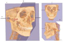
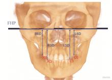
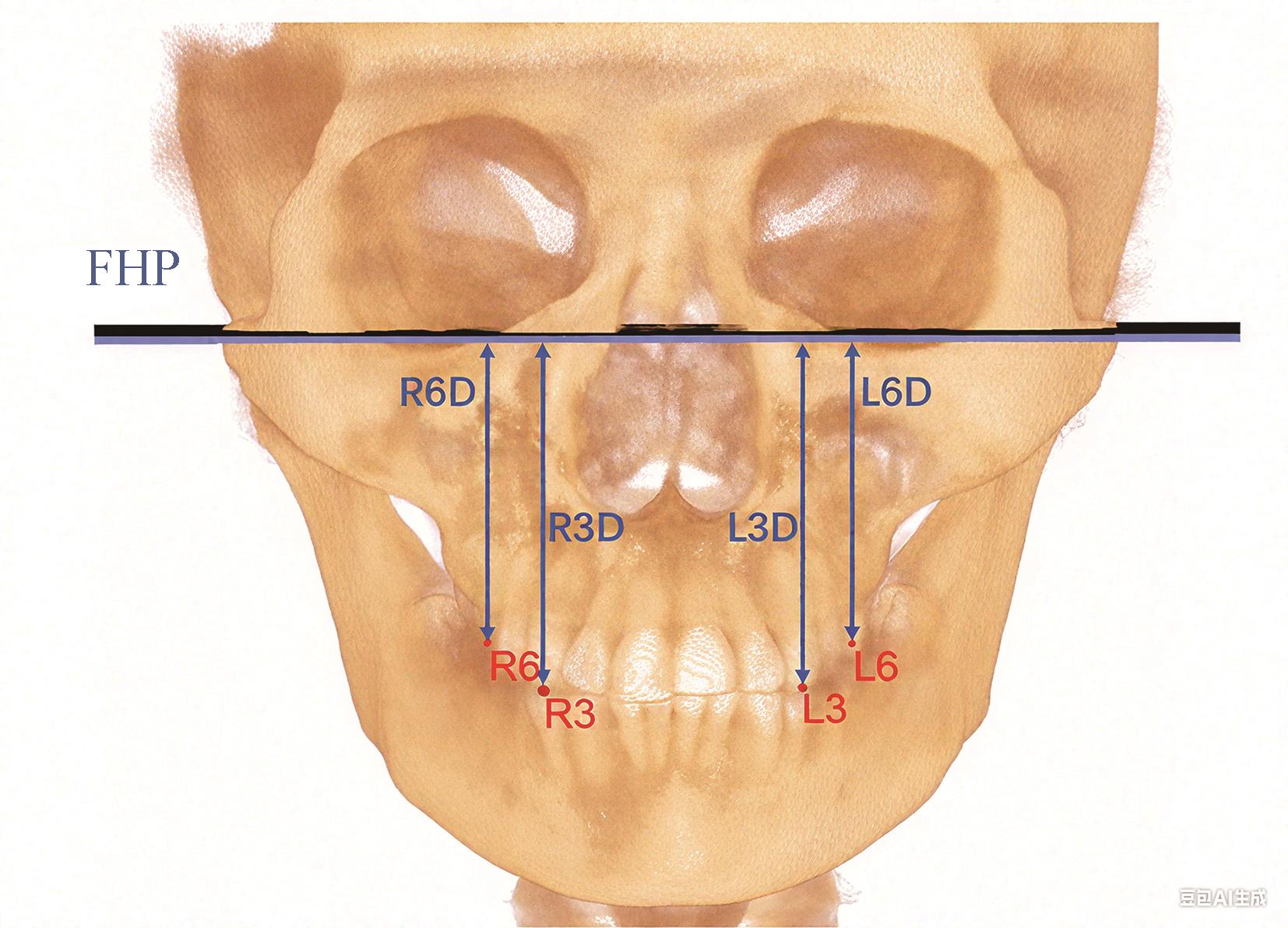
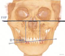
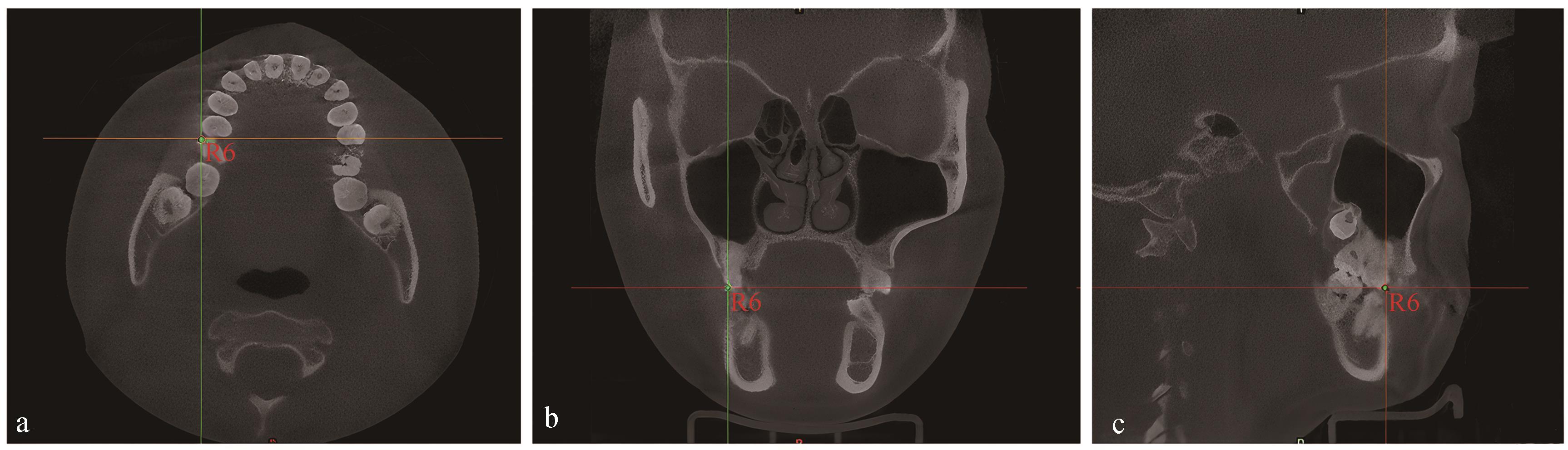
目的 通过研究不同矢状向成人均角患者的上颌尖牙及第一磨牙𬌗平面偏斜量,探讨上颌𬌗平面偏斜量与矢状向之间的相关性,以及安氏亚类患者𬌗平面偏斜方向与磨牙矢状关系间的差异,为此类患者的临床治疗提供参考。 方法 选择180例成人均角患者,其中安氏Ⅰ类60例,安氏Ⅱ1类60例,安氏Ⅲ类60例(其中安氏Ⅱ1、Ⅲ类的亚类与非亚类患者各30例)。应用Mimics软件对患者的锥形束CT(CBCT)成像进行三维重建,分别测量上颌双侧尖牙及第一磨牙至眶耳平面的垂直距离,计算𬌗平面偏斜量;同时记录尖牙及第一磨牙的𬌗平面偏斜方向以及与磨牙的矢状关系。采用SPSS 26.0软件对𬌗平面偏斜量与不同矢状向关系的相关性及安氏亚类的磨牙关系与𬌗平面偏斜方向的相关性进行统计学分析。 结果 尖牙及第一磨牙𬌗平面偏斜量与不同矢状向组别之间存在相关性(P<0.05)。安氏亚类组与非亚类组之间,𬌗平面偏斜量存在相关性(P<0.05)。安氏亚类组中,两侧磨牙关系与𬌗平面偏斜方向存在相关性(P<0.05)。 结论 不同矢状向成人均角患者的𬌗平面偏斜量有一定差异性。临床诊断、分析及治疗安氏Ⅱ类、Ⅲ类及其亚类患者时,不应忽视𬌗平面偏斜的纠正。
中图分类号:
| [1] | Dewey M. Classification of malocclusion[J]. Int J Orthod, 1915, 1(3): 133-147. |
| [2] | Burstone CJ. Diagnosis and treatment planning of patients with asymmetries[J]. Semin Orthod, 1998, 4(3): 153-164. |
| [3] | Janson G, Cruz KS, Woodside DG, et al. Dentoske-letal treatment changes in class Ⅱ subdivision malocclusions in submentovertex and posteroanterior radiographs[J]. Am J Orthod Dentofacial Orthop, 2004, 126(4): 451-463. |
| [4] | Sabri R. The eight components of a balanced smile[J]. J Clin Orthod, 2005, 39(3): 155-167. |
| [5] | Stahl F, Baccetti T, Franchi L, et al. Longitudinal growth changes in untreated subjects with class Ⅱ division 1 malocclusion[J]. Am J Orthod Dentofacial Orthop, 2008, 134(1): 125-137. |
| [6] | Shackelford TK, Larsen RJ. Facial asymmetry as an indicator of psychological, emotional, and physiological distress[J]. J Pers Soc Psychol, 1997, 72(2): 456-466. |
| [7] | Susarla SM, Peacock ZS, Kaban LB. Evaluation and correction of facial asymmetry in the coronal plane[J]. J Craniofac Surg, 2014, 25(4): 1299-1301. |
| [8] | Azevedo AR, Janson G, Henriques JF, et al. Evaluation of asymmetries between subjects with class Ⅱ subdivision and apparent facial asymmetry and those with normal occlusion[J]. Am J Orthod Dentofacial Orthop, 2006, 129(3): 376-383. |
| [9] | 白丁, 赵志河. 口腔正畸策略、控制与技巧[M]. 北京: 人民卫生出版社, 2015: 139-140. |
| Bai D, Zhao ZH. Advanced strategy with positive control in orthodontics[M]. Beijing: People’s Medical Publishing House, 2015: 139-140. | |
| [10] | Lonic D, Sundoro A, Lin HH, et al. Selection of a horizontal reference plane in 3D evaluation: identifying facial asymmetry and occlusal cant in orthognathic surgery planning[J]. Sci Rep, 2017, 7(1): 2157. |
| [11] | Chia MS, Naini FB, Gill DS. The aetiology, diagnosis and management of mandibular asymmetry[J]. Orthod Update, 2008, 1(2): 44-52. |
| [12] | Kurt G, Uysal T, Sisman Y, et al. Mandibular asymmetry in class Ⅱ subdivision malocclusion[J]. Angle Orthod, 2008, 78(1): 32-37. |
| [13] | Rose JM, Sadowsky C, BeGole EA, et al. Mandibular skeletal and dental asymmetry in class Ⅱ subdivision malocclusions[J]. Am J Orthod Dentofacial Orthop, 1994, 105(5): 489-495. |
| [14] | Kanurkova L, Gjorgova J, Dzipunova B, et al. Association between condylar position and tilt of frontal occlusal plane in patients with transversal and vertical dentofacial discrepancy[J]. Balk J Stom, 2012, 16(3): 147-153. |
| [15] | Pluijmers BI, van de Lande LS, Caron CJJM, et al. Part 2: is the maxillary canting and its surgical correction in patients with CFM correlated to the mandibular deformity[J]. J Craniomaxillofac Surg, 2018, 46(9): 1436-1440. |
| [16] | Rhodes G, Sumich A, Byatt G. Are average facial configurations attractive only because of their symmetry[J]. Psychol Sci, 1999, 10(1): 52-58. |
| [17] | Ramirez-Yañez GO, Stewart A, Franken E, et al. Prevalence of mandibular asymmetries in growing patients[J]. Eur J Orthod, 2011, 33(3): 236-242. |
| [18] | Good S, Edler R, Wertheim D, et al. A computerized photographic assessment of the relationship between skeletal discrepancy and mandibular outline asymmetry[J]. Eur J Orthod, 2006, 28(2): 97-102. |
| [19] | Severt TR, Proffit WR. The prevalence of facial asymmetry in the dentofacial deformities population at the University of North Carolina[J]. Int J Adult Orthodon Orthognath Surg, 1997, 12(3): 171-176. |
| [20] | 刘帅. 双侧下颌角点垂直向不调的骨性Ⅲ类偏斜患者上颌骨横向及垂直向发育形态特征的研究[D]. 沈阳: 中国医科大学, 2019. |
| Liu S. A study on the lateral and vertical developmental morphological characteristics of maxilla in patients with skeletal class Ⅲ deviation with vertical imbalance of bilateral mandibular angles[D]. Shenyang: China Medical University, 2019. | |
| [21] | 刘翔, 陈文静. 基于CBCT成像的成人安氏Ⅲ类颅颌面骨性不对称分析[J]. 口腔医学, 2017, 37(3): 233-236. |
| Liu X, Chen WJ. Skeletal and dental analysis of class Ⅲ subdivision malocclusions using cone-beam computed tomography[J]. Stomatology, 2017, 37(3): 233-236. | |
| [22] | Masuoka N, Momoi Y, Ariji Y, et al. Can cephalometric indices and subjective evaluation be consistent for facial asymmetry[J]. Angle Orthod, 2005, 75(4): 651-655. |
| [23] | Cevidanes LHS, Styner MA, Proffit WR. Image analysis and superimposition of 3-dimensional cone-beam computed tomography models[J]. Am J Orthod Dentofacial Orthop, 2006, 129(5): 611-618. |
| [24] | 王玉俏. 安氏Ⅲ类偏颌畸形患者下颌硬组织以及解剖学𬌗平面的三维分析[D]. 青岛: 青岛大学, 2020. |
| Wang YQ. Three-dimensional analysis of mandibular hard tissue and anatomical occlusal plane in patients with Angle Ⅲ malocclusion[D]. Qingdao: Qingdao University, 2020. | |
| [25] | Baek SH, Cho IS, Chang YI, et al. Skeletodental factors affecting chin point deviation in female patients with class Ⅲ malocclusion and facial asymmetry: a three-dimensional analysis using computed tomography[J]. Oral Surg Oral Med Oral Pathol Oral Radiol Endod, 2007, 104(5): 628-639. |
| [26] | 朱玉, 马嘉, 阎秀林, 等. 骨性安氏Ⅲ类错𬌗伴偏颌患者下颌骨偏斜程度与下颌骨形态和肌功能不对称指数相关性研究[J]. 中国实用口腔科杂志, 2015, 8(6): 368-370. |
| Zhu Y, Ma J, Yan XL, et al. Correlationship between mandibular asymmetry and the asymmetry index of mandibular profile and muscle functions in patients with skeletal Angle class Ⅲ malocclusion combined with mandibular asymmetry[J]. Chin J Pract Stomatol, 2015, 8(6): 368-370. | |
| [27] | 李鸿艺, 周诺, 黄旋平, 等. 应用三维有限元分析骨性Ⅲ类偏颌畸形的生物力学研究[C]//中华口腔医学会口腔医学计算机专业委员会. 第十五次全国口腔医学计算机应用学术研讨会会议手册. 南宁: 广西医科大学附属口腔医院, 2017: 77. |
| Li HY, Zhou N, Huang XP, et al. Biomechanical study on skeletal class Ⅲ maxillary/mandibular asymmetry using three-dimensional finite element analysis[C]. Chinese Stomatological Association Computer Professional Committee for Oral Medicine. Handbook for the 15th National Symposium on Computer Applications in Oral Medicine. Nanning: Stomatological Hospital Affiliated to Guangxi Medical University, 2017: 77. | |
| [28] | Haraguchi S, Iguchi Y, Takada K. Asymmetry of the face in orthodontic patients[J]. Angle Orthod, 2008, 78(3): 421-426. |
| [29] | Sanders DA, Rigali PH, Neace WP, et al. Skeletal and dental asymmetries in class Ⅱ subdivision malocclusions using cone-beam computed tomography[J]. Am J Orthod Dentofacial Orthop, 2010, 138(5): 542.e1-542.e20. |
| [30] | 徐静, 朱双林, 潘昱, 等. 安氏Ⅱ类亚类错𬌗颅颌面结构三维形态分析[J]. 中国实用口腔科杂志, 2015, 8(5): 290-293. |
| Xu J, Zhu SL, Pan Y, et al. Three-dimensional dentofacial characteristic analysis in class Ⅱ subdivision malocclusion[J]. Chin J Pract Stomatol, 2015, 8(5): 290-293. | |
| [31] | Al-Khateeb EAA, Al-Khateeb SN. Anteroposterior and vertical components of class Ⅱ division 1 and division 2 malocclusion[J]. Angle Orthod, 2009, 79(5): 859-866. |
| [32] | 段晓媛. 正面𬌗平面倾斜者的牙及牙槽骨形态学研究[D]. 昆明: 昆明医科大学, 2021. |
| Duan XY. Morphological study of teeth and alveolar bone in persons with frontal occlusal plane inclined[D]. Kunming: Kunming Medical University, 2021. | |
| [33] | Janson G, de Lima KJ, Woodside DG, et al. Class Ⅱ subdivision malocclusion types and evaluation of their asymmetries[J]. Am J Orthod Dentofacial Orthop, 2007, 131(1): 57-66. |
| [34] | Uesugi S, Yonemitsu I, Kokai S, et al. Features in subjects with the frontal occlusal plane inclined toward the contralateral side of the mandibular deviation[J]. Am J Orthod Dentofacial Orthop, 2016, 149(1): 46-54. |
| [35] | 贾绮林, 黄金芳. 颜面不对称畸形的颅面骨骼结构及其生长发育的研究[J]. 中华口腔医学杂志, 1994, 29(1): 34-37. |
| Jia QL, Huang JF. Study on the craniofacial skeletal structure and its growth and development in facial asymmetry deformity[J]. Chin J Stomatol, 1994, 29(1): 34-37. | |
| [36] | Linden OE, He JK, Morrison CS, et al. The relationship between age and facial asymmetry[J]. Plast Reconstr Surg, 2018, 142(5): 1145-1152. |
| [1] | 黄美畅,蒋鸿杰,汤亚玲,姚莉洪. 锥形束CT及免疫组织化学染色在根尖周囊肿诊断与鉴别诊断中的应用[J]. 国际口腔医学杂志, 2025, 52(4): 490-497. |
| [2] | 焦明阳,周煜萃,蒋正源,刘雨欣,曲柳. 数字化导板技术在牙髓治疗领域的研究进展[J]. 国际口腔医学杂志, 2024, 51(5): 550-557. |
| [3] | 杨雨楠,刘鹏,王虎,游梦. 上颌窦黏膜增厚的锥形束CT影像分析[J]. 国际口腔医学杂志, 2023, 50(3): 302-307. |
| [4] | 张珊,葛晓磊,李杰,谢新宇,常维维,马文盛. 上颌前方牵引矫治对颌骨生长发育长期影响的Meta分析[J]. 国际口腔医学杂志, 2022, 49(5): 548-555. |
| [5] | 吴文智,冯达兴,陈垂壮,周丽鹃. 海口地区下颌第一恒磨牙近中中央根管发生率及相关因素[J]. 国际口腔医学杂志, 2022, 49(4): 420-425. |
| [6] | 叶泽林,刘璐,龙虎,游梦. 弯曲前牙的影像评价及治疗的研究进展[J]. 国际口腔医学杂志, 2022, 49(2): 173-181. |
| [7] | 田浩楠,林敏,谢丛蔓,任嫒姝. 上颌腭侧阻生尖牙与寰椎后桥相关性的锥形束CT研究[J]. 国际口腔医学杂志, 2021, 48(5): 536-540. |
| [8] | 施丹妮,杨鑫,吴建勇. 锥形束CT三维头影测量参考坐标系的研究进展[J]. 国际口腔医学杂志, 2021, 48(4): 398-404. |
| [9] | 丁张帆,郭陟永,苗诚,李春洁,宣鸣,王晓毅,张壮. 基于锥形束CT的三维可视化技术在颌骨囊性病变手术中的应用[J]. 国际口腔医学杂志, 2021, 48(2): 180-186. |
| [10] | 王奔,许喆桢,韦曦. 数字化微创技术在牙髓根尖周病学中的应用与进展[J]. 国际口腔医学杂志, 2021, 48(1): 110-118. |
| [11] | 唐蓓,赵文俊,王虎,郑广宁,游梦. 根管超填导致下牙槽神经损伤2例[J]. 国际口腔医学杂志, 2020, 47(3): 293-296. |
| [12] | 章婷婷,胡常红,彭燕,周文翘,张慧聪,刘蝶. 300例不同年龄段有牙颌人群上唇软组织侧貌的锥形束CT三维测量分析[J]. 国际口腔医学杂志, 2020, 47(2): 182-188. |
| [13] | 王春林,刘从华,宋思吟,周丽淑,林丽佳. 运用锥形束CT诊断上下颌横向发育不调的研究进展[J]. 国际口腔医学杂志, 2020, 47(1): 121-124. |
| [14] | 黎祺, 黄少宏. 岭南地区广府民系人群下颌第二恒磨牙牙根和根管形态的锥形束CT研究[J]. 国际口腔医学杂志, 2019, 46(6): 640-649. |
| [15] | 曹焜,李家锋,孙玉华,鲍强,卢秋宁,唐巍. 下颌下窝的锥形束CT影像分析[J]. 国际口腔医学杂志, 2019, 46(2): 209-212. |
|
||