国际口腔医学杂志 ›› 2025, Vol. 52 ›› Issue (4): 490-497.doi: 10.7518/gjkq.2025075
黄美畅( ),蒋鸿杰,汤亚玲,姚莉洪()
Meichang Huang(),Hongjie Jiang,Yaling Tang,Lihong Yao()
摘要:
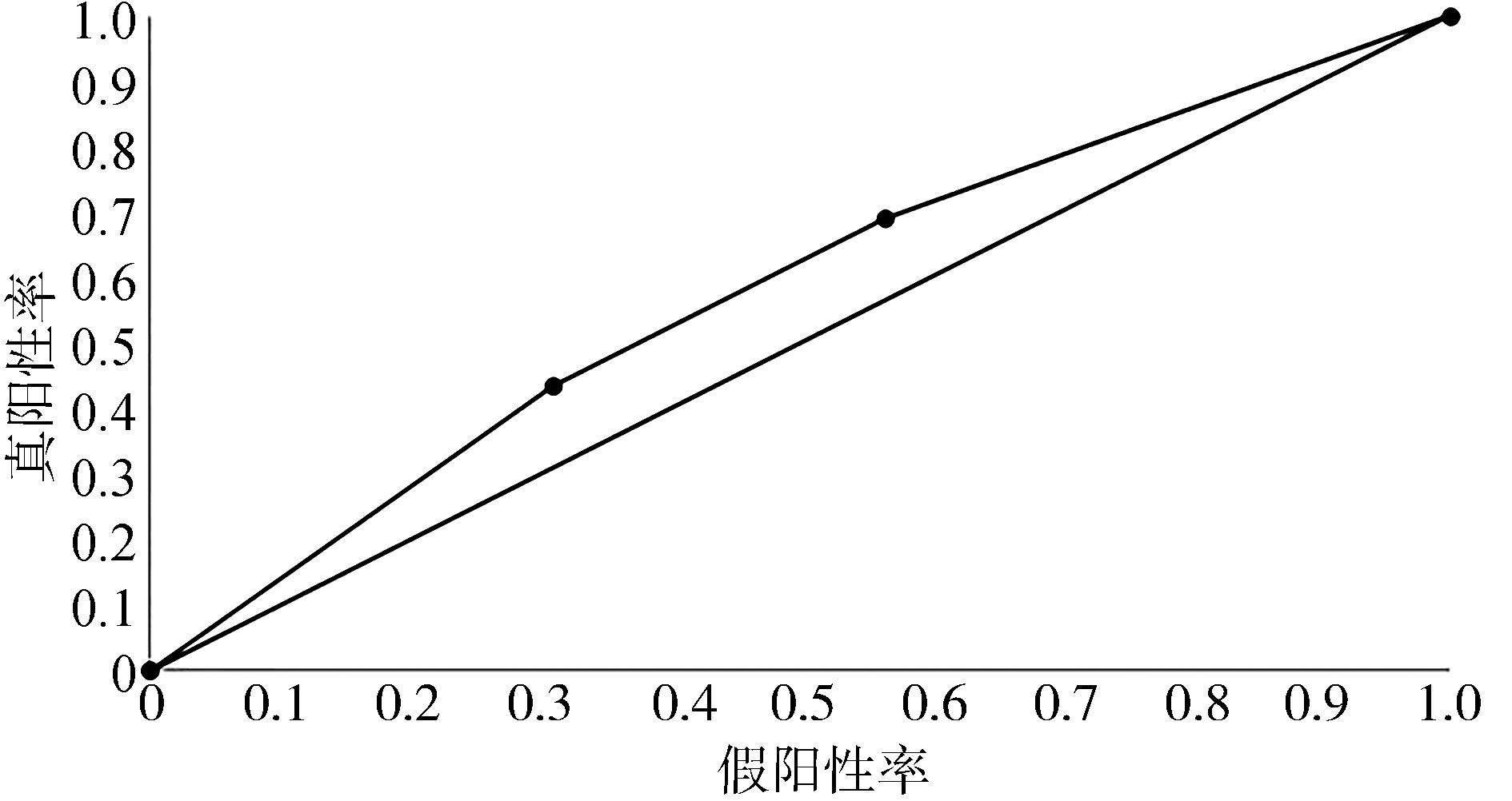
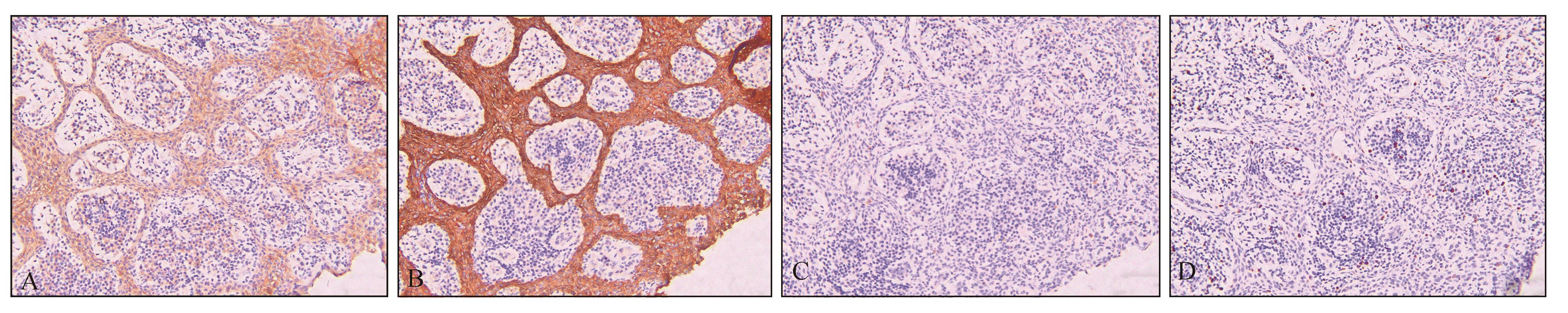
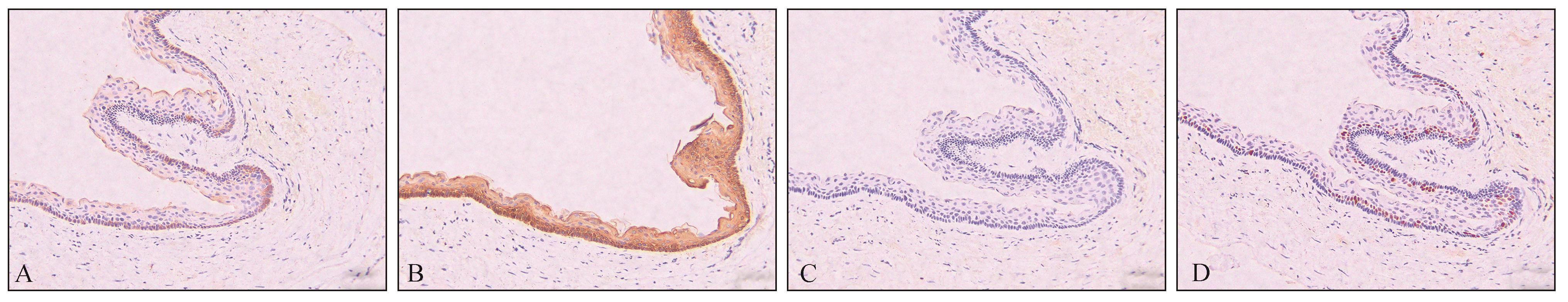
目的 探讨锥形束CT(CBCT)和免疫组织化学染色在根尖周囊肿、根尖周肉芽肿和牙源性角化囊肿鉴别诊断中的应用价值。 方法 收集143例根尖周囊肿和45例根尖周肉芽肿患者的临床病理及影像学资料,分析二者诊断结果的一致性;选取根尖周囊肿29例,牙源性角化囊肿24例,分别进行免疫组织化学染色(CK14、CK19、CD57和Ki-67),分析免疫组织化学染色在根尖周囊肿诊断中的价值。 结果 根尖周囊肿经CBCT诊断准确率为68.5%,根尖周肉芽肿经CBCT诊断准确率为44.4%,但CBCT诊断结果与病理学诊断结果的一致性较差(Kappa<0.4)。CBCT诊断根尖周囊肿的正确性与发病部位(P=0.033)、上皮剩余(P=0.036)和泡沫细胞(P=0.027)有相关性,而与胆固醇裂隙、出血、钙化、急慢性炎症以及炎症程度无相关性(P>0.05)。根尖周囊肿和牙源性角化囊肿中上皮标志物的表达:大部分病例CK14和CK19阳性表达,CD57阴性表达。细胞增殖活性标志物的表达:根尖周囊肿的Ki-67阳性细胞率为1%~3%,严重感染会增加至15%;牙源性角化囊肿Ki-67阳性细胞率为1%~3%或10%~80%。 结论 CBCT诊断根尖周囊肿和根尖周肉芽肿的敏感性较好,而特异性较差。CBCT诊断根尖周囊肿的正确性与发病部位、上皮剩余以及泡沫细胞相关,而与胆固醇裂隙、出血、钙化、急慢性炎症以及炎症程度无关。牙源性角化囊肿患者Ki-67表达阳性细胞数高于根尖周囊肿,且主要定位于副基底细胞层。
中图分类号:
| 1 | Al Khasawnah Q, Hassan F, Malhan D, et al. Nonsurgical clinical management of periapical lesions using calcium hydroxide-iodoform-silicon-oil paste[J]. Biomed Res Int, 2018, 2018: 8198795. |
| 2 | Tsesis I, Krepel G, Koren T, et al. Accuracy for diagnosis of periapical cystic lesions[J]. Sci Rep, 2020, 10(1): 14155. |
| 3 | Howell FV, De la Rosa VM. Cytologic evaluation of cystic lesions of the jaws: a new diagnostic technique[J]. J South Calif Dent Assoc, 1968, 36(4): 161-166. |
| 4 | Cunningham CJ, Penick EC. Use of a roentgenographic contrast medium in the differential diagnosis of periapical lesions[J]. Oral Surg Oral Med Oral Pathol, 1968, 26(1): 96-102. |
| 5 | Cotti E, Schirru E. Present status and future directions: imaging techniques for the detection of periapical lesions[J]. Int Endod J, 2022, 55(): 1085-1099. |
| 6 | Giudice RL, Nicita F, Puleio F, et al. Accuracy of periapical radiography and CBCT in endodontic evaluation[J]. Int J Dent, 2018, 2018: 2514243. |
| 7 | Çalışkan MK, Kaval ME, Tekin U, et al. Radiographic and histological evaluation of persistent periapical lesions associated with endodontic failures after apical microsurgery[J]. Int Endod J, 2016, 49(11): 1011-1019. |
| 8 | Mirković S, Tadić, Durdević Mirković T, et al. Comparative analysis of accuracy of diagnosis of chronic periapical lesions made by clinical and histopatologi-cal examination[J]. Med Pregl, 2012, 65(7/8): 277-280. |
| 9 | Rosenberg PA, Frisbie J, Lee J, et al. Evaluation of pathologists (histopathology) and radiologists (cone beam computed tomography) differentiating radicular cysts from granulomas[J]. J Endod, 2010, 36(3): 423-428. |
| 10 | 纪志勇, 张佳莉, 孟柳燕. 383例根尖周病变的临床病理系统性回顾与分析[J]. 口腔医学研究, 2013, 29(10): 978-979, 981. |
| Ji ZY, Zhang JL, Meng LY. Clinical and pathological analysis of 383 patients with periapcial lesions[J]. J Oral Sci Res, 2013, 29(10): 978-979, 981. | |
| 11 | Bornstein MM, Lauber R, Sendi P, et al. Comparison of periapical radiography and limited cone-beam computed tomography in mandibular molars for analysis of anatomical landmarks before apical surgery[J]. J Endod, 2011, 37(2): 151-157. |
| 12 | Alotaibi O, Alswayyed S, Alshagroud R, et al. Evalua-tion of concordance between clinical and histopa-thological diagnoses in periapical lesions of endo-dontic origin[J]. J Dent Sci, 2020, 15(2): 132-135. |
| 13 | Sönmez G, Kamburoğlu K, Yılmaz F, et al. Versatility of high resolution ultrasonography in the assessment of granulomas and radicular cysts: a comparative in vivo study[J]. Dentomaxillofac Radiol, 2019, 48(6): 20190082. |
| 14 | Lizio G, Salizzoni E, Coe M, et al. Differential diagnosis between a granuloma and radicular cyst: effectiveness of magnetic resonance imaging[J]. Int Endod J, 2018, 51(10): 1077-1087. |
| 15 | Alam H, Sehgal L, Kundu ST, et al. Novel function of keratins 5 and 14 in proliferation and differentiation of stratified epithelial cells[J]. Mol Biol Cell, 2011, 22(21): 4068-4078. |
| 16 | Živković ND, Mihailović DS, Kostić MS, et al. Markers of proliferation and cytokeratins in the differential diagnosis of jaw cysts[J]. Ear Nose Throat J, 2017, 96(9): 376-383. |
| 17 | Shruthi DK, Shivakumar MC, Tegginamani AS, et al. Cytokeratin 14 and cytokeratin 18 expressions in reduced enamel epithelium and dentigerous cyst: possible role in oncofetal transformation and histogenesis- of follicular type of adenomatoid odontogenic tumor[J]. J Oral Maxillofac Pathol, 2014, 18(3): 365-371. |
| 18 | Aristizabal Arboleda P, Sánchez-Romero C, de Almeida OP, et al. Calcifying odontogenic cyst associa-ted with dentigerous cyst in a 15-year-old girl[J]. Int J Surg Pathol, 2018, 26(8): 758-765. |
| 19 | Heatley MK. Keratin expression in human tissues and neoplasms[J]. Histopathology, 2002, 41(4): 365-366. |
| 20 | Tsuji K, Wato M, Hayashi T, et al. The expression of cytokeratin in keratocystic odontogenic tumor, orthokeratinized odontogenic cyst, dentigerous cyst, radicular cyst and dermoid cyst[J]. Med Mol Morphol, 2014, 47(3): 156-161. |
| 21 | Silva LABD, Sá MAR, Melo RA, et al. Analysis of CD57+ natural killer cells and CD8+ T lymphocytes in periapical granulomas and radicular cysts[J]. Braz Oral Res, 2017, 31: e106. |
| 22 | Arai K, Yamamura S, Seki S, et al. Increase of CD57+ T cells in knee joints and adjacent bone marrow of rheumatoid arthritis (RA) patients: implication for an anti-inflammatory role[J]. Clin Exp Immunol, 1998, 111(2): 345-352. |
| 23 | Moreira PR, Santos DF, Martins RD, et al. CD57+ cells in radicular cyst[J]. Int Endod J, 2000, 33(2): 99-102. |
| 24 | Gerdes J, Lemke H, Baisch H, et al. Cell cycle analy-sis of a cell proliferation-associated human nuclear antigen defined by the monoclonal antibody Ki-67[J]. J Immunol, 1984, 133(4): 1710-1715. |
| 25 | Martins CA, Rivero ER, Dufloth RM, et al. Immunohistochemical detection of factors related to cellular proliferation and apoptosis in radicular and dentigerous cysts[J]. J Endod, 2011, 37(1): 36-39. |
| 26 | Mourão RVC, Pinheiro Júnior EC, Barros Silva PG, et al. Study of the relationship between mononu-clear inflammatory infiltrate and Ki-67 and basement membrane and extracellular matrix protein expression in radicular cysts[J]. Int Endod J, 2016, 49(5): 447-453. |
| 27 | Ayoub MS, Baghdadi HM, El-Kholy M. Immunohistochemical detection of laminin-1 and Ki-67 in radicular cysts and keratocystic odontogenic tumors[J]. BMC Clin Pathol, 2011, 11: 4. |
| 28 | Suzuki T, Kumamoto H, Kunimori K, et al. Immunohistochemical analysis of apoptosis-related factors in lining epithelium of radicular cysts[J]. J Oral Pathol Med, 2005, 34(1): 46-52. |
| 29 | 张权, 郑乾坤, 马会青, 等. Bcl-6和ICOS在根尖周囊肿与根尖周肉芽肿中的表达[J]. 解剖学杂志, 2019, 42(2): 128-131. |
| Zhang Q, Zheng QK, Ma HQ, et al. Expression of B cell lymphoma 6 and inducible costimulator in periapical cysts and periapical granulomas[J]. Chin J Anatom, 2019, 42(2): 128-131. | |
| 30 | Peng YQ, Liu L, Li XF, et al. B cells at the core: immune mechanisms and therapeutic potentials in periapical lesions[J]. J Endod, 2025, 51(1): 4-14. |
| 31 | 王海丞, 章燕. 根尖周囊肿囊壁成纤维样细胞中纤维粘连蛋白基因可变剪接片对诱导破骨细胞形成[C]//中华口腔医学会口腔生物医学专业委员会. 2019第九次全国口腔生物医学学术年会论文汇编. 同济大学附属口腔医院上海牙组织修复与再生工程技术研究中心, 2019: 72. |
| Wang HC, Zhang Y. Alternative splicing of fibronectin gene in fibroblast like cells of periapical cyst wall induces osteoclast formation[C]//Chinese Socie-ty of Stomatology Oral Biomedical Professional Committee. Compilation of Papers from the 9th National Oral Biomedical Academic Annual Confe-rence in 2019. Tongji University Affiliated Stomatological Hospital, Shanghai Dental Tissue Restoration and Regeneration Engineering Technology Research Center, 2019: 72 | |
| 32 |
Shen SQ, Wang R, Huang SG. Expression of the stem cell factor in fibroblasts, endothelial cells, and macrophages in periapical tissues in human chronic periapical diseases[J]. Genet Mol Res, 2017, 16(1). doi: 10.4238/gmr16019394 .
doi: 10.4238/gmr16019394 |
| 33 | Yang JW, Jiang JH, Wang HC, et al. The extra domain A of fibronectin facilitates osteoclastogenesis in radicular cysts through vascular endothelial growth factor[J]. Int Endod J, 2020, 53(4): 478-491.[34] SheethalHS, KnH, SmithaT, et al. Role of mast cells in inflammatory and reactive pathologies of pulp, periapical area and periodontium[J]. J Oral Maxillofac Pathol, 2018, 22(1): 92-97.[35] LiuCY, WangHC. The fibroblast of radicular cyst facilitate osteoclastogenesis via the autocrine of fibronectin containing extra domain A[J]. Oral Dis, 2019, 25(4): 1136-1146. |
| 36 | 张梅华, 于蕴之, 缪羽. 破骨细胞核因子κB受体活化因子配体和骨保护素在根尖周囊肿和肉芽肿中的表达及意义[J]. 华西口腔医学杂志, 2012, 30(4): 360-363. |
| Zhang MH, Yu YZ, Miao Y. The expression and significance of receptor activator of nuclear factor kappa B ligand and osteoprotegerin in periapical cyst and periapical granuloma[J]. West China J Stomatol, 2012, 30(4): 360-363. | |
| 37 | Brito LNS, de Lemos Almeida MMR, de Souza LB, et al. Immunohistochemical analysis of galectins- 1, -3, and-7 in periapical granulomas, radicular cysts, and residual radicular cysts[J]. J Endod, 2018, 44(5): 728-733. |
| [1] | 焦明阳,周煜萃,蒋正源,刘雨欣,曲柳. 数字化导板技术在牙髓治疗领域的研究进展[J]. 国际口腔医学杂志, 2024, 51(5): 550-557. |
| [2] | 杨雨楠,刘鹏,王虎,游梦. 上颌窦黏膜增厚的锥形束CT影像分析[J]. 国际口腔医学杂志, 2023, 50(3): 302-307. |
| [3] | 吴文智,冯达兴,陈垂壮,周丽鹃. 海口地区下颌第一恒磨牙近中中央根管发生率及相关因素[J]. 国际口腔医学杂志, 2022, 49(4): 420-425. |
| [4] | 叶泽林,刘璐,龙虎,游梦. 弯曲前牙的影像评价及治疗的研究进展[J]. 国际口腔医学杂志, 2022, 49(2): 173-181. |
| [5] | 田浩楠,林敏,谢丛蔓,任嫒姝. 上颌腭侧阻生尖牙与寰椎后桥相关性的锥形束CT研究[J]. 国际口腔医学杂志, 2021, 48(5): 536-540. |
| [6] | 施丹妮,杨鑫,吴建勇. 锥形束CT三维头影测量参考坐标系的研究进展[J]. 国际口腔医学杂志, 2021, 48(4): 398-404. |
| [7] | 丁张帆,郭陟永,苗诚,李春洁,宣鸣,王晓毅,张壮. 基于锥形束CT的三维可视化技术在颌骨囊性病变手术中的应用[J]. 国际口腔医学杂志, 2021, 48(2): 180-186. |
| [8] | 王奔,许喆桢,韦曦. 数字化微创技术在牙髓根尖周病学中的应用与进展[J]. 国际口腔医学杂志, 2021, 48(1): 110-118. |
| [9] | 唐蓓,赵文俊,王虎,郑广宁,游梦. 根管超填导致下牙槽神经损伤2例[J]. 国际口腔医学杂志, 2020, 47(3): 293-296. |
| [10] | 章婷婷,胡常红,彭燕,周文翘,张慧聪,刘蝶. 300例不同年龄段有牙颌人群上唇软组织侧貌的锥形束CT三维测量分析[J]. 国际口腔医学杂志, 2020, 47(2): 182-188. |
| [11] | 王春林,刘从华,宋思吟,周丽淑,林丽佳. 运用锥形束CT诊断上下颌横向发育不调的研究进展[J]. 国际口腔医学杂志, 2020, 47(1): 121-124. |
| [12] | 黎祺, 黄少宏. 岭南地区广府民系人群下颌第二恒磨牙牙根和根管形态的锥形束CT研究[J]. 国际口腔医学杂志, 2019, 46(6): 640-649. |
| [13] | 曹焜,李家锋,孙玉华,鲍强,卢秋宁,唐巍. 下颌下窝的锥形束CT影像分析[J]. 国际口腔医学杂志, 2019, 46(2): 209-212. |
| [14] | 孟怡彤,张晓东. 成人个别正常颌上气道不同软件三维测量的比较研究[J]. 国际口腔医学杂志, 2018, 45(6): 690-694. |
| [15] | 徐迅, 黄建生, 甘泽坤, 罗震. 上颌第一磨牙区腭侧骨板的锥形束CT测量结果及其临床意义[J]. 国际口腔医学杂志, 2017, 44(6): 686-690. |
|