国际口腔医学杂志 ›› 2025, Vol. 52 ›› Issue (2): 183-194.doi: 10.7518/gjkq.2025047
张潇月1( ),陈舒泽1,周婕妤1,程磊2,赵蕾1()
Xiaoyue Zhang1(),Shuze Chen1,Jieyu Zhou1,Lei Cheng2,Lei Zhao1()
摘要:
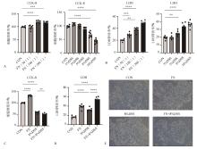
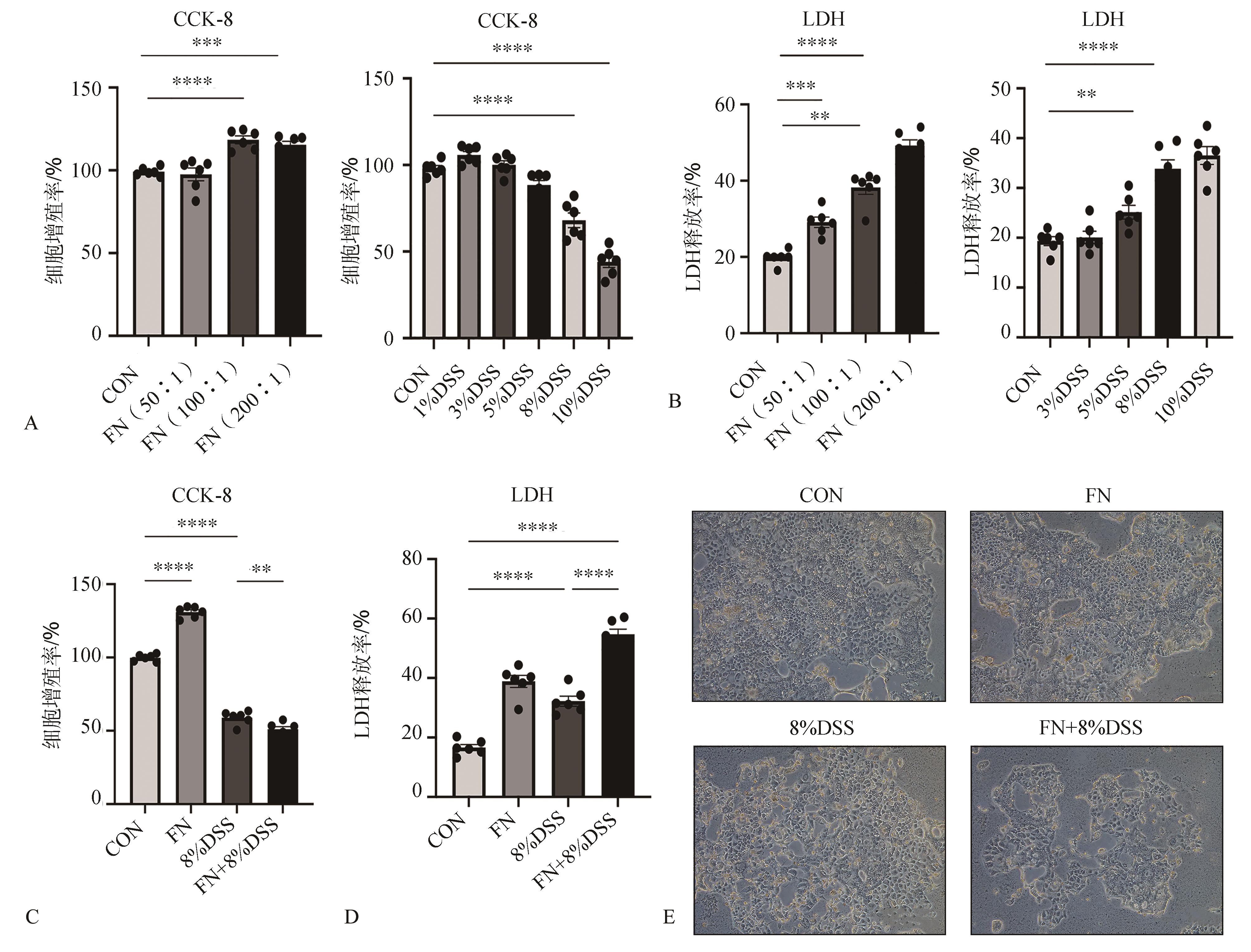
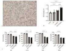
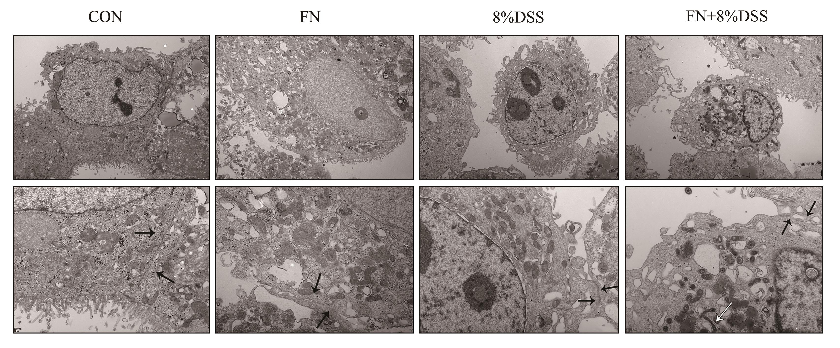
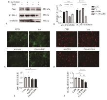
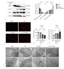
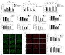
目的 探究具核梭杆菌(F. nucleatum)感染对体外肠道上皮屏障的影响及可能的机制。 方法 人结直肠腺癌细胞Caco-2细胞接种于Transwell小室构建体外肠道上皮屏障模型。采用葡聚糖硫酸钠(DSS)和F. nucleatum建立细胞损伤和感染模型,实验分为CON组、FN组、8%DSS组和FN+8%DSS组,检测F. nucleatum对有无DSS处理的上皮屏障的影响并评估铁死亡在其中的作用;随后引入铁死亡抑制剂铁抑素1(Fer-1)和去铁胺(DFO),探究抑制铁死亡对受损上皮屏障的作用。实验中,采用细胞计数试剂盒8(CCK-8)检测细胞增殖;乳酸脱氢酶(LDH)法检测细胞损伤;跨上皮电阻值(TEER)评估上皮完整性;异硫氰酸荧光素-葡聚糖(FD4)透过率评估上皮通透性;透射电镜观察细胞间连接及线粒体;蛋白免疫印迹和免疫荧光染色检测胞质紧密粘连蛋白ZO-1和紧密连接蛋白CLDN-1的表达;铁死亡检测包括免疫荧光染色检测细胞内亚铁离子(Fe2+)含量;免疫印迹法检测谷胱甘肽过氧化物酶4(GPX4)、铁蛋白重链1(FTH1)、乙酰辅酶A合成酶长链家族4(ACSL4)的表达,以及测量丙二醛(MDA)含量和谷胱甘肽比例(GSH%)评估脂质过氧化水平。 结果 与CON组相比,FN组、8%DSS组和FN+8%DSS组细胞增殖率降低,细胞损伤增加(P<0.05);与CON组相比,FN组TEER值和FD4透过率差异无统计学意义(P>0.05);与8%DSS组相比,FN+8%DSS组的6、12、24 h TEER值下降(P<0.05),FD4透过率增加(P<0.000 1);与CON组相比,FN+8%DSS组ZO-1和CLDN-1蛋白下调(P<0.05);透射电镜观察到8%DSS组细胞间连接破坏,FN+8%DSS组除了细胞间连接破坏还观测到F. nucleatum入侵细胞内部,线粒体呈现铁死亡样改变;GPX4蛋白下调,FTH1和ACSL4蛋白上调,细胞内Fe2+积聚,MDA升高,GSH%降低。引入Fer-1和DFO后,细胞损伤降低,TEER值回升,FD4透过率降低,ZO-1和CLDN-1蛋白表达升高(P<0.05)。 结论 F. nucleatum感染可能通过铁死亡途径促进DSS诱导的体外肠道上皮屏障破坏。
中图分类号:
| 1 | Janakiram C, Dye BA. A public health approach for prevention of periodontal disease[J]. Periodontol 2000, 2020, 84(1): 202-214. |
| 2 | Gurling K. Ulcerative colitis[J]. Postgrad Med J, 1953, 29(327): 2-4. |
| 3 | Kucharzik T, Koletzko S, Kannengiesser K, et al. Ulcerative colitis-diagnostic and therapeutic algorithms[J]. Dtsch Arztebl Int, 2020, 117(33/34): 564-574. |
| 4 | Lin CY, Tseng KS, Liu JM, et al. Increased risk of ulcerative colitis in patients with periodontal disease: a nationwide population-based cohort study[J]. Int J Environ Res Public Health, 2018, 15(11): 2602. |
| 5 | Arenas Rodrigues VA, de Avila ED, Nakano V, et al. Qualitative, quantitative and genotypic evaluation of Aggregatibacter actinomycetemcomitans and Fusobacterium nucleatum isolated from individuals with different periodontal clinical conditions[J]. Anaerobe, 2018, 52: 50-58. |
| 6 | Kitamoto S, Kamada N. Periodontal connection with intestinal inflammation: microbiological and immunological mechanisms[J]. Periodontol 2000, 2022, 89(1): 142-153. |
| 7 | Brennan CA, Garrett WS. Fusobacterium nucleatum-symbiont, opportunist and oncobacterium[J]. Nat Rev Microbiol, 2019, 17(3): 156-166. |
| 8 | Martini E, Krug SM, Siegmund B, et al. Mend your fences: the epithelial barrier and its relationship with mucosal immunity in inflammatory bowel di-sease[J]. Cell Mol Gastroenterol Hepatol, 2017, 4(1): 33-46. |
| 9 | Abraham C, Cho JH. Inflammatory bowel disease[J]. N Engl J Med, 2009, 361(21): 2066-2078. |
| 10 | Lin SL, Zhang XY, Zhu XZ, et al. Fusobacterium nucleatum aggravates ulcerative colitis through promoting gut microbiota dysbiosis and dysmetabolism[J]. J Periodontol, 2023, 94(3): 405-418. |
| 11 | Hidalgo IJ, Raub TJ, Borchardt RT. Characterization of the human colon carcinoma cell line (Caco-2) as a model system for intestinal epithelial permeability[J]. Gastroenterology, 1989, 96(3): 736-749. |
| 12 | Dixon SJ, Lemberg KM, Lamprecht MR, et al. Ferroptosis: an iron-dependent form of nonapoptotic cell death[J]. Cell, 2012, 149(5): 1060-1072. |
| 13 | Ursini F, Maiorino M. Lipid peroxidation and ferroptosis: the role of GSH and GPx4[J]. Free Radic Biol Med, 2020, 152: 175-185. |
| 14 | Doll S, Proneth B, Tyurina YY, et al. ACSL4 dictates ferroptosis sensitivity by shaping cellular lipid composition[J]. Nat Chem Biol, 2017, 13(1): 91-98. |
| 15 | Tang DL, Chen X, Kang R, et al. Ferroptosis: molecular mechanisms and health implications[J]. Cell Res, 2021, 31: 107-125. |
| 16 | Kolenbrander PE. Oral microbial communities: biofilms, interactions, and genetic systems[J]. Annu Rev Microbiol, 2000, 54: 413-437. |
| 17 | Chen YY, Chen Y, Cao P, et al. Fusobacterium nucleatum facilitates ulcerative colitis through activa-ting IL-17F signaling to NF-κB via the upregulation of CARD3 expression[J]. J Pathol, 2020, 250(2): 170-182. |
| 18 | Thurnheer T, Karygianni L, Flury M, et al. Fusobacterium species and subspecies differentially affect the composition and architecture of supra- and subgingival biofilms models[J]. Front Microbiol, 2019, 10: 1716. |
| 19 | Qu H, Zhang WJ, Li JH, et al. A rapid and sensitive CRISPR-Cas12a for the detection of Fusobacterium nucleatum [J]. Microbiol Spectr, 2024, 12(2): e0362923. |
| 20 | Turner JR. Intestinal mucosal barrier function in health and disease[J]. Nat Rev Immunol, 2009, 9(11): 799-809. |
| 21 | Chen YY, Cui WW, Li X, et al. Interaction between commensal bacteria, immune response and the intestinal barrier in inflammatory bowel disease[J]. Front Immunol, 2021, 12: 761981. |
| 22 | Liu H, Hong XL, Sun TT, et al. Fusobacterium nucleatum exacerbates colitis by damaging epithelial barriers and inducing aberrant inflammation[J]. J Dig Dis, 2020, 21(7): 385-398. |
| 23 | Pires CL, Praça C, Martins PAT, et al. Re-use of Caco-2 monolayers in permeability assays-validation regarding cell monolayer integrity[J]. Pharmaceutics, 2021, 13(10): 1563. |
| 24 | Dharmani P, Strauss J, Ambrose C, et al. Fusobacterium nucleatum infection of colonic cells stimulates MUC2 mucin and tumor necrosis factor alpha[J]. Infect Immun, 2011, 79(7): 2597-2607. |
| 25 | Tang B, Wang K, Jia YP, et al. Fusobacterium nucleatum-induced impairment of autophagic flux enhances the expression of proinflammatory cytokines via ROS in Caco-2 cells[J]. PLoS One, 2016, 11(11): e0165701. |
| 26 | Subramanian S, Geng H, Tan XD. Cell death of intestinal epithelial cells in intestinal diseases[J]. Sheng Li Xue Bao, 2020, 72(3): 308-324. |
| 27 | Xu MY, Tao J, Yang YD, et al. Ferroptosis involves in intestinal epithelial cell death in ulcerative colitis[J]. Cell Death Dis, 2020, 11(2): 86. |
| 28 | Ma DL, Jiang PL, Jiang YJ, et al. Effects of lipid peroxidation-mediated ferroptosis on severe acute pancreatitis-induced intestinal barrier injury and bacterial translocation[J]. Oxid Med Cell Longev, 2021, 2021: 6644576. |
| 29 | Tang XM, Liu JQ, Yao S, et al. Ferulic acid allevia-tes alveolar epithelial barrier dysfunction in sepsis-induced acute lung injury by activating the Nrf2/HO-1 pathway and inhibiting ferroptosis[J]. Pharm Biol, 2022, 60(1): 2286-2294. |
| [1] | 李晶,康健. 牙周微创手术中再生材料选择及疗效的研究进展[J]. 国际口腔医学杂志, 2025, 52(2): 161-168. |
| [2] | 钟良军. 数字化技术在重度牙周炎治疗中的应用[J]. 国际口腔医学杂志, 2025, 52(1): 1-10. |
| [3] | 程守正,李太文,赵蕾. 血清淀粉样蛋白A与牙周炎相关性的研究进展[J]. 国际口腔医学杂志, 2025, 52(1): 117-122. |
| [4] | 陈梦洁,徐文华,刘青青,康毓聃,刘蓉,朱丽雷. 全身免疫炎症指数与牙周炎患者分级诊断的相关性研究[J]. 国际口腔医学杂志, 2024, 51(6): 706-712. |
| [5] | 陈蕊,范桢,郝春波. 黑色素瘤缺乏因子2炎症小体在牙周炎及糖尿病中的研究进展[J]. 国际口腔医学杂志, 2024, 51(6): 763-771. |
| [6] | 毛鸿晨,王铮,杨德琴. 牙龈卟啉单胞菌外膜囊泡在口腔疾病中的作用及其机制的研究进展[J]. 国际口腔医学杂志, 2024, 51(5): 608-615. |
| [7] | 漆美瑶,祁星颖,周欣奕,谭震,袁泉. 大麻二酚联合米诺环素对牙周炎治疗作用的实验研究[J]. 国际口腔医学杂志, 2024, 51(4): 392-400. |
| [8] | 陈梦洁, 刘小乐, 朱丽雷. 牙周炎患者牙周支持治疗对血细胞指标影响的回顾性研究[J]. 国际口腔医学杂志, 2024, 51(4): 401-405. |
| [9] | 马玉, 左玉, 刘建华. 抗菌光动力疗法与全身抗菌药物辅助治疗牙周炎疗效比较的Meta分析[J]. 国际口腔医学杂志, 2024, 51(4): 406-415. |
| [10] | 刘诗礼, 赵蕾. 牙周炎与心力衰竭相关性的研究进展[J]. 国际口腔医学杂志, 2024, 51(4): 425-432. |
| [11] | 杨再目,曹沛,刘振华,栾庆先. 血浆无细胞线粒体外线粒体DNA与牙周炎临床指标的相关性研究[J]. 国际口腔医学杂志, 2024, 51(3): 288-295. |
| [12] | 马瑜鸿,赵蕾. 微创非手术牙周治疗技术的临床研究进展[J]. 国际口腔医学杂志, 2024, 51(2): 227-232. |
| [13] | 傅豫, 何薇, 黄兰. 铁死亡在口腔疾病中的研究进展[J]. 国际口腔医学杂志, 2024, 51(1): 36-44. |
| [14] | 罗晓洁,王德续,陈晓涛. 基于生物信息学分析铁死亡调控基因与牙周炎的关系[J]. 国际口腔医学杂志, 2023, 50(6): 661-668. |
| [15] | 黄元鸿,彭显,周学东. 骨碎补在治疗口腔骨相关疾病的研究进展[J]. 国际口腔医学杂志, 2023, 50(6): 679-685. |
|