国际口腔医学杂志 ›› 2025, Vol. 52 ›› Issue (2): 205-209.doi: 10.7518/gjkq.2025030
刘晶晶1,2,3( ),唐睿2,袁丽仙2,刘鑫2()
Jingjing Liu1,2,3(),Rui Tang2,Lixian Yuan2,Xin Liu2()
摘要:
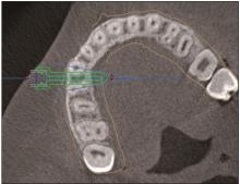
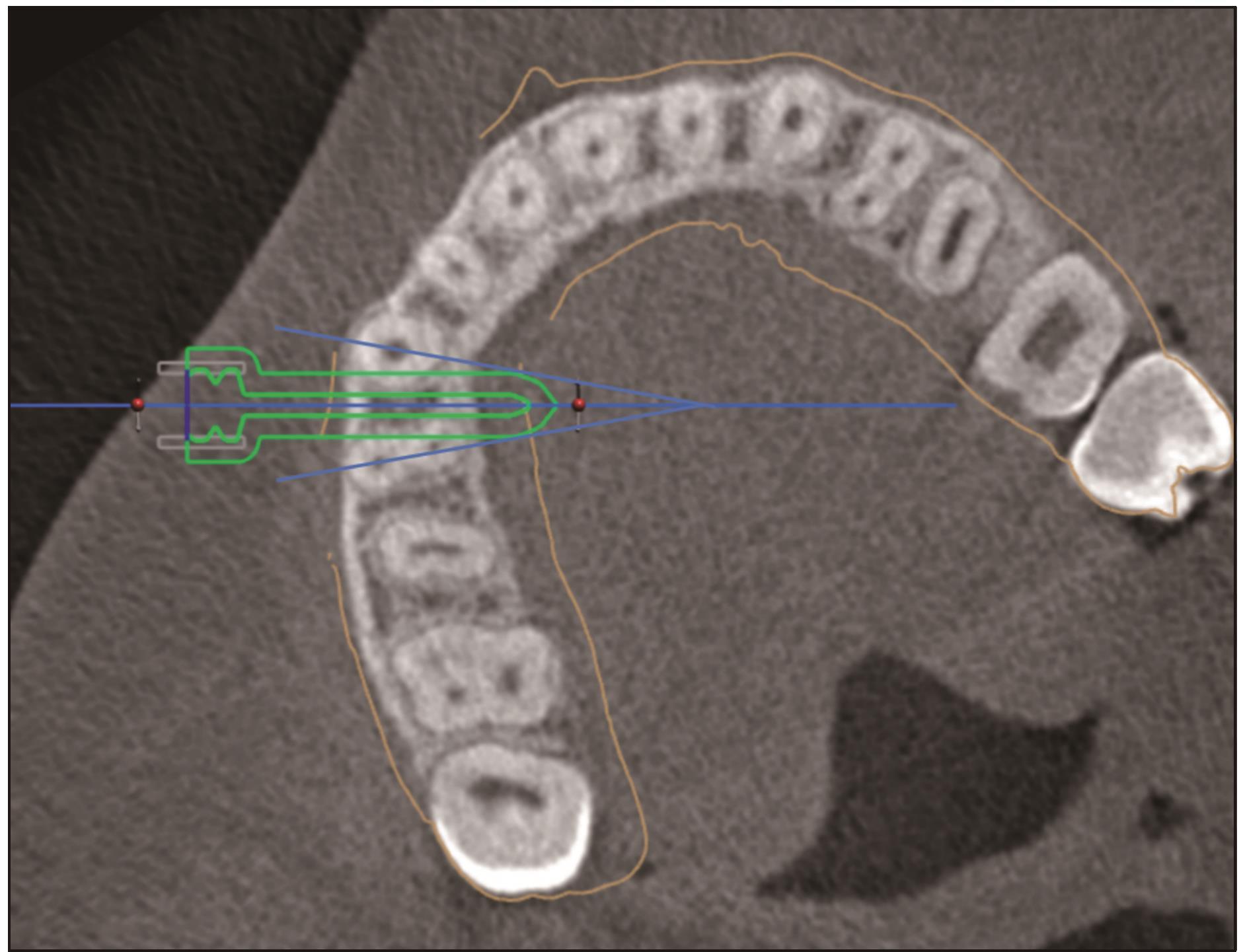
目的 观察本课题组研发的微种植体植入导板的临床应用效果,并比较其与对照组使用传统方式进行微种植体植入的植入效果差异。 方法 按照纳入排除标准共计纳入34名患者,试验侧采用微种植体植入导板辅助微种植体的植入,共计植入34枚微种植体。对照侧参考植入术区影像片,依据操作者的经验进行微种植体的植入,共计植入34枚微种植体,分析导板引导下的微种植体植入精确度,并对2组微种植体的植入成功率进行比较。 结果 试验组植入成功率为97.06%,对照组植入成功率为82.35%,统计分析学结果显示其差异具有统计学意义。 结论 本课题组研发的微种植体植入导板可提高微种植体植入成功率,值得进一步推广临床使用。
中图分类号:
| 1 | Yahya MF, Wang GM, Nimbalkar S. Orthodontic treatment with miniscrew anchorage: analysis of quality of information on YouTube[J]. Am J Orthod Dentofacial Orthop, 2023, 164(1): 97-105. |
| 2 | Papageorgiou SN, Zogakis IP, Papadopoulos MA. Failure rates and associated risk factors of orthodontic miniscrew implants: a meta-analysis[J]. Am J Orthod Dentofacial Orthop, 2012, 142(5): 577-595.e7. |
| 3 | Palone M, Darsiè A, Maino GB, et al. Analysis of bio-logical and structural factors implicated in the cli-nical success of orthodontic miniscrews at posterior maxillary interradicular sites[J]. Clin Oral Investig, 2022, 26(4): 3523-3532. |
| 4 | Xin YL, Wu YK, Chen C, et al. Miniscrews for or-thodontic anchorage: analysis of risk factors correlated with the progressive susceptibility to failure[J]. Am J Orthod Dentofacial Orthop, 2022, 162(4): e192-e202. |
| 5 | Morea C, Dominguez GC, Wuo ADOV, et al. Surgical guide for optimal positioning of mini-implants[J]. J Clin Orthod, 2005, 39(5): 317-321. |
| 6 | Morea C, Hayek JE, Oleskovicz C, et al. Precise insertion of orthodontic miniscrews with a stereolithographic surgical guide based on cone beam compu-ted tomography data: a pilot study[J]. Int J Oral Maxillofac Implants, 2011, 26(4): 860-865. |
| 7 | Cousley RR, Parberry DJ. Surgical stents for accurate miniscrew insertion[J]. J Clin Orthod, 2006, 40(7): 412-417, 419. |
| 8 | 唐睿, 孙建伟, 李成华, 等. 一种新型3D打印正畸微小种植体导板[J]. 口腔医学研究, 2019, 35(12): 1157-1161. |
| Tang R, Sun JW, Li CH, et al. A new type of 3D printed orthodontic microimplant template[J]. J Oral Sci Res, 2019, 35(12): 1157-1161. | |
| 9 | Choi SH, Jeon JY, Lee KJ, et al. Clinical applications of miniscrews that broaden the scope of non-surgical orthodontic treatment[J]. Orthod Craniofac Res, 2021, 24(): 48-58. |
| 10 | Zhang ST, Choi Y, Li W, et al. The effects of cortical bone thickness and miniscrew implant root pro-ximity on the success rate of miniscrew implant: a retrospective study[J]. Orthod Craniofac Res, 2022, 25(3): 342-350. |
| 11 | Oh SH, Lee SR, Choi JY, et al. Geometry of ancho-ring miniscrew in the lateral palate that support a tissue bone borne maxillary expander affects neighbo-ring root damage[J]. Sci Rep, 2021, 11(1): 19880. |
| 12 | Maino BG, Maino G, Mura P. Spider Screw: skeletal anchorage system[J]. Prog Orthod, 2005, 6(1): 70-81. |
| 13 | Kravitz ND, Kusnoto B, Hohlt WF. A simplified stent for anterior miniscrew insertion[J]. J Clin Orthod, 2007, 41(4): 224-226. |
| 14 | Sharma K, Sangwan A. K. s. Micro-implant placement guide[J]. Ann Med Health Sci Res, 2014, 4(): S326-S328. |
| 15 | Suzuki EY, Buranastidporn B. An adjustable surgical guide for miniscrew placement[J]. J Clin Orthod, 2005, 39(10): 588-590. |
| 16 | Takahashi M, Park JH, Uzuka S, et al. Modified surgical stent for accurate TAD placement[J]. J Clin Pediatr Dent, 2018, 42(6): 465-468. |
| 17 | Kim SH, Choi YS, Hwang EH, et al. Surgical positioning of orthodontic mini-implants with guides fabricated on models replicated with cone-beam computed tomography[J]. Am J Orthod Dentofacial Orthop, 2007, 131(4 ): S82-S89. |
| 18 | 陈妍曲, 唐敏, 黄旋平, 等. 高精度三维整合牙颌模型个体化微种植体手术导板的计算机辅助设计与制作[J]. 中国组织工程研究, 2018, 22(10): 1529-1533. |
| Chen YQ, Tang M, Huang XP, et al. The computer-aided design and manufacturing of individualized miniscrew surgical guides based on a high-precision three-dimensional integrated digital maxillodental model[J]. Chin J Tissue Eng Res, 2018, 22(10): 1529-1533. | |
| 19 | 孙应明, 张梦洁, 王晓波. 三维模板定位引导正畸微种植体植入的成功率[J]. 中国组织工程研究与临床康复, 2011, 15(39): 7247-7250. |
| Sun YM, Zhang MJ, Wang XB. Success rate of or-thodontic micro-implant implantation guided by threedimensional template positioning[J]. Chin J Tissue Eng Res, 2011, 15(39): 7247-7250. | |
| 20 | Al-Suleiman M, Shehadah M. AUSOM: a 3D placement guide for orthodontic mini-implants[J]. Orthodontics, 2011, 12(1): 28-37. |
| 21 | 王家艳, 张栋梁, 李梦华. 三维导航微螺钉种植辅助定位装置的设计及临床应用[J]. 中国组织工程研究与临床康复, 2009, 13(13): 2593-2596. |
| Wang JY, Zhang DL, Li MH. Design and clinical application of three-dimensional navigation micro-screw implantation auxiliary positioning device[J]. Chin J Tissue Eng Res, 2009, 13(13): 2593-2596. |
| [1] | 和慧,刘文超,杨锦波. 显微根尖手术术区出血的管理[J]. 国际口腔医学杂志, 2025, 52(1): 99-106. |
| [2] | 蒋佳珍,蒋晓鸽,陈嵩. 颧牙槽嵴区微种植钉成功率的影响因素[J]. 国际口腔医学杂志, 2024, 51(6): 677-686. |
| [3] | 焦明阳,周煜萃,蒋正源,刘雨欣,曲柳. 数字化导板技术在牙髓治疗领域的研究进展[J]. 国际口腔医学杂志, 2024, 51(5): 550-557. |
| [4] | 满毅, 黄定明. 美学区种植骨增量与邻牙慢性根尖周病的联合治疗策略(下):临床诊治流程及实践病例[J]. 国际口腔医学杂志, 2022, 49(6): 621-632. |
| [5] | 汤芝伟,高莺. 靶向牙髓显微外科技术的应用与进展[J]. 国际口腔医学杂志, 2022, 49(6): 678-683. |
| [6] | 蔡娉娉,卓盈颖,林捷,郑志强. 计算机辅助技术在纤维桩拆除中的应用[J]. 国际口腔医学杂志, 2022, 49(6): 731-736. |
| [7] | 庞瑜,刘显,王了. 数字化导板在埋伏多生牙拔除中的应用[J]. 国际口腔医学杂志, 2022, 49(4): 448-452. |
| [8] | 王奔,许喆桢,韦曦. 数字化微创技术在牙髓根尖周病学中的应用与进展[J]. 国际口腔医学杂志, 2021, 48(1): 110-118. |
| [9] | 颜丹,张锡忠,王建国. 螺纹深度对支抗微种植体和颌骨影响的三维有限元分析[J]. 国际口腔医学杂志, 2019, 46(4): 387-392. |
| [10] | 张婷婷,胡建. 数字化导板与动态导航在口腔种植应用中的研究进展[J]. 国际口腔医学杂志, 2019, 46(1): 99-104. |
| [11] | 吕晶, 凌均棨. 根管定位数字化导板的研究进展[J]. 国际口腔医学杂志, 2018, 45(2): 233-238. |
| [12] | 赵静子1 张文君2 张晓东2. 上颌后牙区正畸微种植体支抗植入安全区的测量研究[J]. 国际口腔医学杂志, 2015, 42(6): 659-663. |
| [13] | 颜丹,张锡忠,王增全,王建国,关泽建. 螺距对支抗微种植体—骨界面影响的三维有限元分析[J]. 国际口腔医学杂志, 2015, 42(5): 557-561. |
| [14] | 陆轩,陈小冬,邢文忠,李振春. 全瓷贴面修复的临床效果评估[J]. 国际口腔医学杂志, 2015, 42(2): 170-172. |
| [15] | 贺涵 贺红. 种植体支抗在正畸治疗中垂直向控制的应用进展[J]. 国际口腔医学杂志, 2013, 40(5): 648-652. |
|