国际口腔医学杂志 ›› 2026, Vol. 53 ›› Issue (3): 328-334.doi: 10.7518/gjkq.2026218
• 数字化专栏 • 上一篇
周文媛1( ),范娟1,熊再道1,朱林1,余泽正1,王璐1,金龙2,张盼盼3,顾永春1()
Wenyuan Zhou1(),Juan Fan1,Zaidao Xiong1,Lin Zhu1,Zezheng Yu1,Lu Wang1,Long Jin2,Panpan Zhang3,Yongchun Gu1()
摘要:
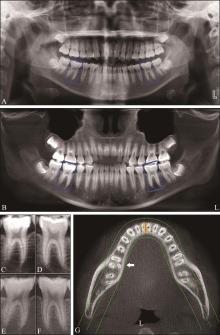
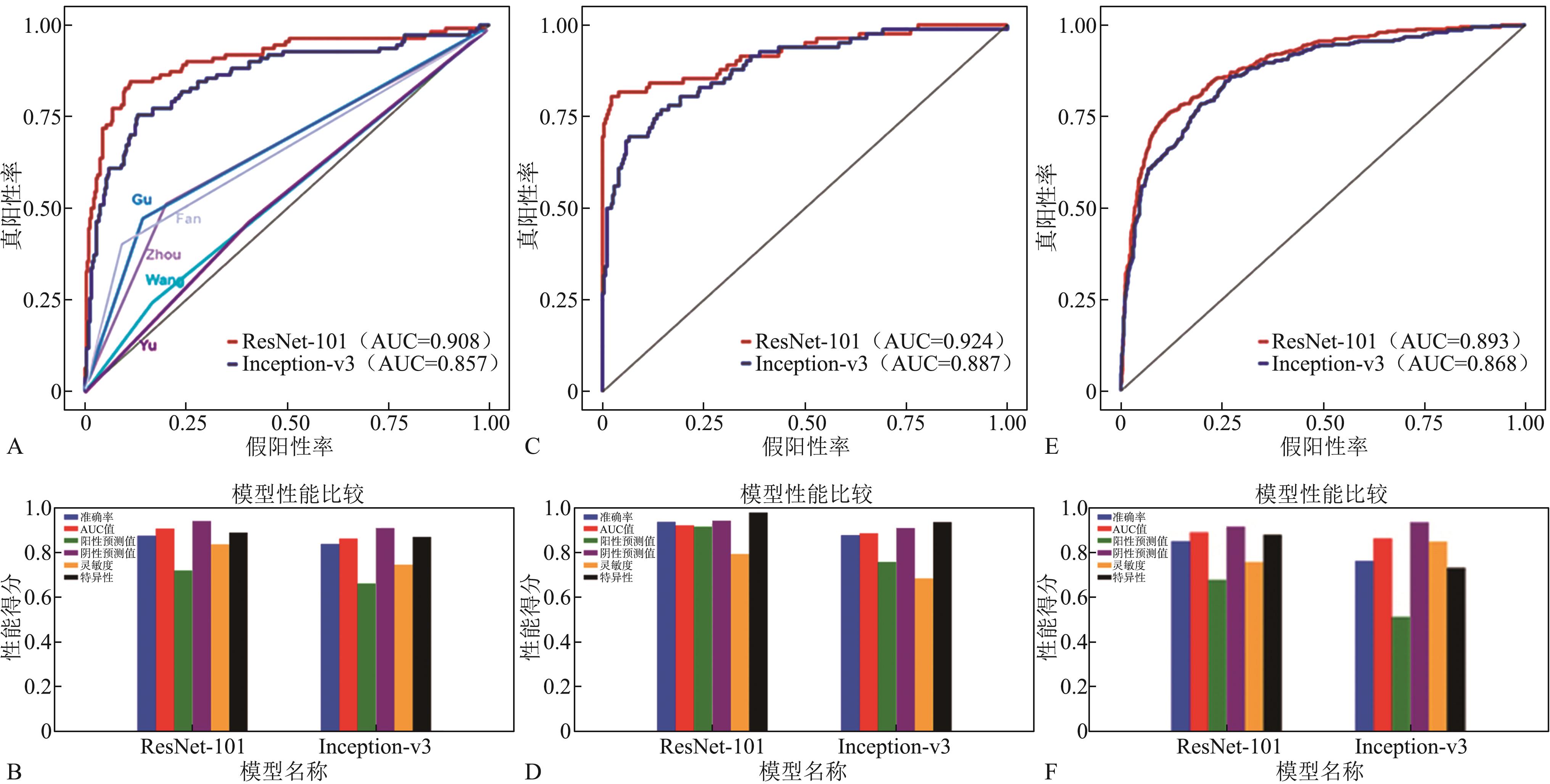
目的 基于深度卷积神经网络(CNN)ResNet-101和Inception-v3对口腔曲面体层片中下颌第一磨牙远舌根变异进行诊断,并评估其准确性。 方法 从口腔影像科采集锥形束CT(CBCT)和口腔曲面体层片。患者被分为2组:A组CBCT图像和口腔曲面体层片来自相同患者,通过切割曲面体层片,制备了1 444个下颌第一磨牙的图像块(其中367个为三根型);B组患者无常规口腔曲面体层片,基于其拍摄的CBCT影像,重建取得模拟的口腔曲面体层片,通过切割获得1 203个下颌第一磨牙的图像块(其中283个为三根型)。采用2个CNN模型(ResNet-101和Inception-v3),基于上述2组图像对模型进行训练与测试,对图像块进行分类(三根及双根型)。以锥形束CT检查为金标准,受试者工作特征(ROC)曲线分析CNN模型的诊断效能,并与5位口腔医学专业人员的诊断效能进行比较。 结果 2个CNN模型的诊断性能良好,其中ResNet-101的诊断效能更佳。在A组,其准确度、灵敏度、特异性分别为87.5%、83.6%和88.9%;AUC为0.908,明显高于Inception-v3的0.857(P<0.01)。采用B组图像训练CNN模型,再用A组图像进行测试,可以取得和A组图像训练相当的诊断效能,对于表现较好的ResNet-101,其准确度、灵敏度、特异性和曲线下面积(AUC)值分别为85.1%、75.8%、88.1%和0.893。而5位口腔医学专业人员的诊断效能均较低,AUC值仅为0.532~0.668。 结论 CNN模型对口腔曲面体层片中三根型下颌第一磨牙的诊断具有较高的准确性。口腔曲面体层片训练集在图片数量和质量上存在不足时,利用CBCT生成的模拟口腔曲面体层片训练CNN模型,可获得相似的诊断效果。
中图分类号:
| [1] | Hatipoğlu FP, Mağat G, Hatipoğlu Ö, et al. Assessment of the prevalence of radix entomolaris and distolingual canal in mandibular first molars in 15 countries: a multinational cross-sectional study with meta-analysis[J]. J Endod, 2023, 49(10): 1308-1318. |
| [2] | Martins JNR, Gu Y, Marques D, et al. Differences on the root and root canal morphologies between Asian and white ethnic groups analyzed by cone-beam computed tomography[J]. J Endod, 2018, 44(7): 1096-1104. |
| [3] | Zhang R, Wang H, Tian YY, et al. Use of cone-beam computed tomography to evaluate root and canal morphology of mandibular molars in Chinese individuals[J]. Int Endod J, 2011, 44(11): 990-999. |
| [4] | 汤颖, 裴凡, 陈秀春, 等. 儿童三根型下颌磨牙的CBCT影像学分析[J]. 实用口腔医学杂志, 2024, 40(4): 60-64. |
| Tang Y, Pei F, Chen XC, et al. CBCT imaging analysis of three-rooted mandibular molars in children[J]. J Pract Stomatol, 2024, 40(4): 60-64. | |
| [5] | Gu Y, Lu Q, Wang P, et al. Root canal morphology of permanent three-rooted mandibular first molars: partⅡ—Measurement of root canal curvatures[J]. J Endod, 2010, 36(8): 1341-1346. |
| [6] | Jiang C, Pei F, Wu Y, et al. Investigation of three-rooted deciduous mandibular second molars in a Chinese population using cone-beam computed tomography[J]. BMC Oral Health, 2022, 22(1): 329. |
| [7] | Tu MG, Huang HL, Hsue SS, et al. Detection of permanent three-rooted mandibular first molars by cone-beam computed tomography imaging in Taiwanese individuals[J]. J Endod, 2009, 35(4): 503-507. |
| [8] | Bai B, Tang Y, Wu Y, et al. Ex vivo detection of mandibular incisors’ root canal morphology using cone-beam computed tomography with 4 different voxel sizes and micro-computed tomography[J]. BMC Oral Health, 2023, 23(1): 656. |
| [9] | Singh NK, Raza K. Progress in deep learning-based dental and maxillofacial image analysis: a systema-tic review[J]. Expert Syst Appl, 2022, 199: 116968. |
| [10] | Schwendicke F, Samek W, Krois J. Artificial intelligence in dentistry: chances and challenges[J]. J Dent Res, 2020, 99(7): 769-774. |
| [11] | Lee JH, Kim DH, Jeong SN, et al. Detection and dia-gnosis of dental caries using a deep learning-based convolutional neural network algorithm[J]. J Dent, 2018, 77: 106-111. |
| [12] | Fukuda M, Inamoto K, Shibata N, et al. Evaluation of an artificial intelligence system for detecting vertical root fracture on panoramic radiography[J]. Oral Radiol, 2020, 36(4): 337-343. |
| [13] | Orhan K, Bayrakdar IS, Ezhov M, et al. Evaluation of artificial intelligence for detecting periapical pathosis on cone-beam computed tomography scans[J]. Int Endod J, 2020, 53(5): 680-689. |
| [14] | Hiraiwa T, Ariji Y, Fukuda M, et al. A deep-learning artificial intelligence system for assessment of root morphology of the mandibular first molar on pano-ramic radiography[J]. Dentomaxillofac Radiol, 2019, 48(3): 20180218. |
| [15] | Yang S, Lee H, Jang B, et al. Development and validation of a visually explainable deep learning model for classification of C-shaped canals of the mandibular second molars in periapical and panoramic dental radiographs[J]. J Endod, 2022, 48(7): 914-921. |
| [16] | Jeon SJ, Yun JP, Yeom HG, et al. Deep-learning for predicting C-shaped canals in mandibular second molars on panoramic radiographs[J]. Dentomaxillofac Radiol, 2021, 50(5): 20200513. |
| [17] |
He K, Zhang X, Ren S, et al. Deep residual learning for image recognition[J]. IEEE, 2016. doi: 10.1109/CVPR.2016.90 .
doi: 10.1109/CVPR.2016.90 |
| [18] |
Szegedy C, Ioffe S, Vanhoucke V, et al. Inception-v4, Inception-ResNet and the impact of residual connections on learning[J]. arXiv, 2016. doi: 10.48550/arXiv.1602.07261 .
doi: 10.48550/arXiv.1602.07261 |
| [19] | Deng J, Dong W, Socher R, et al. ImageNet: a large-scale hierarchical image database[C]//2009 IEEE Conference on Computer Vision and Pattern Recognition. Miami, USA: IEEE, 2009: 248-255. |
| [20] | Jin L, Zhou W, Tang Y, et al. Detection of C-shaped mandibular second molars on panoramic radiographs using deep convolutional neural networks[J]. Clin Oral Investig, 2024, 28(12): 646. |
| [21] | 齐帅, 张旗. 卷积神经网络在牙体牙髓病影像诊断中的研究和应用[J]. 口腔医学研究, 2023, 39(11): 960-964. |
| Qi S, Zhang Q. Research and application of convolutional neural network in endodontic imaging diagnosis[J]. J Oral Sci Res, 2023, 39(11): 960-964. |
| [1] | 兰菁,伍军. 成人均角安氏Ⅰ类、Ⅱ1类及Ⅲ类患者 平面偏斜量的对比研究[J]. 国际口腔医学杂志, 2026, 53(1): 26-35. 平面偏斜量的对比研究[J]. 国际口腔医学杂志, 2026, 53(1): 26-35. |
| [2] | 林超英,张岚,黄定明. 人工智能在根管治疗中的研究进展[J]. 国际口腔医学杂志, 2025, 52(5): 572-578. |
| [3] | 周小洁,侯本祥. 基于深度学习技术诊断龋病方法的研究进展[J]. 国际口腔医学杂志, 2025, 52(5): 579-585. |
| [4] | 黄美畅,蒋鸿杰,汤亚玲,姚莉洪. 锥形束CT及免疫组织化学染色在根尖周囊肿诊断与鉴别诊断中的应用[J]. 国际口腔医学杂志, 2025, 52(4): 490-497. |
| [5] | 焦明阳,周煜萃,蒋正源,刘雨欣,曲柳. 数字化导板技术在牙髓治疗领域的研究进展[J]. 国际口腔医学杂志, 2024, 51(5): 550-557. |
| [6] | 汪云毅,朱珠,张峰. 人工智能在头影测量自动定点算法上的研究进展[J]. 国际口腔医学杂志, 2024, 51(5): 630-641. |
| [7] | 陈新月,潘晓予,杨燕,贾媛媛,陈亮. 卷积神经网络在牙体牙髓病学中的应用进展[J]. 国际口腔医学杂志, 2024, 51(4): 483-488. |
| [8] | 杨雨楠,刘鹏,王虎,游梦. 上颌窦黏膜增厚的锥形束CT影像分析[J]. 国际口腔医学杂志, 2023, 50(3): 302-307. |
| [9] | 吴文智,冯达兴,陈垂壮,周丽鹃. 海口地区下颌第一恒磨牙近中中央根管发生率及相关因素[J]. 国际口腔医学杂志, 2022, 49(4): 420-425. |
| [10] | 叶泽林,刘璐,龙虎,游梦. 弯曲前牙的影像评价及治疗的研究进展[J]. 国际口腔医学杂志, 2022, 49(2): 173-181. |
| [11] | 田浩楠,林敏,谢丛蔓,任嫒姝. 上颌腭侧阻生尖牙与寰椎后桥相关性的锥形束CT研究[J]. 国际口腔医学杂志, 2021, 48(5): 536-540. |
| [12] | 施丹妮,杨鑫,吴建勇. 锥形束CT三维头影测量参考坐标系的研究进展[J]. 国际口腔医学杂志, 2021, 48(4): 398-404. |
| [13] | 丁张帆,郭陟永,苗诚,李春洁,宣鸣,王晓毅,张壮. 基于锥形束CT的三维可视化技术在颌骨囊性病变手术中的应用[J]. 国际口腔医学杂志, 2021, 48(2): 180-186. |
| [14] | 王奔,许喆桢,韦曦. 数字化微创技术在牙髓根尖周病学中的应用与进展[J]. 国际口腔医学杂志, 2021, 48(1): 110-118. |
| [15] | 唐蓓,赵文俊,王虎,郑广宁,游梦. 根管超填导致下牙槽神经损伤2例[J]. 国际口腔医学杂志, 2020, 47(3): 293-296. |
|
||