国际口腔医学杂志 ›› 2023, Vol. 50 ›› Issue (3): 251-262.doi: 10.7518/gjkq.2023048
• 专家笔谈 • 下一篇
黄定明1( ),张岚1,满毅2()
Huang Dingming1(),Zhang Lan1,Man Yi2()
摘要:
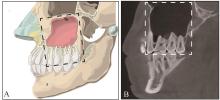
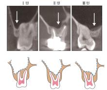
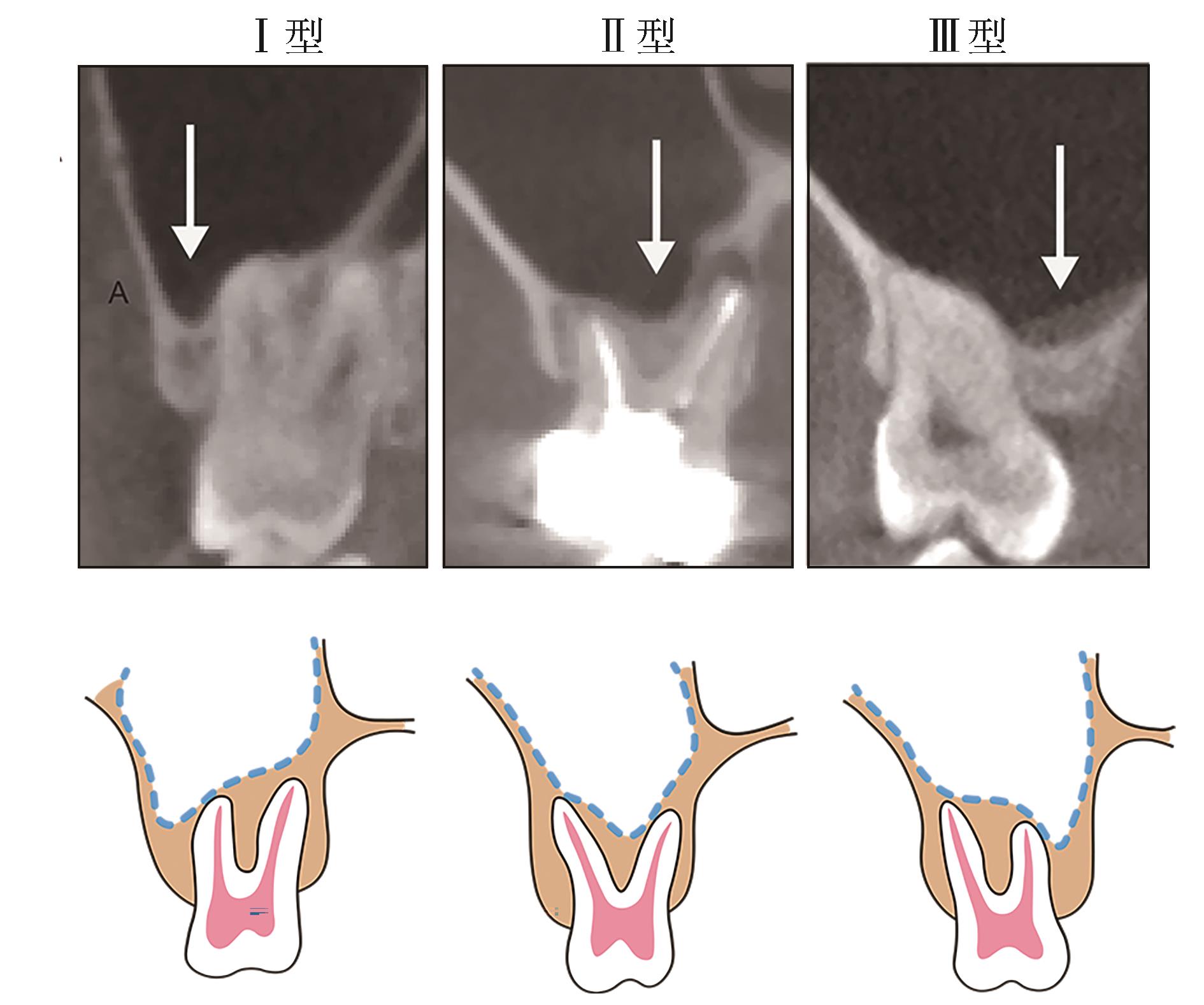
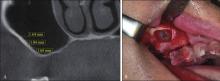
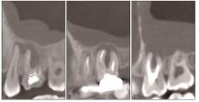
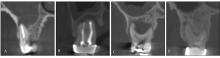
上颌后牙缺失需行种植修复时,如果骨量不足,临床上常规采取上颌窦底提升术进行骨增量,以满足种植体初期稳定性的要求。上颌后牙的根管系统非常复杂,出现牙髓根尖周疾病时,根管治疗有可能无法彻底控制根管内感染,治疗后仍可能再度发生慢性根尖周病变。上颌牙、牙槽骨、上颌窦解剖位置及其功能关系紧密,笔者将其命名为“上颌牙-牙槽骨-上颌窦复合体”。当上颌后牙发生慢性根尖周病变时,牙根进入上颌窦内或者病变扩散至上颌窦可形成牙源性上颌窦炎。采用显微根尖手术治疗这类患牙时,常通过上颌窦底提升术建立手术入路。笔者将这种为治疗牙源性上颌窦炎并保存疑难根尖周病患牙而采取的显微根尖手术联合上颌窦底提升术命名为“牙保存相关上颌窦底提升术”。该技术与牙种植相关上颌窦底提升术不同,是一种全新的治疗术式。本文通过文献回顾分析,结合临床开展该技术的经验和认识,从解剖学、病因学和病理学三方面阐述了该技术的生物学基础:上颌牙-牙槽骨-上颌窦复合体是该技术的解剖学基础,牙源性感染引起上颌窦疾病是其病因学基础,根尖周术区和上颌窦黏骨膜的感染性炎症反应是其病理学基础。本文详细解析上述三方面生物学基础,为牙保存相关上颌窦底提升术临床术式的设计、诊治流程的规范和诊疗路径的建立提供了理论依据。
中图分类号:
| 1 | Zheng QH, Wang Y, Zhou XD, et al. A cone-beam computed tomography study of maxillary first permanent molar root and canal morphology in a Chinese population[J]. J Endod, 2010, 36(9): 1480-1484. |
| 2 | Meirinhos J, Martins JNR, Pereira B, et al. Prevalence of apical periodontitis and its association with previous root canal treatment, root canal filling length and type of coronal restoration-a cross-sectional study[J]. Int Endod J, 2020, 53(4): 573-584. |
| 3 | Huumonen S, Suominen AL, Vehkalahti MM. Pre-valence of apical periodontitis in root filled teeth: findings from a nationwide survey in Finland[J]. Int Endod J, 2017, 50(3): 229-236. |
| 4 | Zhou W, Zheng QH, Tan XL, et al. Comparison of mineral trioxide aggregate and iRoot BP plus root repair material as root-end filling materials in en-dodontic microsurgery: a prospective randomized controlled study[J]. J Endod, 2017, 43(1): 1-6. |
| 5 | Safi C, Kohli MR, Kratchman SI, et al. Outcome of endodontic microsurgery using mineral trioxide aggregate or root repair material as root-end filling material: a randomized controlled trial with cone-beam computed tomographic evaluation[J]. J Endod, 2019, 45(7): 831-839. |
| 6 | Tatum H Jr. Maxillary and sinus implant reconstructions[J]. Dent Clin North Am, 1986, 30(2): 207-229. |
| 7 | Summers RB. A new concept in maxillary implant surgery: the osteotome technique[J]. Compendium, 1994, 15(2): 152, 154-152, 156, 158, 162. |
| 8 | Chao YL, Chen HH, Mei CC, et al. Meta-regression analysis of the initial bone height for predicting implant survival rates of two sinus elevation procedures[J]. J Clin Periodontol, 2010, 37(5): 456-465. |
| 9 | Pjetursson BE, Tan WC, Zwahlen M, et al. A systematic review of the success of sinus floor elevation and survival of implants inserted in combination with sinus floor elevation[J]. J Clin Periodontol, 2008, 35(8 ): 216-240. |
| 10 | Albu S, Baciut M. Failures in endoscopic surgery of the maxillary sinus[J]. Otolaryngol Head Neck Surg, 2010, 142(2): 196-201. |
| 11 | Melén I, Lindahl L, Andréasson L, et al. Chronic maxillary sinusitis. Definition, diagnosis and relation to dental infections and nasal polyposis[J]. Acta Otolaryngol, 1986, 101(3/4): 320-327. |
| 12 | Kale TP, Urolagin S, Khurana V, et al. Treatment of oroantral fistula using palatal flap-a case report and technical note[J]. J Int Oral Health, 2010, 2(3): 77-82. |
| 13 | Cortes AR, Pinheiro LR, Cavalcanti MG, et al. Sinus floor bone failures in maxillary sinus floor augmentation: a case-control study[J]. Clin Implant Dent Relat Res, 2015, 17(2): 335-342. |
| 14 | Lavasani SA, Tyler C, Roach SH, et al. Cone-beam computed tomography: anatomic analysis of maxillary posterior teeth-impact on endodontic microsurgery[J]. J Endod, 2016, 42(6): 890-895. |
| 15 | Clark D, Levin L. In the dental implant era, why do we still bother saving teeth[J]. Dent Traumatol, 2019, 35(6): 368-375. |
| 16 | Oberli K, Bornstein MM, von Arx T. Periapical surgery and the maxillary sinus: radiographic parameters for clinical outcome[J]. Oral Surg Oral Med Oral Pathol Oral Radiol Endod, 2007, 103(6): 848-853. |
| 17 | von Arx T, Käch S, Suter VGA, et al. Perforation of the maxillary sinus floor during apical surgery of maxillary molars: a retrospective analysis using cone beam computed tomography[J]. Aust Endod J, 2020, 46(2): 176-183. |
| 18 | Silberman JJ, Moldauer BI, Torres J, et al. Palatal root surgery of a maxillary molar using a piezosurgery transantral approach with simultaneous sinus lift grafting: a case report[J]. Int Endod J, 2021, 54(3): 464-475. |
| 19 | Azim AA, Wang HH, Serebro M. Selective retreatment and sinus lift: an alternative approach to surgically manage the palatal roots of maxillary molars[J]. J Endod, 2021, 47(4): 648-657. |
| 20 | Kfir E, Goldstein M, Abramovitz I, et al. The effects of sinus membrane pathology on bone augmentation and procedural outcome using minimal invasive antral membrane balloon elevation[J]. J Oral Implantol, 2014, 40(3): 285-293. |
| 21 | Kwak HH, Park HD, Yoon HR, et al. Topographic anatomy of the inferior wall of the maxillary sinus in Koreans[J]. Int J Oral Maxillofac Surg, 2004, 33(4): 382-388. |
| 22 | Lu Y, Liu ZJ, Zhang L, et al. Associations between maxillary sinus mucosal thickening and apical pe-riodontitis using cone-beam computed tomography scanning: a retrospective study[J]. J Endod, 2012, 38(8): 1069-1074. |
| 23 | Watzek G, Bernhart T, Ulm C. Complications of sinus perforations and their management in endodontics[J]. Dent Clin North Am, 1997, 41(3): 563-583. |
| 24 | Freedman A, Horowitz I. Complications after apicoectomy in maxillary premolar and molar teeth[J]. Int J Oral Maxillofac Surg, 1999, 28(3): 192-194. |
| 25 | Lin L, Chance K, Shovlin F, et al. Oroantral communication in periapical surgery of maxillary posterior teeth[J]. J Endod, 1985, 11(1): 40-44. |
| 26 | Rud J, Rud V. Surgical endodontics of upper molars: relation to the maxillary sinus and operation in acute state of infection[J]. J Endod, 1998, 24(4): 260-261. |
| 27 | van den Bergh JP, ten Bruggenkate CM, Disch FJ, et al. Anatomical aspects of sinus floor elevations[J]. Clin Oral Implants Res, 2000, 11(3): 256-265. |
| 28 | Sharan A, Madjar D. Maxillary sinus pneumatization following extractions: a radiographic study[J]. Int J Oral Maxillofac Implants, 2008, 23(1): 48-56. |
| 29 | Zijderveld SA, van den Bergh JP, Schulten EA, et al. Anatomical and surgical findings and complications in 100 consecutive maxillary sinus floor elevation procedures[J]. J Oral Maxillofac Surg, 2008, 66(7): 1426-1438. |
| 30 | Shams N, Dabbaghi A, Shams B, et al. Anatomy of the posterior superior alveolar artery: a cone-beam computed tomographic study[J]. J Maxillofac Oral Surg, 2022, 21(1): 203-210. |
| 31 | Danesh-Sani SA, Movahed A, ElChaar ES, et al. Radiographic evaluation of maxillary sinus lateral wall and posterior superior alveolar artery anatomy: a cone-beam computed tomographic study[J]. Clin Implant Dent Relat Res, 2017, 19(1): 151-160. |
| 32 | Taleghani F, Tehranchi M, Shahab S, et al. Prevalence, location, and size of maxillary sinus septa: computed tomography scan analysis[J]. J Contemp Dent Pract, 2017, 18(1): 11-15. |
| 33 | Gandhi KR, Wabale RN, Siddiqui AU, et al. The incidence and morphology of maxillary sinus septa in dentate and edentulous maxillae: a cadaveric study with a brief review of the literature[J]. J Korean Assoc Oral Maxillofac Surg, 2015, 41(1): 30-36. |
| 34 | Alqahtani S, Alsheraimi A, Alshareef A, et al. Maxillary sinus pneumatization following extractions in Riyadh, Saudi Arabia: a cross-sectional study[J]. Cureus, 2020, 12(1): e6611. |
| 35 | Krennmair G, Ulm CW, Lugmayr H, et al. The incidence, location, and height of maxillary sinus septa in the edentulous and dentate maxilla[J]. J Oral Ma-xillofac Surg, 1999, 57(6): 667-672. |
| 36 | Çakur B, Sümbüllü MA, Durna D. Relationship among Schneiderian membrane, Underwood’s septa, and the maxillary sinus inferior border[J]. Clin Implant Dent Relat Res, 2013, 15(1): 83-87. |
| 37 | Siqueira JF Jr, Rôças IN. Diversity of endodontic microbiota revisited[J]. J Dent Res, 2009, 88(11): 969-981. |
| 38 | Ricucci D, Siqueira JF Jr, Lopes WS, et al. Extraradicular infection as the cause of persistent symptoms: a case series[J]. J Endod, 2015, 41(2): 265-273. |
| 39 | Signoretti FG, Endo MS, Gomes BP, et al. Persistent extraradicular infection in root-filled asymp-tomatic human tooth: scanning electron microscopic analysis and microbial investigation after apical microsurgery[J]. J Endod, 2011, 37(12): 1696-1700. |
| 40 | Ricucci D, Rôças IN, Hernández S, et al. “true” versus “bay” apical cysts: clinical, radiographic, histopathologic, and histobacteriologic features[J]. J Endod, 2020, 46(9): 1217-1227. |
| 41 | Curi FR, Pelegrine RA, Nascimento MDCC, et al. Odontogenic infection as a predisposing factor for pathologic disorder development in maxillary sinus[J]. Oral Dis, 2020, 26(8): 1727-1735. |
| 42 | Sakir M, Ercalik Yalcinkaya S. Associations between periapical health of maxillary molars and mucosal thickening of maxillary sinuses in cone-beam computed tomographic images: a retrospective study[J]. J Endod, 2020, 46(3): 397-403. |
| 43 | Maillet M, Bowles WR, McClanahan SL, et al. Cone-beam computed tomography evaluation of ma-xillary sinusitis[J]. J Endod, 2011, 37(6): 753-757. |
| 44 | Brook I. Microbiology of acute and chronic maxillary sinusitis associated with an odontogenic origin[J]. Laryngoscope, 2005, 115(5): 823-825. |
| 45 | Yassin-Kassab A, Bhargava P, Tibbetts RJ, et al. Comparison of bacterial maxillary sinus cultures between odontogenic sinusitis and chronic rhinosinu-sitis[J]. Int Forum Allergy Rhinol, 2021, 11(1): 40-47. |
| 46 | Lin YH, Yang YC, Wen SC, et al. The influence of sinus membrane thickness upon membrane perforation during lateral window sinus augmentation[J]. Clin Oral Implants Res, 2016, 27(5): 612-617. |
| 47 | Insua A, Monje A, Urban I, et al. The sinus membrane-maxillary lateral wall complex: histologic description and clinical implications for maxillary sinus floor elevation[J]. Int J Periodontics Restorative Dent, 2017, 37(6): e328-e336. |
| 48 | Munakata M, Yamaguchi K, Sato D, et al. Factors influencing the sinus membrane thickness in edentulous regions: a cone-beam computed tomography study[J]. Int J Implant Dent, 2021, 7(1): 16. |
| 49 | Block MS, Dastoury K. Prevalence of sinus membrane thickening and association with unhealthy teeth: a retrospective review of 831 consecutive patients with 1, 662 cone-beam scans[J]. J Oral Maxillofac Surg, 2014, 72(12): 2454-2460. |
| 50 | Yoo JY, Pi SH, Kim YS, et al. Healing pattern of the mucous membrane after tooth extraction in the ma-xillary sinus[J]. J Periodontal Implant Sci, 2011, 41(1): 23-29. |
| 51 | Yilmaz HG, Tözüm TF. Are gingival phenotype, residual ridge height, and membrane thickness critical for the perforation of maxillary sinus[J]. J Periodontol, 2012, 83(4): 420-425. |
| 52 | Dumitrescu A, Martu MA, Nemtoi A, et al. Association between cone-beam computed tomography and histological and immunohistochemical features in periapical lesions correlated with thickened maxillary sinus mucosa[J]. Medicina (Kaunas), 2021, 57(8): 840. |
| 53 | Tassoker M. What are the risk factors for maxillary sinus pathologies? A CBCT study[J]. Oral Radiol, 2020, 36(1): 80-84. |
| 54 | Testori T, Yu SH, Tavelli L, et al. Perforation risk assessment in maxillary sinus augmentation with late-ral wall technique[J]. Int J Periodontics Restorative Dent, 2020, 40(3): 373-380. |
| 55 | 满毅, 袁珊珊, 赵磊, 等. 上颌窦提升术的历史、现状和发展[J]. 国际口腔医学杂志, 2014, 41(6): 621-627. |
| Man Y, Yuan SS, Zhao L, et al. The history, present situation and development of the maxillary sinus lifting[J]. Int J Stomatol, 2014, 41(6): 621-627. | |
| 56 | Amid R, Kadkhodazadeh M, Moscowchi A, et al. Effect of schneiderian membrane thickening on the maxillary sinus augmentation and implantation outcomes: a systematic review[J]. J Maxillofac Oral Surg, 2021, 20(4): 534-544. |
| 57 | Pizzini A, Basma HS, Li P, et al. The impact of anatomic, patient and surgical factors on membrane perforation during lateral wall sinus floor elevation[J]. Clin Oral Implants Res, 2021, 32(3): 274-284. |
| [1] | 戢晓,张岚,黄定明. 牙源性与非牙源性上颌窦炎鉴别诊断及其治疗方案的研究进展[J]. 国际口腔医学杂志, 2023, 50(5): 566-572. |
| [2] | 吴思佳,舒畅,王洋,王媛,邓淑丽,王慧明. 根管内感染控制对年轻恒牙牙髓再生治疗的影响及研究进展[J]. 国际口腔医学杂志, 2023, 50(4): 388-394. |
| [3] | 王铝亚,张竞心,林洁. 口腔术后聚维酮碘与氯己定控制感染效果的系统评价分析[J]. 国际口腔医学杂志, 2023, 50(4): 438-444. |
| [4] | 杨雨楠,刘鹏,王虎,游梦. 上颌窦黏膜增厚的锥形束CT影像分析[J]. 国际口腔医学杂志, 2023, 50(3): 302-307. |
| [5] | 朱秋艳,吴道敏,鲍济波,谢志刚. 上颌窦宽度与角度对上颌窦底提升术后成骨效果的影响[J]. 国际口腔医学杂志, 2023, 50(2): 159-165. |
| [6] | 朱嘉妮,苏勤. 难治性根尖周炎根管内及根尖外菌群的研究现状[J]. 国际口腔医学杂志, 2022, 49(3): 283-289. |
| [7] | 林洁,刘帆. 基于多酶清洗剂控制的口腔专科器械清洗消毒模式的探索[J]. 国际口腔医学杂志, 2022, 49(3): 324-327. |
| [8] | 王璐璇,侯本祥. 根管内氢氧化钙残留对根管治疗的影响[J]. 国际口腔医学杂志, 2022, 49(3): 367-372. |
| [9] | 杨加震,张颖,刘育含,李帆,曾飞,李修珍,马玉莹,杨芳. 口腔诊疗环境细菌群落的时间变化趋势研究[J]. 国际口腔医学杂志, 2022, 49(2): 132-137. |
| [10] | 余舒星,邹静,李雨庆. 基于唾液检测病毒感染性生物标志物的研究进展[J]. 国际口腔医学杂志, 2022, 49(2): 189-196. |
| [11] | 刘程程, 丁一. 妊娠期常见口腔感染性疾病的临床诊疗和管理策略[J]. 国际口腔医学杂志, 2021, 48(6): 621-628. |
| [12] | 付琢惠,谭学莲,黄定明. 牙源性上颌窦炎的诊疗策略[J]. 国际口腔医学杂志, 2021, 48(3): 367-372. |
| [13] | 易俭如,罗梦奇,尹一佳,刘治清,柳茜,石永乐,杨征,刘帆,韩向龙. 新型冠状病毒肺炎流行期降低口腔诊疗中气溶胶传播风险的策略[J]. 国际口腔医学杂志, 2020, 47(3): 362-365. |
| [14] | 税钰森,吕潇颖,李静雅,杨燃. 粪肠球菌在口腔及全身系统性疾病中的致病相关因素及其机制的研究进展[J]. 国际口腔医学杂志, 2020, 47(2): 225-234. |
| [15] | 毕小琴,熊茂婧,陈丽先,白沅艳,田莉,杨晖. 新型冠状病毒肺炎疫情下口腔颌面外科的护理防控[J]. 国际口腔医学杂志, 2020, 47(2): 244-248. |
|