国际口腔医学杂志 ›› 2022, Vol. 49 ›› Issue (5): 497-505.doi: 10.7518/gjkq.2022084
• 专家笔谈 • 下一篇
满毅1,黄定明2( )
)
Man Yi1,Huang Dingming2()
摘要:
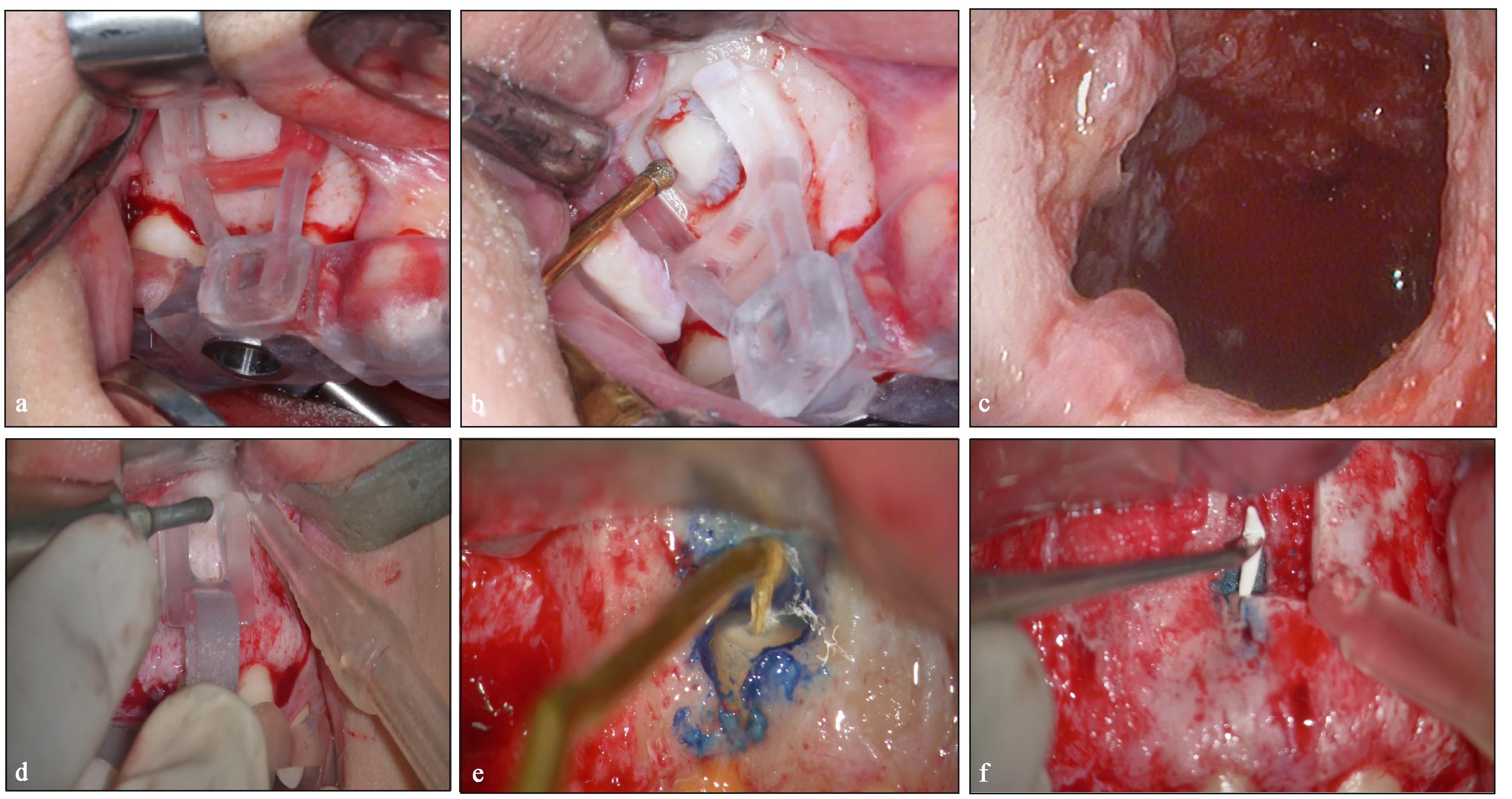
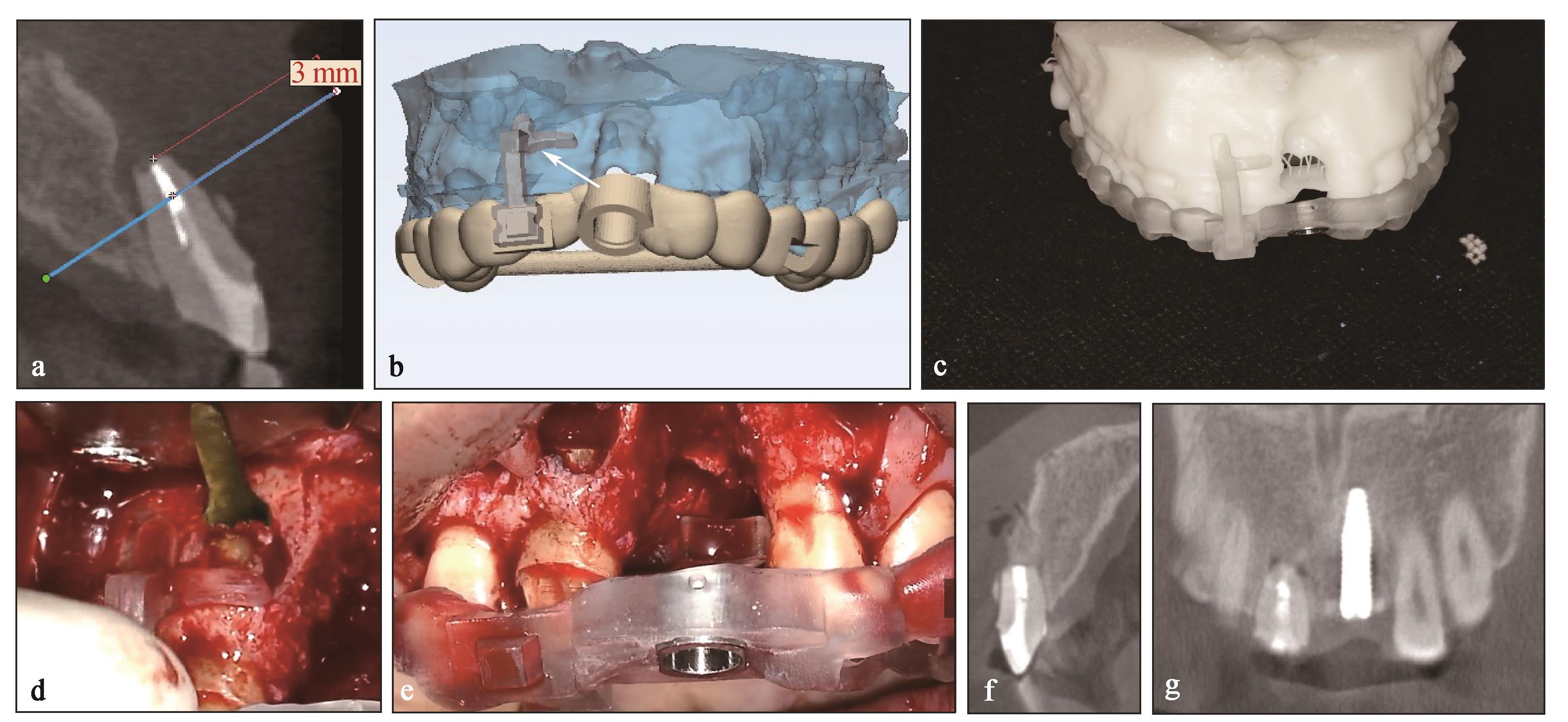
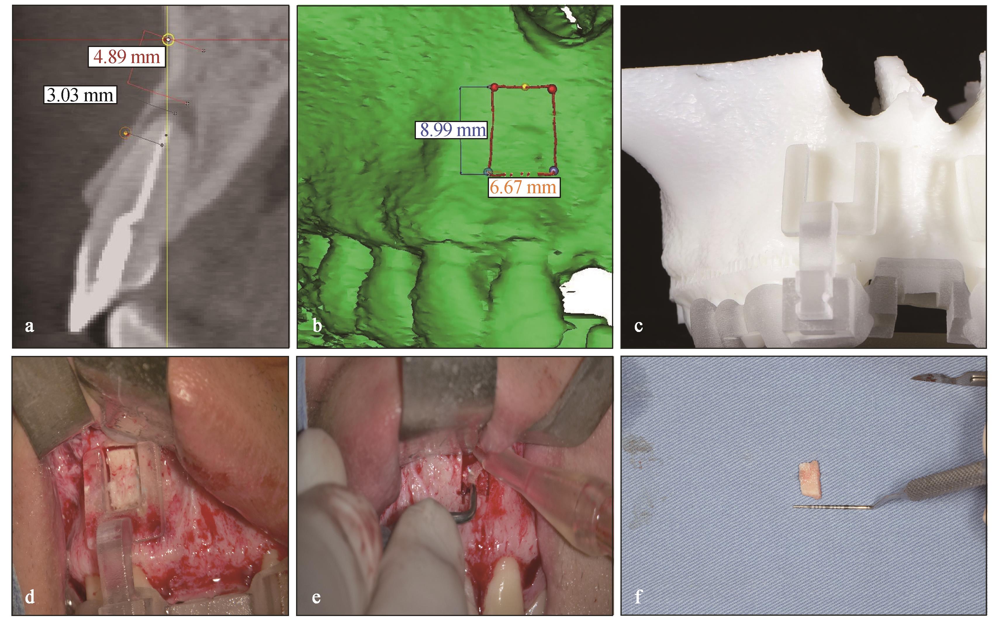
在口腔种植治疗的临床实践中,种植相关治疗术区骨量不足同时合并邻牙慢性根尖周炎的临床现象特殊而又普遍,邻牙慢性根尖周炎可成为骨增量手术中潜在的感染源而引起早期治疗失败/或延伸至种植体根尖周引起逆行性种植体周炎。不同于传统分科诊疗的模式,笔者基于两个学科的发展基础,提出了邻牙显微根尖手术与种植相关治疗同期完成的联合治疗新模式。目的是在实施种植相关手术同期消除邻牙潜在感染源;同时根尖手术中保留的自体骨可以避免种植相关骨增量手术中开辟第二术区取骨,大大提高了手术效率,并进一步减小了患者创伤,节省了医疗费用。本文针对这一新的联合治疗策略的应用基础及临床适应证进行探讨。
中图分类号:
| 1 | Penarrocha-Diago M, Maestre-Ferrín L, Penarrocha-Oltra D, et al. Inflammatory implant periapical lesion prior to osseointegration: a case series study[J]. Int J Oral Maxillofac Implants, 2013, 28(1): 158-162. |
| 2 | Mohamed JB, Alam MN, Singh G, et al. The mana-gement of retrograde peri-implantitis: a case report[J]. J Clin Diagn Res, 2012, 6(9): 1600-1602. |
| 3 | Lefever D, van Assche N, Temmerman A, et al. Ae-tiology, microbiology and therapy of periapical lesions around oral implants: a retrospective analysis[J]. J Clin Periodontol, 2013, 40(3): 296-302. |
| 4 | Jakovljevic A, Nikolic N, Jacimovic J, et al. Prevalence of apical periodontitis and conventional nonsurgical root canal treatment in general adult population: an updated systematic review and meta-analysis of cross-sectional studies published between 2012 and 2020[J]. J Endod, 2020, 46(10): 1371-1386.e8. |
| 5 | Moy PK, Aghaloo T. Risk factors in bone augmentation procedures[J]. Periodontol 2000, 2019, 81(1): 76-90. |
| 6 | Dammaschke T, Steven D, Kaup M, et al. Long-term survival of root-canal-treated teeth: a retrospective study over 10 years[J]. J Endod, 2003, 29(10): 638-643. |
| 7 | Danesh N, Ljunggren AC, Wolf E, et al. Development of criteria for investigation of periapical tissue from root-filled teeth[J]. Acta Odontol Scand, 2019, 77(4): 269-274. |
| 8 | Zhou W, Zheng QH, Tan XL, et al. Comparison of mineral trioxide aggregate and iRoot BP plus root repair material as root-end filling materials in en-dodontic microsurgery: a prospective randomized controlled study[J]. J Endod, 2017, 43(1): 1-6. |
| 9 | Moraschini V, Poubel LA, Ferreira VF, et al. Evaluation of survival and success rates of dental implants reported in longitudinal studies with a follow-up period of at least 10 years: a systematic review[J]. Int J Oral Maxillofac Surg, 2015, 44(3): 377-388. |
| 10 | McAllister BS, Masters D, Meffert RM. Treatment of implants demonstrating periapical radiolucencies[J]. Pract Periodontics Aesthet Dent, 1992, 4(9): 37-41. |
| 11 | Green TL, Walton RE, Taylor JK, et al. Radiogra-phic and histologic periapical findings of root canal treated teeth in cadaver[J]. Oral Surg Oral Med Oral Pathol Oral Radiol Endod, 1997, 83(6): 707-711. |
| 12 | Rowe AH, Binnie WH. Correlation between radiological and histological inflammatory changes following root canal treatment[J]. J Br Endod Soc, 1974, 7(2): 57-63. |
| 13 | Seltzer S. Long-term radiographic and histological observations of endodontically treated teeth[J]. J Endod, 1999, 25(12): 818-822. |
| 14 | Schwarz F, Derks J, Monje A, et al. Peri-implantitis[J]. J Periodontol, 2018, 89(): S267-S290. |
| 15 | Sarmast ND, Wang HH, Soldatos NK, et al. A novel treatment decision tree and literature review of retrograde peri-implantitis[J]. J Periodontol, 2016, 87(12): 1458-1467. |
| 16 | Ramanauskaite A, Juodzbalys G, Tözüm TF. Apical/retrograde periimplantitis/implant periapical lesion: etiology, risk factors, and treatment options: a syste-matic review[J]. Implant Dent, 2016, 25(5): 684-697. |
| 17 | Marshall G, Canullo L, Logan RM, et al. Histopa-thological and microbiological findings associated with retrograde peri-implantitis of extra-radicular en-dodontic origin: a systematic and critical review[J]. Int J Oral Maxillofac Surg, 2019, 48(11): 1475-1484. |
| 18 | di Murro B, Papi P, di Murro C, et al. Correlation between endodontic pulpal/periapical disease and retrograde peri-implantitis: a case series[J]. Aust Endod J, 2021, 47(2): 358-364. |
| 19 | Zhou W, Han C, Li DH, et al. Endodontic treatment of teeth induces retrograde peri-implantitis[J]. Clin Oral Implants Res, 2009, 20(12): 1326-1332. |
| 20 | Fernandez de Grado G, Keller L, Idoux-Gillet Y, et al. Bone substitutes: a review of their characteristics, clinical use, and perspectives for large bone defects management[J]. J Tissue Eng, 2018, 9: 204173-1418776819. |
| 21 | Antoun H, Sitbon JM, Martinez H, et al. A prospective randomized study comparing two techniques of bone augmentation: onlay graft alone or associated with a membrane[J]. Clin Oral Implants Res, 2001, 12(6): 632-639. |
| 22 | Meijndert CM, Raghoebar GM, Meijndert L, et al. Single implants in the aesthetic region preceded by local ridge augmentation; a 10-year randomized controlled trial[J]. Clin Oral Implants Res, 2017, 28(4): 388-395. |
| 23 | Sbordone L, Toti P, Menchini-Fabris G, et al. Implant survival in maxillary and mandibular osseous onlay grafts and native bone: a 3-year clinical and computerized tomographic follow-up[J]. Int J Oral Maxillofac Implants, 2009, 24(4): 695-703. |
| 24 | Setzer FC, Shah SB, Kohli MR, et al. Outcome of endodontic surgery: a meta-analysis of the literature: part 1: comparison of traditional root-end surgery and endodontic microsurgery[J]. J Endod, 2010, 36(11): 1757-1765. |
| 25 | Friedman S. Outcome of endodontic surgery: a meta-analysis of the literature-part 1: comparison of traditional root-end surgery and endodontic microsurgery[J]. J Endod, 2011, 37(5): 577-580. |
| 26 | Kim D, Kim S, Song MJ, et al. Outcome of endo-dontic micro-resurgery: a retrospective study based on propensity score-matched survival analysis[J]. J Endod, 2018, 44(11): 1632-1640. |
| 27 | Huang SY, Chen NN, Yu VSH, et al. Long-term success and survival of endodontic microsurgery[J]. J Endod, 2020, 46(2): 149-157.e4. |
| 28 | Conejo J, Atria PJ, Schweitzer D, et al. Digital implant planning and surgical guides: tools for clinical success[J]. Compend Contin Educ Dent, 2021, 42(7): 400-401. |
| 29 | Michelinakis G, Apostolakis D, Kamposiora P, et al. The direct digital workflow in fixed implant pros-thodontics: a narrative review[J]. BMC Oral Health, 2021, 21(1): 37. |
| 30 | Buser D, Sennerby L, de Bruyn H. Modern implant dentistry based on osseointegration: 50 years of pro-gress, current trends and open questions[J]. Perio-dontol 2000, 2017, 73(1): 7-21. |
| 31 | Hawkins TK, Wealleans JA, Pratt AM, et al. Targe-ted endodontic microsurgery and endodontic microsurgery: a surgical simulation comparison[J]. Int Endod J, 2020, 53(5): 715-722. |
| 32 | Ray JJ, Giacomino CM, Wealleans JA, et al. Targe-ted endodontic microsurgery: digital workflow options[J]. J Endod, 2020, 46(6): 863-871. |
| 33 | Dianat O, Gupta S, Price JB, et al. Guided endodontic access in a maxillary molar using a dynamic na-vigation system[J]. J Endod, 2021, 47(4): 658-662. |
| 34 | Joda T, Zarone F, Ferrari M. The complete digital workflow in fixed prosthodontics: a systematic review[J]. BMC Oral Health, 2017, 17(1): 124. |
| 35 | Stanley M, Paz AG, Miguel I, et al. Fully digital workflow, integrating dental scan, smile design and CAD-CAM: case report[J]. BMC Oral Health, 2018, 18(1): 134. |
| 36 | Blatz MB, Conejo J. The current state of chairside digital dentistry and materials[J]. Dent Clin North Am, 2019, 63(2): 175-197. |
| 37 | Coachman C, Sesma N, Blatz MB. The complete digital workflow in interdisciplinary dentistry[J]. Int J Esthet Dent, 2021, 16(1): 34-49. |
| 38 | Wakimoto M, Matsumura T, Ueno T, et al. Bone quality and quantity of the anterior maxillary trabe-cular bone in dental implant sites[J]. Clin Oral Implants Res, 2012, 23(11): 1314-1319. |
| 39 | Ribeiro CG, Bittencourt TC, Ferreira CF, et al. An alternative approach for augmenting the anterior maxilla using autogenous free gingival bone graft for implant retained prosthesis[J]. J Oral Implantol, 2014, 40(2): 183-187. |
| 40 | Chen ST, Buser D. Esthetic outcomes following immediate and early implant placement in the anterior maxilla: a systematic review[J]. Int J Oral Maxillofac Implants, 2014, 29(): 186-215. |
| 41 | Cha HS, Kim JW, Hwang JH, et al. Frequency of bone graft in implant surgery[J]. Maxillofac Plast Reconstr Surg, 2016, 38(1): 19. |
| 42 | Wang J, Luo YL, Tan XL, et al. Horizontal bone augmentation of the edentulous area with simulta-neous endodontic microsurgery of the adjacent too-th: a digitally-driven multidisciplinary case report with a 1-year follow-up[J]. Int J Oral Implantol (Berl), 2021, 14(4): 435-451. |
| 43 | Daubert D, Black RM, Chrepa V, et al. Endodontic peri-implant defects: a new disease entity[J]. J Endod, 2020, 46(3): 444-448. |
| [1] | 常欣楠,刘磊. 生物可降解镁基材料在颅颌面外科的应用及其研究进展[J]. 国际口腔医学杂志, 2024, 51(1): 107-115. |
| [2] | 韩冲,何东宁,余飞燕,吴东潮. 口腔种植术后疼痛机制及治疗的研究进展[J]. 国际口腔医学杂志, 2024, 51(1): 99-106. |
| [3] | 朱可石,廖安琪,余优成. 机器学习在口腔种植学中的应用研究进展[J]. 国际口腔医学杂志, 2023, 50(4): 491-498. |
| [4] | 满毅, 黄定明. 美学区种植骨增量与邻牙慢性根尖周病的联合治疗策略(下):临床诊治流程及实践病例[J]. 国际口腔医学杂志, 2022, 49(6): 621-632. |
| [5] | 赵文俊,陈宇. 引导组织/骨再生牙周功能梯度膜的研究进展[J]. 国际口腔医学杂志, 2021, 48(4): 391-397. |
| [6] | 汪是琦,常雅琴,陈斌,谭葆春,泥艳红. 植骨术与植骨联用屏障膜在牙周再生治疗中临床疗效对比的系统评价与Meta分析[J]. 国际口腔医学杂志, 2020, 47(6): 644-651. |
| [7] | 刘梦齐,盖阔,蒋丽. 抗菌性口腔种植材料的研究进展[J]. 国际口腔医学杂志, 2018, 45(5): 516-521. |
| [8] | 陈青娅, 黄茜, 王黎. 口腔种植患者牙科焦虑的调查分析[J]. 国际口腔医学杂志, 2018, 45(1): 14-19. |
| [9] | 万双全, 邓飞龙. 上皮下结缔组织瓣在种植软组织缺陷中的应用[J]. 国际口腔医学杂志, 2018, 45(1): 68-73. |
| [10] | 王晓娜 赵静辉 储顺礼 周延民. 骨替代材料在口腔种植领域中的成骨效果[J]. 国际口腔医学杂志, 2016, 43(1): 113-. |
| [11] | 雷文龙1 施斌1,2. 血小板衍生生长因子-BB在口腔种植领域中的作用[J]. 国际口腔医学杂志, 2014, 41(2): 199-203. |
| [12] | 陈红亮 赵承初 赵峰 钟科 孙勇. 国产异种脱细胞真皮基质在引导骨再生术治疗种植义齿受植区骨缺损中的成骨效果评价[J]. 国际口腔医学杂志, 2013, 40(1): 33-36. |
| [13] | 汪池 朱慧勇. 基因修饰的纳米纤维支架的研究进展[J]. 国际口腔医学杂志, 2013, 40(1): 64-67. |
| [14] | 杨世茂1综述 王明国2审校. 富血小板纤维蛋白的研究进展[J]. 国际口腔医学杂志, 2012, 39(3): 373-375. |
| [15] | 郭大伟1,2综述 梁星1 陈悦1审校. 发达国家口腔种植学的教育现状[J]. 国际口腔医学杂志, 2011, 38(3): 326-328. |
|