国际口腔医学杂志 ›› 2023, Vol. 50 ›› Issue (3): 302-307.doi: 10.7518/gjkq.2023027
杨雨楠1( ),刘鹏1,王虎2(),游梦2
Yang Yunan1(),Liu Peng1,Wang Hu2(),You Meng2
摘要:
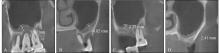
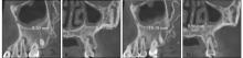
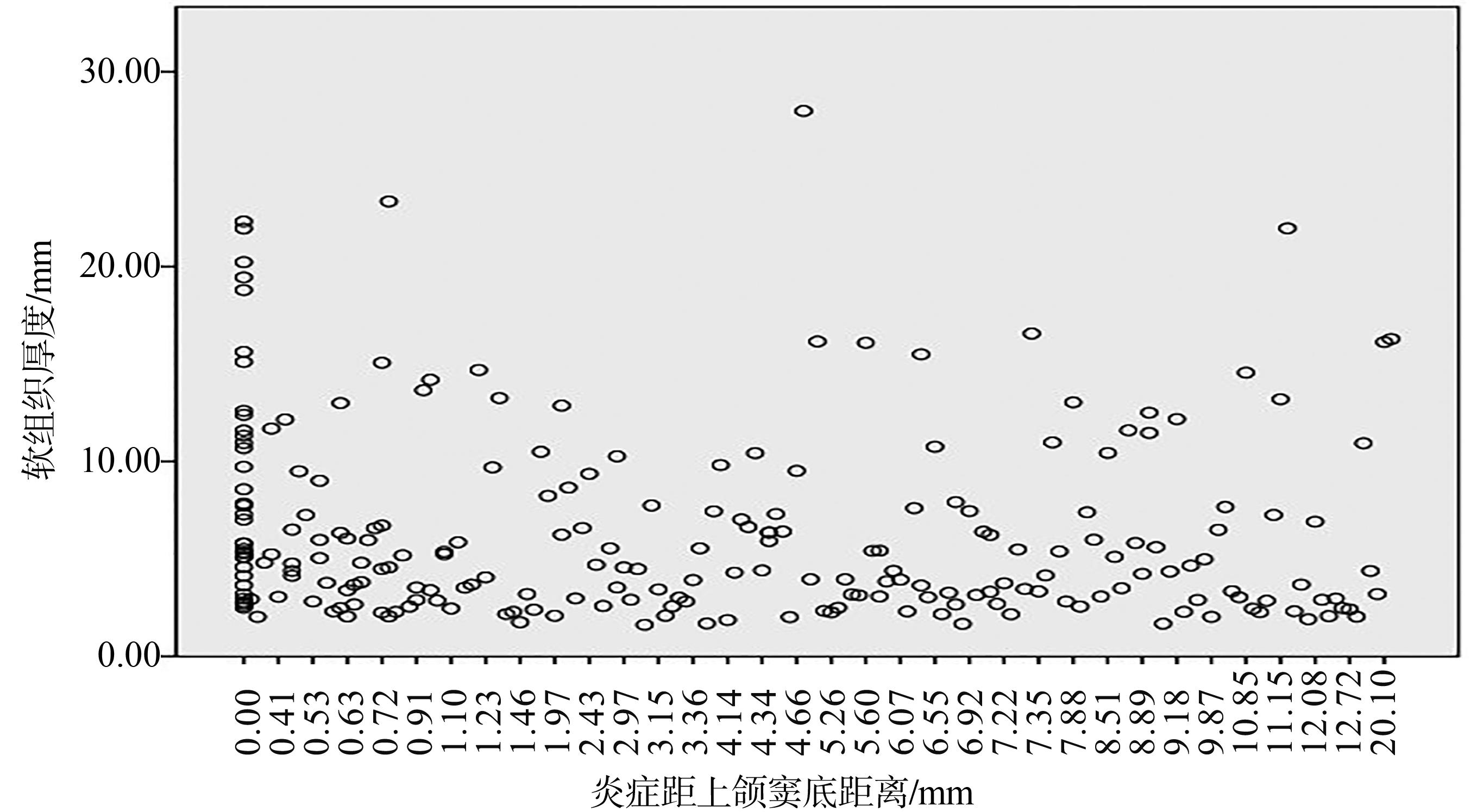
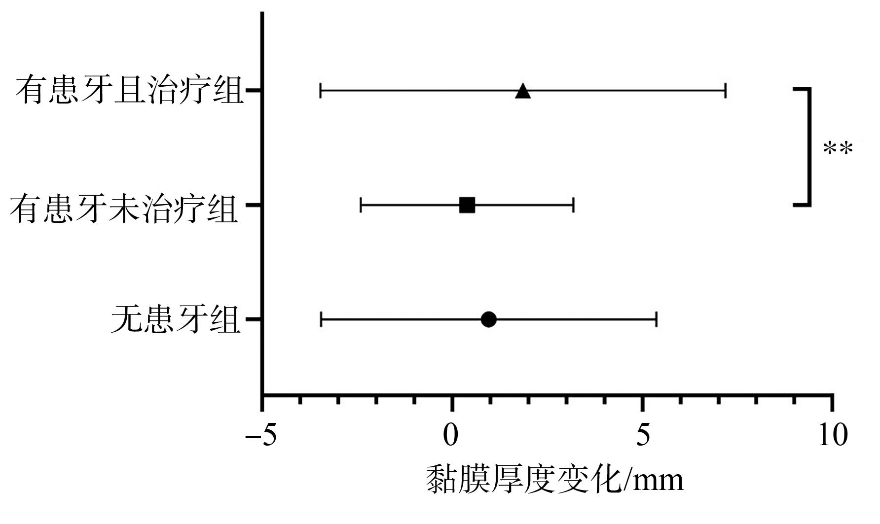
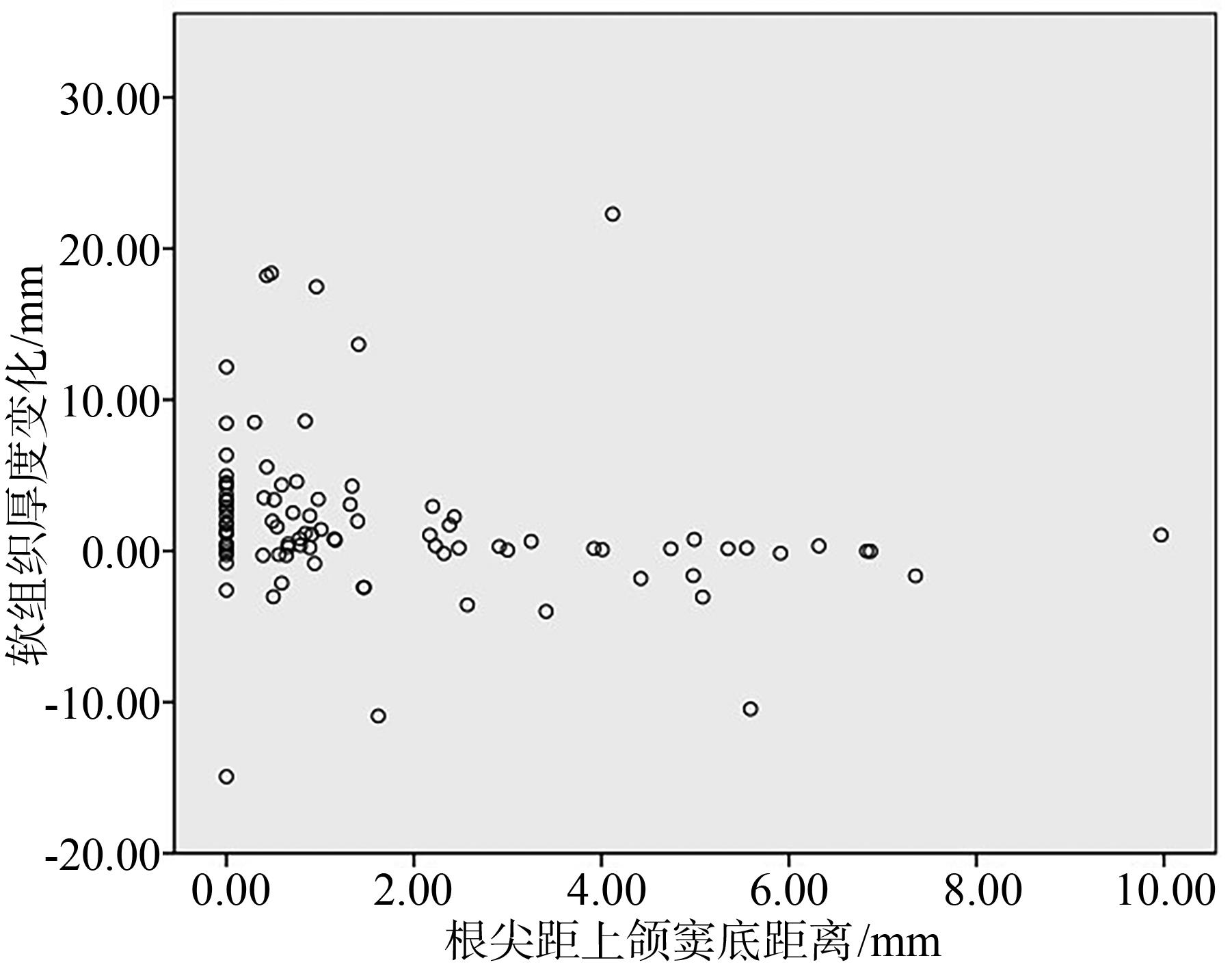
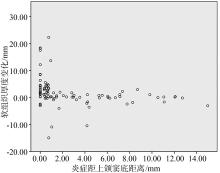
目的 运用锥形束CT(CBCT)研究牙源性因素对上颌窦黏膜增厚的影响,同时,通过对上颌窦黏膜增厚的随访,研究口腔治疗的作用。 方法 回顾性分析2017年8—12月四川大学华西口腔医院就诊的患者,通过CBCT图像筛选出上颌窦最大黏膜厚度>2 mm的患者,评估上颌窦底及上颌窦黏膜增厚与患牙的关系,同时,记录患牙及其治疗的情况,观察随访前后上颌窦黏膜增厚的变化。 结果 黏膜厚度与炎症距窦底的距离呈极弱负相关(P<0.05,r=-0.154),而根尖距窦底的距离与黏膜厚度无直接相关性(P>0.05)。在治疗组中,窦底破坏组的黏膜厚度变化较窦底连续组大,黏膜厚度变化与根尖距窦底的距离呈弱负相关(P<0.01,r=-0.382),黏膜厚度变化与炎症距窦底的距离呈中等程度负相关(P<0.001,r=-0.524)。 结论 上颌窦炎的严重程度可能更大程度上取决于根尖周炎症边缘距窦底的远近而与单纯的根尖位置没有明显的关系。但从治疗效果来讲,炎症病变离上颌窦越近,上颌窦底越易被破坏,上颌窦炎受牙源性因素影响越大,口腔治疗的效果也越好。
中图分类号:
| 1 | Kim SM. Definition and management of odontoge-nic maxillary sinusitis[J]. Maxillofac Plast Reconstr Surg, 2019, 41(1): 13. |
| 2 | Lu Y, Liu ZJ, Zhang L, et al. Associations between maxillary sinus mucosal thickening and apical pe-riodontitis using cone-beam computed tomography scanning: a retrospective study[J]. J Endod, 2012, 38(8): 1069-1074. |
| 3 | Maillet M, Bowles WR, McClanahan SL, et al. Cone-beam computed tomography evaluation of ma-xillary sinusitis[J]. J Endod, 2011, 37(6): 753-757. |
| 4 | Fokkens WJ, Lund VJ, Mullol J, et al. European position paper on rhinosinusitis and nasal polyps 2012[J]. Rhinol Suppl, 2012, 23: 1-298. |
| 5 | Capelli M, Gatti P. Radiological study of maxillary sinus using CBCT: relationship between mucosal thickening and common anatomic variants in chro-nic rhinosinusitis[J]. J Clin Diagn Res, 2016, 10(11): MC07-MC10. |
| 6 | Shanbhag S, Karnik P, Shirke P, et al. Association between periapical lesions and maxillary sinus mucosal thickening: a retrospective cone-beam compu-ted tomographic study[J]. J Endod, 2013, 39(7): 853-857. |
| 7 | Cagici CA, Yilmazer C, Hurcan C, et al. Appropria-te interslice gap for screening coronal paranasal sinus tomography for mucosal thickening[J]. Eur Arch Otorhinolaryngol, 2009, 266(4): 519-525. |
| 8 | Psillas G, Papaioannou D, Petsali S, et al. Odontogenic maxillary sinusitis: a comprehensive review[J]. J Dent Sci, 2021, 16(1): 474-481. |
| 9 | Peñarrocha-Oltra S, Soto-Peñaloza D, Bagán-Debón L, et al. Association between maxillary sinus patho-logy and odontogenic lesions in patients evaluated by cone beam computed tomography. A systematic review and meta-analysis[J]. Med Oral Patol Oral Cir Bucal, 2020, 25(1): e34-e48. |
| 10 | Ferguson M. Rhinosinusitis in oral medicine and dentistry[J]. Aust Dent J, 2014, 59(3): 289-295. |
| 11 | Simuntis R, Kubilius R, Vaitkus S. Odontogenic maxillary sinusitis: a review[J]. Stomatologija, 2014, 16(2): 39-43. |
| 12 | Patel NA, Ferguson BJ. Odontogenic sinusitis: an ancient but under-appreciated cause of maxillary sinusitis[J]. Curr Opin Otolaryngol Head Neck Surg, 2012, 20(1): 24-28. |
| 13 | Shahbazian M, Jacobs R. Diagnostic value of 2D and 3D imaging in odontogenic maxillary sinusitis: a review of literature[J]. J Oral Rehabil, 2012, 39(4): 294-300. |
| 14 | Phothikhun S, Suphanantachat S, Chuenchompoonut V, et al. Cone-beam computed tomographic evidence of the association between periodontal bone loss and mucosal thickening of the maxillary sinus[J]. J Periodontol, 2012, 83(5): 557-564. |
| 15 | Bajoria AA, Sarkar S, Sinha P. Evaluation of odontogenic maxillary sinusitis with cone beam compu-ted tomography: a retrospective study with review of literature[J]. J Int Soc Prev Community Dent, 2019, 9(2): 194-204. |
| 16 | Dobroś K, Zarzecka J. Dental assessment of odontogenic maxillary sinusitis, aided by cone beam computed tomography[J]. Folia Med Cracov, 2020, 60(1): 85-96. |
| 17 | Gomes CC, Pinto LC, Victor FL, et al. Aspergillus in endodontic infection near the maxillary sinus[J]. Braz J Otorhinolaryngol, 2015, 81(5): 527-532. |
| 18 | Workman AD, Granquist EJ, Adappa ND. Odontogenic sinusitis: developments in diagnosis, micro-biology, and treatment[J]. Curr Opin Otolaryngol Head Neck Surg, 2018, 26(1): 27-33. |
| 19 | Patel NA, Ferguson BJ. Odontogenic sinusitis: an ancient but under-appreciated cause of maxillary sinusitis[J]. Curr Opin Otolaryngol Head Neck Surg, 2012, 20(1): 24-28. |
| 20 | Nunes CA, Guedes OA, Alencar AH, et al. Evaluation of periapical lesions and their association with maxillary sinus abnormalities on cone-beam computed tomographic images[J]. J Endod, 2016, 42(1): 42-46. |
| [1] | 刘盼明,李政泽,李军鹤,崔淑霞. 成人骨性Ⅱ类患者不同垂直骨面型上颌窦容积及口咽气道体积的锥形束计算机断层扫描研究[J]. 国际口腔医学杂志, 2023, 50(5): 528-537. |
| [2] | Huangphattarakul Vicha,满毅. 上颌窦提升中上颌窦黏骨膜穿孔的研究进展[J]. 国际口腔医学杂志, 2023, 50(5): 552-557. |
| [3] | 戢晓,张岚,黄定明. 牙源性与非牙源性上颌窦炎鉴别诊断及其治疗方案的研究进展[J]. 国际口腔医学杂志, 2023, 50(5): 566-572. |
| [4] | 黄定明, 张岚, 满毅. 牙保存相关上颌窦底提升术的生物学基础[J]. 国际口腔医学杂志, 2023, 50(3): 251-262. |
| [5] | 朱秋艳,吴道敏,鲍济波,谢志刚. 上颌窦宽度与角度对上颌窦底提升术后成骨效果的影响[J]. 国际口腔医学杂志, 2023, 50(2): 159-165. |
| [6] | 吴文智,冯达兴,陈垂壮,周丽鹃. 海口地区下颌第一恒磨牙近中中央根管发生率及相关因素[J]. 国际口腔医学杂志, 2022, 49(4): 420-425. |
| [7] | 叶泽林,刘璐,龙虎,游梦. 弯曲前牙的影像评价及治疗的研究进展[J]. 国际口腔医学杂志, 2022, 49(2): 173-181. |
| [8] | 吴兴胜,黄迪,石连水. 上颌窦过度气化及其影响因素的研究进展[J]. 国际口腔医学杂志, 2022, 49(2): 204-211. |
| [9] | 田浩楠,林敏,谢丛蔓,任嫒姝. 上颌腭侧阻生尖牙与寰椎后桥相关性的锥形束CT研究[J]. 国际口腔医学杂志, 2021, 48(5): 536-540. |
| [10] | 施丹妮,杨鑫,吴建勇. 锥形束CT三维头影测量参考坐标系的研究进展[J]. 国际口腔医学杂志, 2021, 48(4): 398-404. |
| [11] | 叶冠琛,余晓雯,赵飞亚,俞梦飞,王柏翔,王慧明. 上颌窦提升术前上颌窦病变评估和处理的研究进展[J]. 国际口腔医学杂志, 2021, 48(4): 468-474. |
| [12] | 付琢惠,谭学莲,黄定明. 牙源性上颌窦炎的诊疗策略[J]. 国际口腔医学杂志, 2021, 48(3): 367-372. |
| [13] | 丁张帆,郭陟永,苗诚,李春洁,宣鸣,王晓毅,张壮. 基于锥形束CT的三维可视化技术在颌骨囊性病变手术中的应用[J]. 国际口腔医学杂志, 2021, 48(2): 180-186. |
| [14] | 王奔,许喆桢,韦曦. 数字化微创技术在牙髓根尖周病学中的应用与进展[J]. 国际口腔医学杂志, 2021, 48(1): 110-118. |
| [15] | 石海涛,黄金霞,潘剑. 内镜技术在上颌窦异物取出术中的应用进展[J]. 国际口腔医学杂志, 2020, 47(4): 452-457. |
|