国际口腔医学杂志 ›› 2023, Vol. 50 ›› Issue (5): 528-537.doi: 10.7518/gjkq.2023062
刘盼明( ),李政泽,李军鹤,崔淑霞()
Liu Panming(),Li Zhengze,Li Junhe,Cui Shuxia.()
摘要:
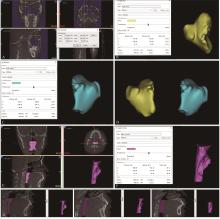
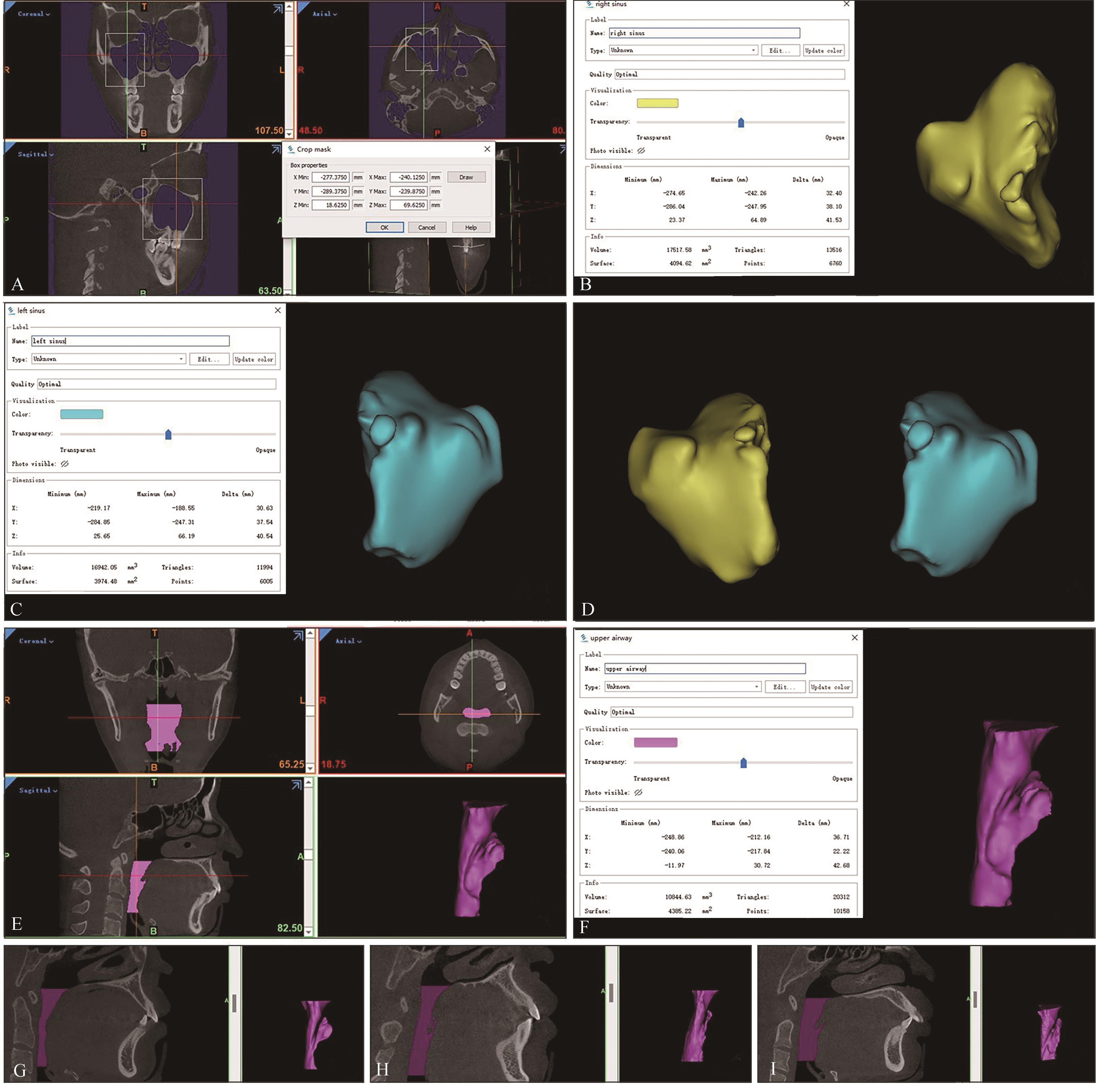
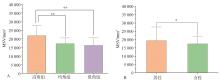
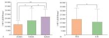
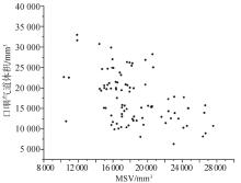
目的 评估成人骨性Ⅱ类患者不同垂直骨面型上颌窦容积(MSV)及口咽气道体积的差异,并探索MSV与口咽气道之间的相关性。 方法 选取成人骨性Ⅱ类患者90例,年龄、性别严格匹配。应用Mimics 21.0软件对所有样本的锥形束计算机断层扫描(CBCT)影像进行双侧上颌窦及口咽段气道的三维重建,分别计算出其体积。使用SPSS 21.0软件进行统计学分析,比较不同垂直骨面型、不同性别的MSV及口咽气道体积差异,并探索MSV与口咽气道体积的相关性。 结果 成人骨性Ⅱ类患者平均MSV为(18 360.42±3 747.41) mm3,左右侧MSV组间差异无统计学意义;男性明显大于女性,差异具有统计学意义(P<0.05);在垂直骨面型分组中,高角组MSV明显大于均角组和低角组,差异具有统计学意义(P<0.01),均角组大于低角组,差异无统计学意义(P>0.05)。成人骨性Ⅱ类患者平均口咽气道体积为(17 517.80±6 056.33) mm3;男性明显大于女性,差异具有统计学意义(P<0.01);从高角组、均角组至低角组口咽气道体积逐渐增大,差异具有统计学意义(P<0.05)。MSV与口咽气道体积呈负相关,相关系数为-0.458(P<0.01)。 结论 在成人骨性Ⅱ类错𬌗畸形患者中,下颌平面角较大者,MSV较大,口咽气道体积较小,且MSV与口咽气道体积呈显著负相关。
中图分类号:
| 1 | 伍军, 徐宝华. 安氏Ⅱ类Ⅰ分类错𬌗的分型及其高低面角的颅面特征[J]. 口腔医学纵横, 1999, 15(3): 151-153. |
| Wu J, Xu BH. The subtypes of Angle Class Ⅱ division Ⅰ malocclusion and the maxillofical morphologic features of high and low mandibular angle[J]. J Compr Stomatol, 1999, 15(3): 151-153. | |
| 2 | 翁嘉华, 蔡斌, 麦理想, 等. 锥形束CT扫描测量不同矢状骨面型青少年上颌窦大小[J]. 中华口腔医学研究杂志(电子版), 2012, 6(1): 65-72. |
| Weng JH, Cai B, Mai LX, et al. Evaluation of maxillary sinus sizes in adolescence of different sagittal skeletal patterns on cone-beam computed tomography images[J]. Chin J Stomatol Res (Electron Ed), 2012, 6(1): 65-72. | |
| 3 | El H, Palomo JM. Airway volume for different dentofacial skeletal patterns[J]. Am J Orthod Dentofac Orthop, 2011, 139(6): e511-e521. |
| 4 | Emirzeoglu M, Sahin B, Bilgic S, et al. Volumetric evaluation of the paranasal sinuses in normal subjects using computer tomography images: a stereological study[J]. Auris Nasus Larynx, 2007, 34(2): 191-195. |
| 5 | Scuderi AJ, Harnsberger HR, Boyer RS. Pneumatization of the paranasal sinuses: normal features of importance to the accurate interpretation of CT scans and MR images[J]. Am J Roentgenol, 1993, 160(5): 1101-1104. |
| 6 | Sharan A, Madjar D. Maxillary sinus pneumatization following extractions: a radiographic study[J]. Int J Oral Maxillofac Implants, 2008, 23(1): 48-56. |
| 7 | Ariji Y, Kuroki T, Moriguchi S, et al. Age changes in the volume of the human maxillary sinus: a study using computed tomography[J]. Dentomaxillofac Ra-diol, 1994, 23(3): 163-168. |
| 8 | Woo I, Le BT. Maxillary sinus floor elevation: review of anatomy and two techniques[J]. Implant Dent, 2004, 13(1): 28-32. |
| 9 | Zeynep OA, Alper OA, Hakan E, et al. Maxillary sinus volume in patients with impacted canines[J]. Angle Orthod, 2017, 87(1): 25-32. |
| 10 | Schendel SA, Jacobson R, Khalessi S. Airway grow-th and development: a computerized 3-dimensional analysis[J]. J Oral Maxillofac Surg, 2012, 70(9): 2174-2183. |
| 11 | Li L, Liu H, Cheng HJ, et al. CBCT evaluation of the upper airway morphological changes in growing patients of Class Ⅱ division 1 malocclusion with mandibular retrusion using twin block appliance: a comparative research[J]. PLoS One, 2014, 9(4): e94378. |
| 12 | Banno K, Kryger MH. Sleep apnea: clinical investigations in humans[J]. Sleep Med, 2007, 8(4): 400-426. |
| 13 | Li M, Li XY, Lu Y. Obstructive sleep apnea syndrome and metabolic diseases[J]. Endocrinology, 2018, 159(7): 2670-2675. |
| 14 | Maestre-Ferrín L, Galán-Gil S, Carrillo-García C, et al. Radiographic findings in the maxillary sinus: comparison of panoramic radiography with compu-ted tomography[J]. Int J Oral Maxillofac Implants, 2011, 26(2): 341-346. |
| 15 | Ryan CF, Lowe AA, Li D, et al. Magnetic resonance imaging of the upper airway in obstructive sleep apnea before and after chronic nasal continuous positive airway pressure therapy[J]. Am Rev Respir Dis, 1991, 144(4): 939-944. |
| 16 | Schwab RJ, Goldberg AN. Upper airway assessment: radiographic and other imaging techniques[J]. Otolaryngol Clin North Am, 1998, 31(6): 931-968. |
| 17 | Schulze R, Heil U, Gross D, et al. Artefacts in CBCT: a review[J]. Dentomaxillofac Radiol, 2011, 40(5): 265-273. |
| 18 | Moss ML. The functional matrix hypothesis revisi-ted. 3. The genomic thesis[J]. Am J Orthod Dentofac Orthop, 1997, 112(3): 338-342. |
| 19 | 傅民魁. 口腔正畸专科教程[M]. 北京: 人民卫生出版社, 2007: 68-71. |
| Fu MK. Orthodontic course[M]. Beijing: People’s Medical Publishing House, 2007: 68-71. | |
| 20 | Shrestha B, Shrestha R, Lin TW, et al. Evaluation of maxillary sinus volume in different craniofacial patterns: a CBCT study[J]. Oral Radiol, 2021, 37(4): 647-652. |
| 21 | Okşayan R, Sökücü O, Yeşildal S. Evaluation of maxillary sinus volume and dimensions in different vertical face growth patterns: a study of cone-beam computed tomography[J]. Acta Odontol Scand, 2017, 75(5): 345-349. |
| 22 | Rani SU, Rao GV, Kumar DR, et al. Age and gender assessment through three-dimensional morphome-tric analysis of maxillary sinus using magnetic resonance imaging[J]. J Forensic Dent Sci, 2017, 9(1): 46. |
| 23 | Favato MN, Vidigal BC, Cosso MG, et al. Impact of human maxillary sinus volume on grafts dimensio-nal changes used in maxillary sinus augmentation: a multislice tomographic study[J]. Clin Oral Implants Res, 2015, 26(12): 1450-1455. |
| 24 | Daimaruya T, Takahashi I, Nagasaka H, et al. Effects of maxillary molar intrusion on the nasal floor and tooth root using the skeletal anchorage system in dogs[J]. Angle Orthod, 2003, 73(2): 158-166. |
| 25 | Heravi F, Bayani S, Madani AS, et al. Intrusion of supra-erupted molars using miniscrews: clinical success and root resorption[J]. Am J Orthod Dentofacial Orthop, 2011, 139(4 ): S170-S175. |
| 26 | Park JH, Tai K, Kanao A, et al. Space closure in the maxillary posterior area through the maxillary sinus[J]. Am J Orthod Dentofacial Orthop, 2014, 145(1): 95-102. |
| 27 | Chung KR, Kim YS, Linton JL, et al. The miniplate with tube for skeletal anchorage[J]. J Clin Orthod, 2002, 36(7): 407-412. |
| 28 | Junqueira RB, Souza-Nunes LA, Scalioni FAR, et al. Anatomical evaluation of the relationship between the maxillary posterior teeth and maxillary sinus[J]. Gen Dent, 2020, 68(1): 66-71. |
| 29 | Anandarajah S, Dudhia R, Sandham A, et al. Risk factors for small pharyngeal airway dimensions in preorthodontic children: a three-dimensional study[J]. Angle Orthod, 2017, 87(1): 138-146. |
| 30 | Nath M, Ahmed J, Ongole R, et al. CBCT analysis of pharyngeal airway volume and comparison of airway volume among patients with skeletal Class Ⅰ, Class Ⅱ, and Class Ⅲ malocclusion: a retrospective study[J]. Cranio, 2021, 39(5): 379-390. |
| 31 | Ucar FI, Uysal T. Comparision of orofacial airway dimensions in subject with different breathing pattern[J]. Prog Orthod, 2012, 13(3): 210-217. |
| 32 | Zhong Z, Tang ZH, Gao XM, et al. A comparison study of upper airway among different skeletal craniofacial patterns in nonsnoring Chinese children[J]. Angle Orthod, 2010, 80(2): 267-274. |
| 33 | de Freitas MR, Alcazar NM, Janson G, et al. Upper and lower pharyngeal airways in subjects with Class Ⅰ and Class Ⅱ malocclusions and different growth patterns[J]. Am J Orthod Dentofacial Orthop, 2006, 130(6): 742-745. |
| 34 | Silva NN, Lacerda RH, Silva AW, et al. Assessment of upper airways measurements in patients with mandibular skeletal Class Ⅱ malocclusion[J]. Dental Press J Orthod, 2015, 20(5): 86-93. |
| 35 | Donner MW, Bosma JF, Robertson DL. Anatomy and physiology of the pharynx[J]. Gastrointest Ra-diol, 1985, 10(3): 196-212. |
| 36 | Ricketts RM. Respiratory obstruction syndrome[J]. Am J Orthod, 1968, 54(7): 495-507. |
| 37 | Dunn GF, Green LJ, Cunat JJ. Relationships between variation of mandibular morphology and va-riation of nasopharyngeal airway size in monozygotic twins[J]. Angle Orthod, 1973, 43(2): 129-135. |
| 38 | Joseph AA, Elbaum J, Cisneros GJ, et al. A cephalometric comparative study of the soft tissue airway dimensions in persons with hyperdivergent and normodivergent facial patterns[J]. J Oral Maxillofac Surg, 1998, 56(2): 135-140. |
| 39 | Grauer D, Cevidanes LS, Styner MA, et al. Pharyngeal airway volume and shape from cone-beam computed tomography: relationship to facial morphology[J]. Am J Orthod Dentofacial Orthop, 2009, 136(6): 805-814. |
| 40 | Hui DS, Ko FW, Chu AS, et al. Cephalometric assessment of craniofacial morphology in Chinese patients with obstructive sleep apnoea[J]. Respir Med, 2003, 97(6): 640-646. |
| 41 | Yamaoka M, Furusawa K, Uematsu T, et al. Relationship of the hyoid bone and posterior surface of the tongue in prognathism and micrognathia[J]. J Oral Rehabil, 2003, 30(9): 914-920. |
| 42 | Fernandes P, Pinto J, Ustrell-Torrent J. Relationship between oro and nasopharynx permeability and the direction of facial growth[J]. Eur J Paediatr Dent, 2017, 18(1): 37-40. |
| 43 | Proffit WR, Fields HW, Nixon WL. Occlusal forces in normal- and long-face adults[J]. J Dent Res, 1983, 62(5): 566-570. |
| 44 | Ingervall B, Minder C. Correlation between maximum bite force and facial morphology in children[J]. Angle Orthod, 1997, 67(6): 415-424. |
| 45 | Braun S, Bantleon HP, Hnat WP, et al. A study of bite force, part 2: relationship to various cephalometric measurements[J]. Angle Orthod, 1995, 65(5): 373-377. |
| 46 | Moscarino S, Kötter F, Brandt M, et al. Influence of different surgical concepts for moderate skeletal Class Ⅱ and Ⅲ treatment on the nasopharyngeal airway space[J]. J Craniomaxillofac Surg, 2019, 47(10): 1489-1497. |
| [1] | 韩婧文,任诗琦,刘星宇,郎鑫,储梦诗,Waseem Saleh Abdo Kaid Algumaei,郑艳. 成人不同垂直及矢状骨面型髁突特征的研究[J]. 国际口腔医学杂志, 2022, 49(2): 153-162. |
| [2] | 陈玉,姜欢,刘楠,陆晨萌,唐中元,韩茹钰,胡敏. 正畸治疗对骨性Ⅱ类错畸形患者上气道及周围结构变化的影响[J]. 国际口腔医学杂志, 2019, 46(5): 578-584. |
| [3] | 李婧,樊永杰. 女性青少年不同垂直骨面型颏部形态的研究[J]. 国际口腔医学杂志, 2016, 43(4): 387-390. |
| [4] | 李放,王建国. 不同垂直骨面型安氏Ⅰ类成年患者颞下颌关节形态特征的锥形束CT研究[J]. 国际口腔医学杂志, 2015, 42(5): 538-541. |
| [5] | 李正明1 刘新强2 黄永谦1 刘学1. 骨性Ⅱ类高角型开牙合拔除上颌第一前磨牙及下颌第二磨牙矫治的临床研究[J]. 国际口腔医学杂志, 2011, 38(5): 511-514. |
| [6] | 聂盼 李婧 李伟. Activator 治疗骨性Ⅱ类畸形的临床应用[J]. 国际口腔医学杂志, 2011, 38(4): 380-383. |
|