国际口腔医学杂志 ›› 2025, Vol. 52 ›› Issue (6): 722-729.doi: 10.7518/gjkq.2025092
陈睿桢( ),姜醒,沈纪元,林玲,郑志强,林捷()
Ruizhen Chen(),Xing Jiang,Jiyuan Shen,Ling Lin,Zhiqiang Zheng,Jie Lin()
摘要:
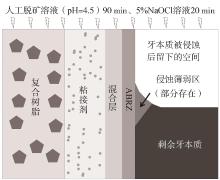
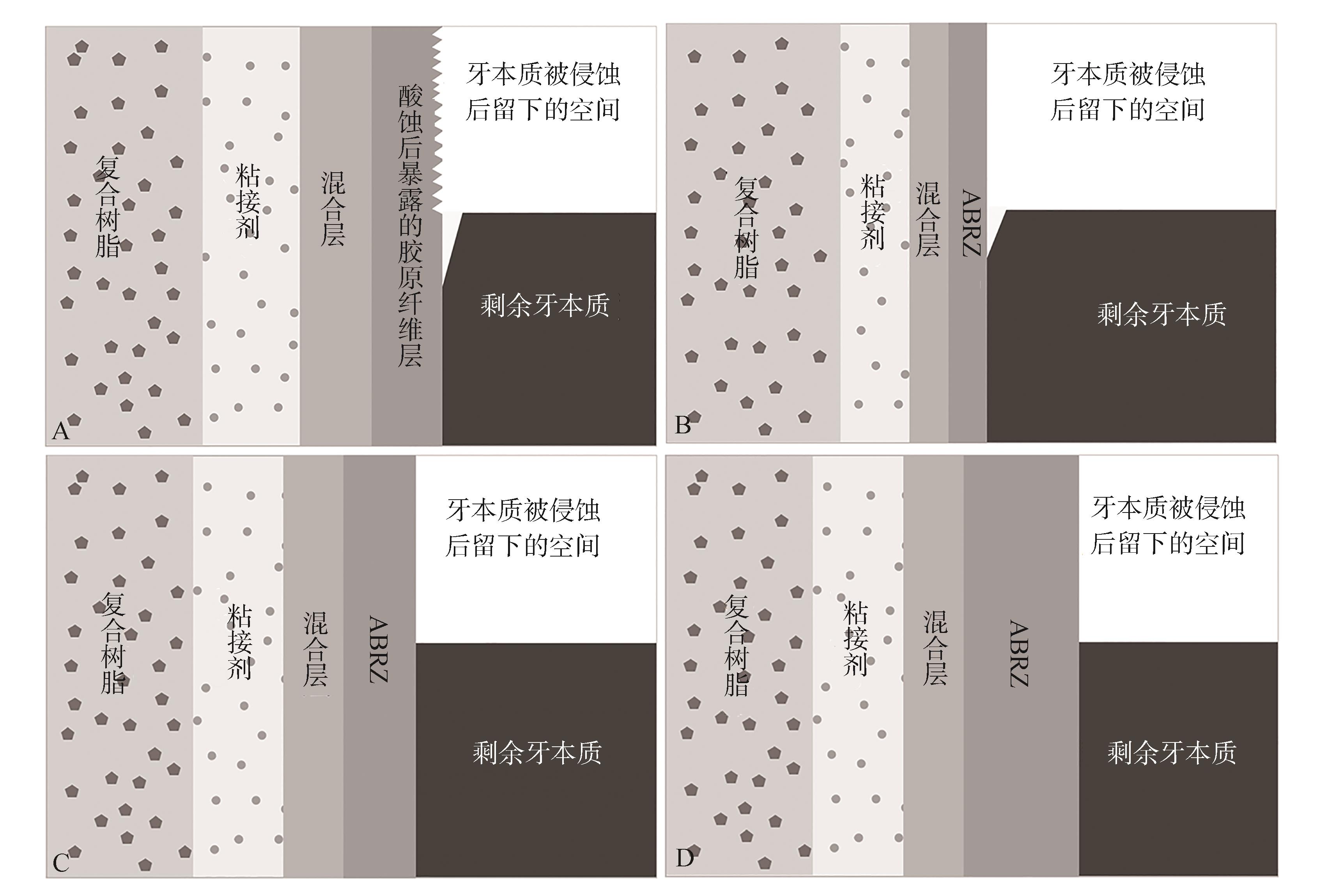
扫描电子显微镜观察证实牙体粘接界面的混合层之下存在抗酸碱层(ABRZ),其形成与酸性功能性单体与牙体中的羟磷灰石发生化学反应有关。与传统混合层不同,ABRZ以树脂包裹的部分脱矿羟磷灰石为特征,可提升粘接界面的抗酸碱侵蚀能力及长期粘接耐久性。ABRZ在自酸蚀粘接系统中更易形成,其结构与粘接剂的化学组成密切相关。本文对ABRZ的微观结构、影响因素及形成机制作一综述,为牙体粘接的临床和科研提供参考。
中图分类号:
| [1] | van Meerbeek B, Yoshihara K, van Landuyt K, et al. From buonocore’s pioneering acid-etch technique to self-adhering restoratives. A status perspective of rapidly advancing dental adhesive technology[J]. J Adhes Dent, 2020, 22(1): 7-34. |
| [2] | Tsuchiya S, Nikaido T, Sonoda H, et al. Ultrastructure of the dentin-adhesive interface after acid-base challenge[J]. J Adhes Dent, 2004, 6(3): 183-190. |
| [3] | Yoshihara K, Yoshida Y, Nagaoka N, et al. Nano-controlled molecular interaction at adhesive interfa-ces for hard tissue reconstruction[J]. Acta Biomater, 2010, 6(9): 3573-3582. |
| [4] | Yoshihara K, Yoshida Y, Hayakawa S, et al. Nanolayering of phosphoric acid ester monomer on ena-mel and dentin[J]. Acta Biomater, 2011, 7(8): 3187-3195. |
| [5] | Yoshida Y, Van Meerbeek B, Nakayama Y, et al. Adhesion to and decalcification of hydroxyapatite by carboxylic acids[J]. J Dent Res, 2001, 80(6): 1565-1569. |
| [6] | Yoshioka M, Yoshida Y, Inoue S, et al. Adhesion/decalcification mechanisms of acid interactions with human hard tissues[J]. J Biomed Mater Res, 2002, 59(1): 56-62. |
| [7] | Inoue G, Tsuchiya S, Nikaido T, et al. Morphological and mechanical characterization of the acid-base resistant zone at the adhesive-dentin interface of intact and caries-affected dentin[J]. Oper Dent, 2006, 31(4): 466-472. |
| [8] | Guan R, Takagaki T, Matsui N, et al. Dentin bon-ding performance using Weibull statistics and eva-luation of acid-base resistant zone formation of recen-tly introduced adhesives[J]. Dent Mater J, 2016, 35(4): 684-693. |
| [9] | Van Landuyt KL, Yoshida Y, Hirata I, et al. Influen-ce of the chemical structure of functional monomers on their adhesive performance[J]. J Dent Res, 2008, 87(8): 757-761. |
| [10] | Tichy A, Hosaka K, Yang Y, et al. Can a new HEMA-free two-step self-etch adhesive improve dentin bonding durability and marginal adaptation[J]. J Adhes Dent, 2021, 23(6): 505-512. |
| [11] | Kakiuchi Y, Takagaki T, Ikeda M, et al. Evaluation of MDP and NaF in two-step self-etch adhesives on enamel microshear bond strength and morphology of the adhesive-enamel interface[J]. J Adhes Dent, 2018, 20(6): 527-534. |
| [12] | Carrilho E, Cardoso M, Marques Ferreira M, et al. 10-MDP based dental adhesives: adhesive interface characterization and adhesive stability-a systematic review[J]. Materials, 2019, 12(5): 790. |
| [13] | Nurrohman H, Nikaido T, Takagaki T, et al. Apatite crystal protection against acid-attack beneath resin-dentin interface with four adhesives: TEM and crystallography evidence[J]. Dent Mater, 2012, 28(7): e89-e98. |
| [14] | Yoshida Y, Nagakane K, Fukuda R, et al. Comparative study on adhesive performance of functional monomers[J]. J Dent Res, 2004, 83(6): 454-458. |
| [15] | Waidyasekera K, Nikaido T, Weerasinghe DS, et al. Reinforcement of dentin in self-etch adhesive technology: a new concept[J]. J Dent, 2009, 37(8): 604-609. |
| [16] | Nikaido T, Nurrohman H, Takagaki T, et al. Nanolea-kage in hybrid layer and acid-base resistant zone at the adhesive/dentin interface[J]. Microsc Microanal, 2015, 21(5): 1271-1277. |
| [17] | Nikaido T, Inoue G, Takagaki T, et al. New strategy to create “Super Dentin” using adhesive technology: reinforcement of adhesive-dentin interface and protection of tooth structures[J]. Jpn Dent Sci Rev, 2011, 47(1): 31-42. |
| [18] | Matsui N, Takagaki T, Sadr A, et al. The role of MDP in a bonding resin of a two-step self-etching adhesive system[J]. Dent Mater J, 2015, 34(2): 227-233. |
| [19] | Vicheva M, Sato T, Takagaki T, et al. Effect of repair systems on dentin bonding performance[J]. Dent Mater J, 2021, 40(4): 903-910. |
| [20] | Ko AK, Matsui N, Nakamoto A, et al. Effect of silver diammine fluoride application on dentin bon-ding performance[J]. Dent Mater J, 2020, 39(3): 407-414. |
| [21] | Ochiai Y, Inoue G, Nikaido T, et al. Evaluation of experimental calcium-containing primer in adhesive system on micro-tensile bond strength and acid resistance[J]. Dent Mater J, 2019, 38(4): 565-572. |
| [22] | Nikaido T, Takagaki T, Sato T, et al. Fluoride-relea-sing self-etch adhesives create thick ABRZ at the interface[J]. Biomed Res Int, 2021, 2021: 9731280. |
| [23] | Aung SSMP, Takagaki T, Ko AK, et al. Adhesion durability of dual-cure resin cements and acid-base resistant zone formation on human dentin[J]. Dent Mater, 2019, 35(7): 945-952. |
| [24] | Giannini M, Makishi P, Ayres AP, et al. Self-etch adhesive systems: a literature review[J]. Braz Dent J, 2015, 26(1): 3-10. |
| [25] | Yang Y, Inoue G, Hosaka K, et al. The effect of a deproteinizing pretreatment on the bonding performance and acid resistance of a two-step self-etch adhesive on eroded dentin[J]. Oper Dent, 2024, 49(1): 65-75. |
| [26] | Li N, Nikaido T, Takagaki T, et al. The role of functional monomers in bonding to enamel: acid-base resistant zone and bonding performance[J]. J Dent, 2010, 38(9): 722-730. |
| [27] | Kumagai RY, Takagaki T, Sato T, et al. Resin cement/enamel interface: a morphological evaluation of the acid-base resistant zone, enamel etching pattern, and effect of thermocycling on the microshear bond strength[J]. J Adhes Dent, 2023, 25: 71-78. |
| [28] | Sato T, Takagaki T, Ikeda M, et al. Effects of selective phosphoric acid etching on enamel using “no-wait” self-etching adhesives[J]. J Adhes Dent, 2018, 20(5): 407-415. |
| [29] | Sato A, Sato T, Ikeda M, et al. Influence of different tooth etchants on bur-cut and uncut enamel[J]. Dent Mater J, 2023, 42(3): 311-318. |
| [30] | Sato T, Takagaki T, Baba YT, et al. Effects of diffe-rent tooth conditioners on the bonding of universal self-etching adhesive to dentin[J]. J Adhes Dent, 2019, 21(1): 77-85. |
| [31] | Nikaido T, Takagaki T, Sato T, et al. The concept of super enamel formation-relationship between chemical interaction and enamel acid-base resistant zone at the self-etch adhesive/enamel interface[J]. Dent Mater J, 2020, 39(4): 534-538. |
| [32] | Nakamoto A, Sato T, Matsui N, et al. Effect of fluoride mouthrinse and fluoride concentration on bon-ding of a one-step self-etch adhesive to bovine root dentin[J]. J Oral Sci, 2019, 61(1): 125-132. |
| [33] | Sato T, Nikaido T, Takagaki T, et al. Influence of primer contamination on the bonding interface of enamel pre-etched with phosphoric acid[J]. Dent Mater J, 2021, 40(5): 1086-1093. |
| [34] | Kirihara M, Inoue G, Nikaido T, et al. Effect of fluoride concentration in adhesives on morphology of acid-base resistant zones[J]. Dent Mater J, 2013, 32(4): 578-584. |
| [35] | Nikaido T, Ichikawa C, Li N, et al. Effect of functional monomers in all-in-one adhesive systems on formation of enamel/dentin acid-base resistant zone[J]. Dent Mater J, 2011, 30(5): 576-582. |
| [36] | Aung SSMP, Takagaki T, Ikeda M, et al. Ultra-morphological studies on enamel-universal adhesive interface[J]. J Dent, 2021, 104: 103527. |
| [37] | Yoshihara K, Nagaoka N, Nakamura A, et al. Nano-layering adds strength to the adhesive interface[J]. J Dent Res, 2021, 100(5): 515-521. |
| [1] | 陈阿璇,戴雯玉,韩向龙. 应用于牙釉质的抗菌再矿化材料的研究进展[J]. 国际口腔医学杂志, 2025, 52(5): 606-613. |
| [2] | 雷彬,陈柯. 牙本质发育不良Ⅰ型及其分型治疗[J]. 国际口腔医学杂志, 2022, 49(3): 332-336. |
| [3] | 李嫣斐,张新春. 牙本质作为骨修复材料的研究进展[J]. 国际口腔医学杂志, 2022, 49(2): 197-203. |
| [4] | 丁景瑜,田子璐,王惠敏,朱轩言,杨宇斌,朱松. 即刻牙本质封闭的研究进展[J]. 国际口腔医学杂志, 2022, 49(1): 121-124. |
| [5] | 章善,沈树平,张舫,杨卫东. Er: YAG激光光子激活光声流技术对根管壁牙本质失水状况及牙根抗压强度的影响[J]. 国际口腔医学杂志, 2022, 49(1): 55-59. |
| [6] | 何蓉,刘学军,周宇琨. 光子引导的光声流效应在根管荡洗中应用的系统评价[J]. 国际口腔医学杂志, 2021, 48(6): 644-655. |
| [7] | 赵彬彬,仲维剑,马国武. 牙本质作为骨移植材料的研究进展[J]. 国际口腔医学杂志, 2021, 48(1): 82-89. |
| [8] | 刘恩言,李明云. 茶多酚类化合物在牙本质粘接中应用的研究进展[J]. 国际口腔医学杂志, 2020, 47(6): 732-738. |
| [9] | 张凯莹,房付春,吴补领. 非编码RNA在牙源性干细胞成牙本质向分化中作用的研究进展[J]. 国际口腔医学杂志, 2019, 46(5): 540-545. |
| [10] | 秦娇娇,焦珊,王成坤. Er:YAG和Nd:YAG激光对牙本质与瓷修复体粘接面粘接强度影响的研究进展[J]. 国际口腔医学杂志, 2019, 46(3): 361-366. |
| [11] | 谭欣,于海洋. 模拟髓腔压力在牙本质粘接强度研究中的应用[J]. 国际口腔医学杂志, 2018, 45(6): 723-727. |
| [12] | 陈燕活, 安少锋, 高燕. 硅酸钙类盖髓剂生物学性能的研究进展[J]. 国际口腔医学杂志, 2018, 45(4): 459-464. |
| [13] | 廖文婷, 李彦. 半胱氨酸组织蛋白酶对牙本质粘接耐久性的影响[J]. 国际口腔医学杂志, 2017, 44(3): 340-343. |
| [14] | 陈尽欢,孙建勋,陈新梅. 转化生长因子-β超家族成员在牙本质发生发育中的作用[J]. 国际口腔医学杂志, 2016, 43(4): 477-481. |
| [15] | 周凤,赵玉鸣. 磨牙-切牙釉质矿化不全的研究进展[J]. 国际口腔医学杂志, 2016, 43(4): 456-461. |
|