国际口腔医学杂志 ›› 2023, Vol. 50 ›› Issue (6): 653-660.doi: 10.7518/gjkq.2023098
 畸形的关系
畸形的关系
王罗丹1( ),范红1,2()
Wang Luodan1(),Fan Hong1,2()
摘要:
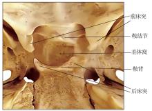
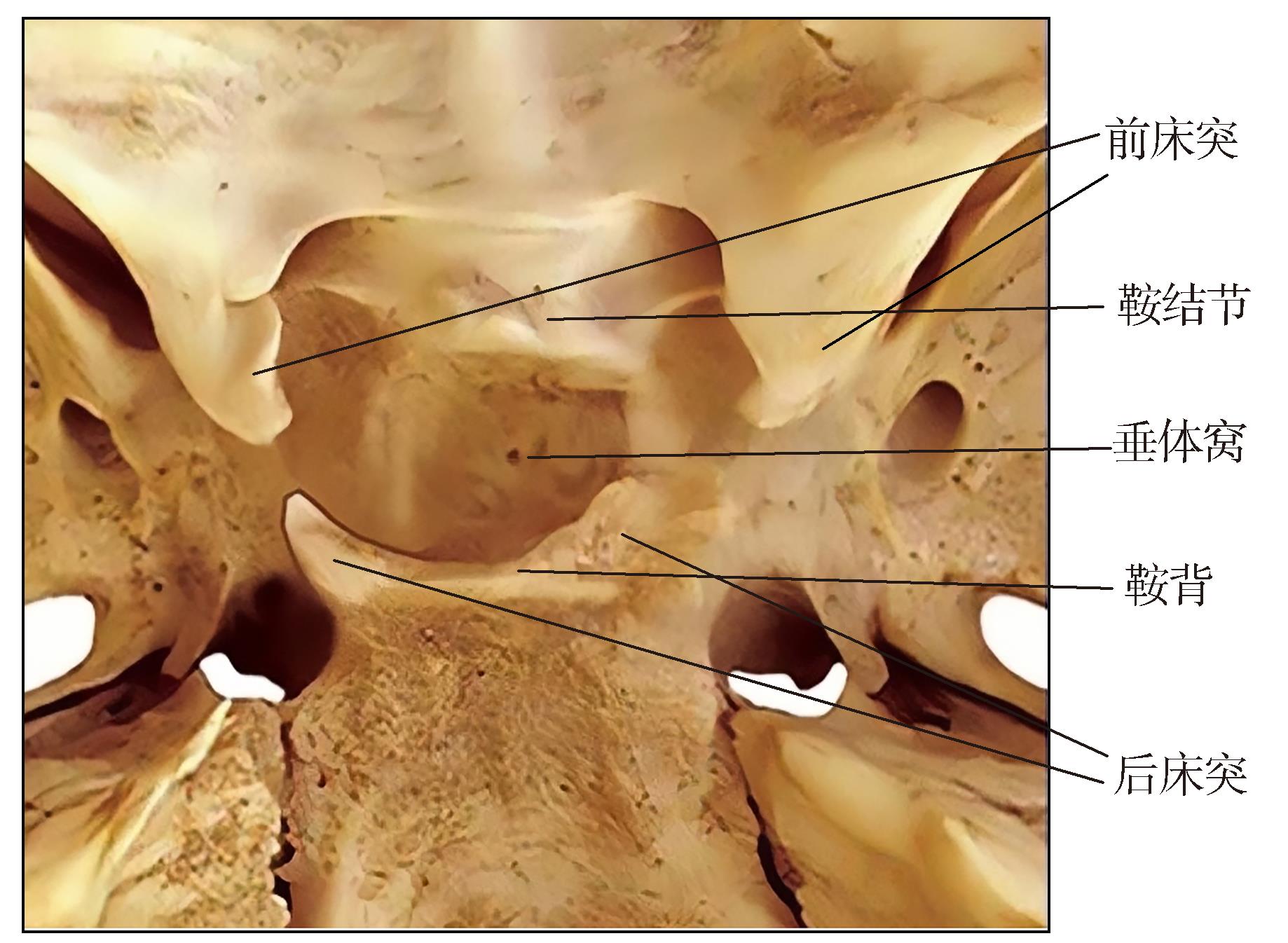
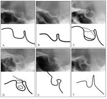
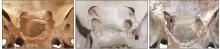
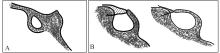
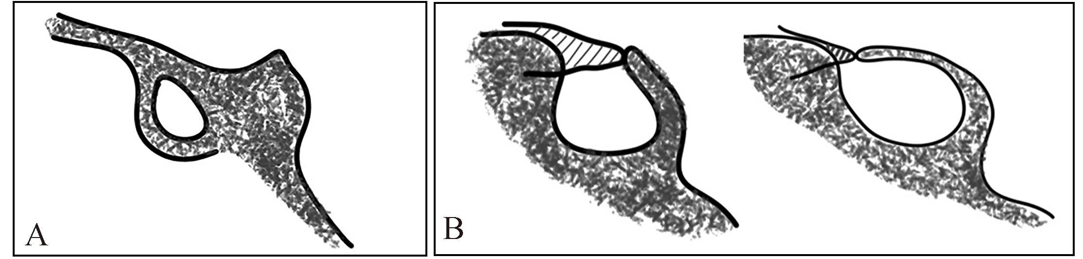
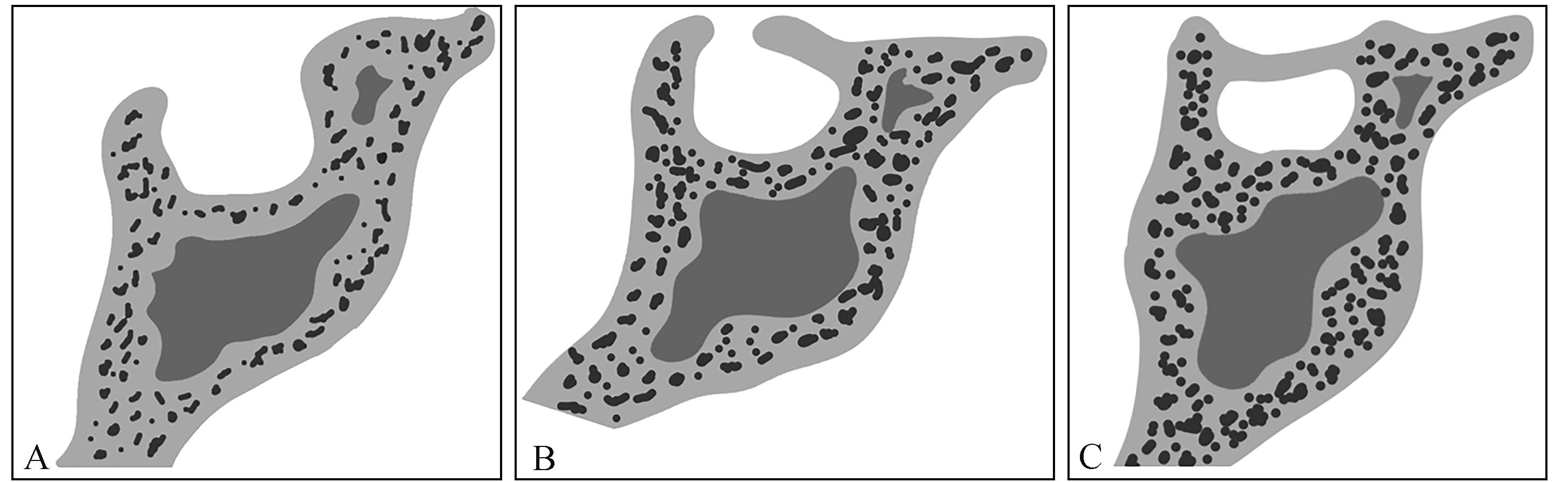
蝶鞍是位于蝶骨体内表面的鞍形骨结构。在正畸领域中,蝶鞍中心的蝶鞍点是头影测量分析时最常用的标志点之一。它的形态学常用于描述颅面形态,进而评估颅面形态与上下颌的关系。蝶鞍的大小、形态变化,尤其是鞍桥常常与颅颌面畸形、牙齿发育异常存在密切关系。正畸医生应熟悉蝶鞍区域的正常解剖结构特点及形态变异,以便识别和研究与之相关的病理情况。本文主要对蝶鞍的形态学特点,以及蝶鞍形态与各类错 畸形、牙齿发育异常的相关性进行总结,为临床诊疗工作提供辅助诊断依据。
中图分类号:
| 1 | Roomaney IA, Chetty M. Sella turcica morphology in patients with genetic syndromes: a systematic review[J]. Orthod Craniofac Res, 2021, 24(2): 194-205. |
| 2 | Alam MK, Alfawzan AA. Evaluation of sella turcica bridging and morphology in different types of cleft patients[J]. Front Cell Dev Biol, 2020, 8: 656. |
| 3 | Sathyanarayana HP, Kailasam V, Chitharanjan AB. Sella turcica-its importance in orthodontics and craniofacial morphology[J]. Dent Res J (Isfahan), 2013, 10(5): 571-575. |
| 4 | 郑博文, 边慧慧, 刘奕, 等. 蝶鞍的类型和鞍桥的发生率[J]. 口腔医学, 2016, 36(8): 756-760. |
| Zheng BW, Bian HH, Liu Y, et al. Different morphological types of sella turcica and incidence rate of sella turcica bridge[J]. Stomatology, 2016, 36(8): 756-760. | |
| 5 | Jankowski T, Jedliński M, Grocholewicz K, et al. Sella turcica morphology on cephalometric radiographs and dental abnormalities-is there any association-systematic review[J]. Int J Environ Res Public Health, 2021, 18(9): 4456. |
| 6 | 边慧慧, 郑颖, 安娜, 等. 蝶鞍的线性测量和形态分析[J]. 口腔医学, 2017, 37(4): 341-345. |
| Bian HH, Zheng Y, An N, et al. Linear measurement and morphological analysis of sella turcica[J]. Stomatology, 2017, 37(4): 341-345. | |
| 7 | Ouaknine GE, Hardy J. Microsurgical anatomy of the pituitary gland and the sellar region. 1. The pi-tuitary gland[J]. Am Surg, 1987, 53(5): 285-290. |
| 8 | Yasa Y, Ocak A, Bayrakdar IS, et al. Morphometric analysis of sella turcica using cone beam computed tomography[J]. J Craniofac Surg, 2017, 28(1): e70-e74. |
| 9 | Shrestha GK, Pokharel PR, Gyawali R, et al. The morphology and bridging of the sella turcica in adult orthodontic patients[J]. BMC Oral Health, 2018, 18(1): 45. |
| 10 | Scribante A, Sfondrini MF, Cassani M, et al. Sella tur-cica bridging and dental anomalies: is there an association[J]. Int J Paediatr Dent, 2017, 27(6): 568-573. |
| 11 | Chou ST, Chen CM, Chen PH, et al. Morphology of sella turcica and bridging prevalence correlated with sex and craniofacial skeletal pattern in eastern Asia population: CBCT study[J]. Biomed Res Int, 2021, 2021: 6646406. |
| 12 | Alkofide EA. The shape and size of the sella turcica in skeletal class Ⅰ, class Ⅱ, and class Ⅲ Saudi subjects[J]. Eur J Orthod, 2007, 29(5): 457-463. |
| 13 | Camp JD. The normal and pathologic anatomy of the sella turcica as revealed at necropsy[J]. Radiology, 1923, 1(2): 65-73. |
| 14 | Ruiz CR, Wafae N, Wafae GC. Sella turcica morphometry usingcomputed tomography[J]. Eur J Anat, 2008, 12(1): 47-50. |
| 15 | Axelsson S, Storhaug K, Kjaer I. Post-natal size and morphology of the sella turcica. Longitudinal cephalometric standards for Norwegians between 6 and 21 years of age[J]. Eur J Orthod, 2004, 26(6): 597-604. |
| 16 | Valizadeh S, Shahbeig S, Mohseni S, et al. Correlation of shape and size of sella turcica with the type of facial skeletal class in an Iranian group[J]. Iran J Radiol, 2015, 12(3): e16059. |
| 17 | Kucia A, Jankowski T, Siewniak M, et al. Sella turcica anomalies on lateral cephalometric radiographs of Polish children[J]. Dentomaxillofac Radiol, 2014, 43(8): 20140165. |
| 18 | Becktor JP, Einersen S, Kjaer I. A sella turcica bridge in subjects with severe craniofacial deviations[J]. Eur J Orthod, 2000, 22(1): 69-74. |
| 19 | Acevedo AM, Lagravere-Vich M, Al-Jewair T. Diagnostic accuracy of lateral cephalograms and cone-beam computed tomography for the assessment of sella turcica bridging[J]. Am J Orthod Dentofacial Orthop, 2021, 160(2): 231-239. |
| 20 | Cuschieri A, Cuschieri S, Zammit C. Sella turcica bridging: a systematic review[J]. Surg Radiol Anat, 2022, 44(3): 381-389. |
| 21 | Ali B, Shaikh A, Fida M. Association between sella turcica bridging and palatal canine impaction[J]. Am J Orthod Dentofacial Orthop, 2014, 146(4): 437-441. |
| 22 | Skrzat J, Szewczyk R, Walocha J. The ossified interclinoid ligament[J]. Folia Morphol (Warsz), 2006, 65(3): 242-245. |
| 23 | Leonardi R, Barbato E, Vichi M, et al. A sella turcica bridge in subjects with dental anomalies[J]. Eur J Orthod, 2006, 28(6): 580-585. |
| 24 | O’Rahilly R, Müller F. Minireview: summary of the initial development of the human nervous system[J]. Teratology, 1999, 60(1): 39-41. |
| 25 | Haji Ghadimi M, Amini F, Hamedi S, et al. Associations among sella turcica bridging, atlas arcuate foramen (ponticulus posticus) development, atlas posterior arch deficiency, and the occurrence of palatally displaced canine impaction[J]. Am J Orthod Dentofacial Orthop, 2017, 151(3): 513-520. |
| 26 | 田浩楠. 上颌腭侧阻生尖牙与蝶鞍鞍桥和寰椎骨骼异常相关性的锥形束CT研究[D]. 重庆: 重庆医科大学, 2021. |
| Tian HN. A cone-beam computed tomography study on the maxillary palatal impacted canine’s relationship with sella turcica bridge and atlas skeletal abnormalities[D]. Chongqing: Chongqing Medical Uni-versity, 2021. | |
| 27 | Kjær I. Sella turcica morphology and the pituitary gland-a new contribution to craniofacial diagnostics based on histology and neuroradiology[J]. Eur J Orthod, 2015, 37(1): 28-36. |
| 28 | Tepedino M, Laurenziello M, Guida L, et al. Morphometric analysis of sella turcica in growing patients: an observational study on shape and dimensions in different sagittal craniofacial patterns[J]. Sci Rep, 2019, 9(1): 19309. |
| 29 | Kjaer I, Fischer-Hansen B. The adenohypophysis and the cranial base in early human development[J]. J Craniofac Genet Dev Biol, 1995, 15(3): 157-161. |
| 30 | Jankowski T, Jedliński M, Schmeidl K, et al. Sella turcica abnormalities, dental age and dental abnormalities in Polish children[J]. Int J Environ Res Public Health, 2021, 18(19): 10101. |
| 31 | 李真真. 不同骨面型患者蝶鞍形状尺寸的测量分析[D]. 长春: 吉林大学, 2022. |
| Li ZZ. Measurement and analysis of the shape and size of sella turcica in patients with different skeletal facial types[D]. Changchun: Jilin University, 2022. | |
| 32 | 王雪慎. 332例正畸患者蝶鞍与颅面结构的相关性[D]. 大连: 大连医科大学, 2020. |
| Wang XS. Correlation of sella turcica and craniofacial structure for 332 orthodontic patients[D]. Dalian: Dalian Medical University, 2020. | |
| 33 | 牛磊, 郑颖, 边慧慧, 等. 中国东北地区3~24岁正畸患者的蝶鞍形态[J]. 口腔医学, 2016, 36(12): 1104-1107. |
| Niu L, Zheng Y, Bian HH, et al. Sella turcica morphology of 3-24 year-old patients by orthodontic treatment in northeast of China[J]. Stomatology, 2016, 36(12): 1104-1107. | |
| 34 | Silverman FN. Roentgen standards fo-size of the pituitary fossa from infancy through adolescence[J]. Am J Roentgenol Radium Ther Nucl Med, 1957, 78(3): 451-460. |
| 35 | Choi WJ, Hwang EH, Lee SR. The study of shape and size of normal sella turcica in cephalometric radiographs[J]. Imag Sci Dent, 2001, 31(1): 43-49. |
| 36 | Jones RM, Faqir A, Millett DT, et al. Bridging and dimensions of sella turcica in subjects treated by surgical-orthodontic means or orthodontics only[J]. Angle Orthod, 2005, 75(5): 714-718. |
| 37 | Abdel-Kader HM. Sella turcica bridges in orthodontic and orthognathic surgery patients. A retrospective cephalometric study[J]. Aust Orthod J, 2007, 23(1): 30-35. |
| 38 | Sobuti F, Dadgar S, Seifi A, et al. Relationship between bridging and dimensions of sella turcica with classification of craniofacial skeleton[J]. Pol J Ra-diol, 2018, 83: e120-e126. |
| 39 | El Wak T, Akl R, Mati M, et al. Association between sella turcica bridging and palatal canine impaction: evaluation using lateral cephalograms and CBCT[J]. Int Orthod, 2018, 16(2): 338-348. |
| 40 | Silveira BT, Fernandes KS, Trivino T, et al. Assessment of the relationship between size, shape and volume of the sella turcica in class Ⅱ and Ⅲ patients prior to orthognathic surgery[J]. Surg Radiol Anat, 2020, 42(5): 577-582. |
| 41 | Yan S, Huang S, Wu Z, et al. A CBCT investigation of the sella turcica dimension and sella turcica bri-dging in different vertical growth patterns[J]. J Clin Med, 2023, 12(5): 1890. |
| 42 | Yasa Y, Bayrakdar IS, Ocak A, et al. Evaluation of sella turcica shape and dimensions in cleft subjects using cone-beam computed tomography[J]. Med Princ Pract, 2017, 26(3): 280-285. |
| 43 | Sinha SP, Shetty A, Nayak USK. The morphology of sella turcica in cleft and non-cleft individuals[J]. Saudi Dent J, 2020, 32(2): 86-92. |
| 44 | Alkofide EA. Sella turcica morphology and dimensions in cleft subjects[J]. Cleft Palate Craniofac J, 2008, 45(6): 647-653. |
| 45 | Sundareswaran S, Nipun CA. Bridging the gap: sella turcica in unilateral cleft lip and palate patients[J]. Cleft Palate Craniofac J, 2015, 52(5): 597-604. |
| 46 | Yalcin ED. Morphometric analysis of sella turcica using cone-beam computed tomography in patients with cleft lip and palate[J]. J Craniofac Surg, 2020, 31(1): 306-309. |
| 47 | 李亚其, 王梓千, 刘家琦, 等. 恒牙先天缺失患者蝶鞍和颅底的头影测量研究[J]. 华西口腔医学杂志, 2022, 40(5): 582-588. |
| Li YQ, Wang ZQ, Liu JQ, et al. Morphometric eva-luation of sella turcica and cranial base in patients with congenital absence of teeth[J]. West China J Stomatol, 2022, 40(5): 582-588. | |
| 48 | 斯贝尔, 陈澜月, 陈艳娜, 等. 牙齿异位与鞍桥之间的关系[J]. 临床口腔医学杂志, 2017, 33(2): 95-98. |
| Shbair MFS, Chen LY, Chen YN, et al. The study on the relationship between sella bridge and dental trans-portation[J]. J Clin Stomatol, 2017, 33(2): 95-98. | |
| 49 | Leonardi R, Farella M, Cobourne MT. An association between sella turcica bridging and dental transposition[J]. Eur J Orthod, 2011, 33(4): 461-465. |
| 50 | Alqahtani H. Association between sella turcica bridging and congenitally missing maxillary lateral incisors[J]. J Dent Sci, 2020, 15(1): 59-64. |
| 51 | Saokar PC, Dinesh MR, Shetty A. A correlative study of sella turcica bridging and dental anomalies related to size, shape, structure, number and eruption of teeth[J]. J Orthod Sci, 2022, 11: 2. |
| [1] | 徐书奎,张珊,谢新宇,马文盛. 上颌前方牵引矫治骨性Ⅲ类错 畸形远期疗效稳定性的研究进展[J]. 国际口腔医学杂志, 2023, 50(6): 646-652. 畸形远期疗效稳定性的研究进展[J]. 国际口腔医学杂志, 2023, 50(6): 646-652. |
| [2] | 于冬洋,李绍东,韩雷,单奔,柳勇,赵正宇. CT形态特征、性别联合放射组学鉴别腮腺多形性腺瘤与腺淋巴瘤[J]. 国际口腔医学杂志, 2023, 50(5): 506-513. |
| [3] | 彭佳美. 下前牙区多生牙2例[J]. 国际口腔医学杂志, 2016, 43(1): 34-. |
| [4] | 宗弋 王敤 王虎. 腭裂修复术后软腭形态多样性的研究[J]. 国际口腔医学杂志, 2015, 42(3): 281-284. |
| [5] | 姜世同,许崇开,刘洪杰,焦广军,安忠军,姜良. 8 例成人安氏Ⅱ类1 分类错(牙合)畸形患者的二次矫治[J]. 国际口腔医学杂志, 2009, 36(1): 21-21~22,26. |
| [6] | 鲁大为,石冰,. 唇腭裂患者亲属颅面形态学特征的研究进展[J]. 国际口腔医学杂志, 2007, 34(04): 290-292. |
| [7] | 刘波,徐芸,. 后牙单侧反He与下颌偏斜[J]. 国际口腔医学杂志, 2006, 33(06): 474-476. |
|