国际口腔医学杂志 ›› 2023, Vol. 50 ›› Issue (5): 506-513.doi: 10.7518/gjkq.2023090
于冬洋1,2( ),李绍东1(),韩雷2,单奔2,柳勇2,赵正宇2
Yu Dongyang1,2(),Li Shaodong1(),Han Lei2,Shan Ben2,Liu Yong2,Zhao Zhengyu2
摘要:
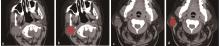
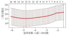
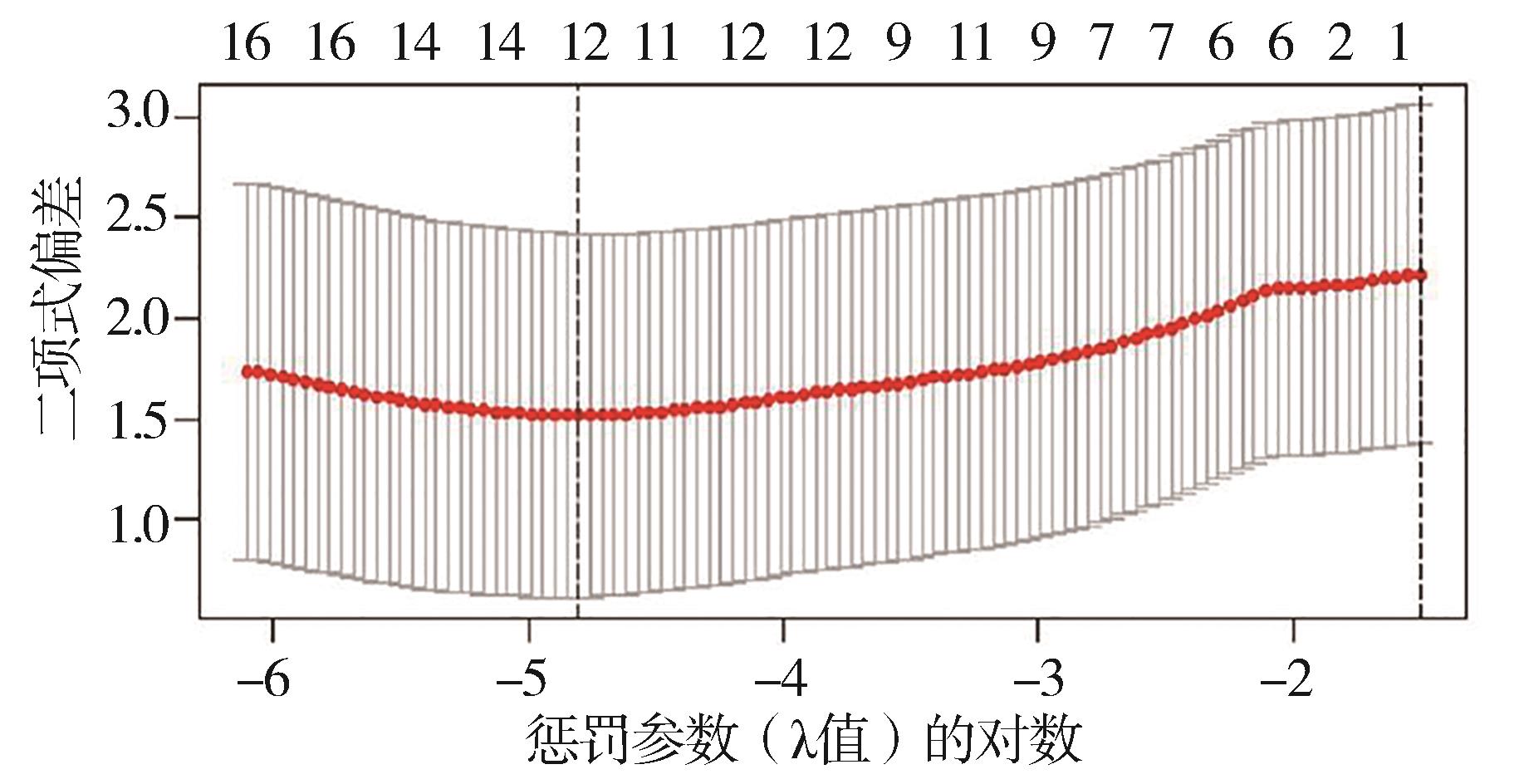
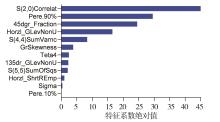
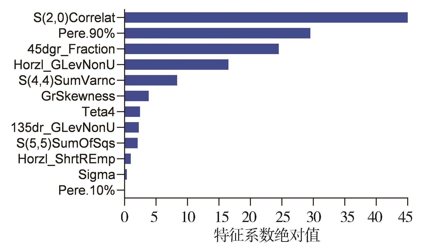
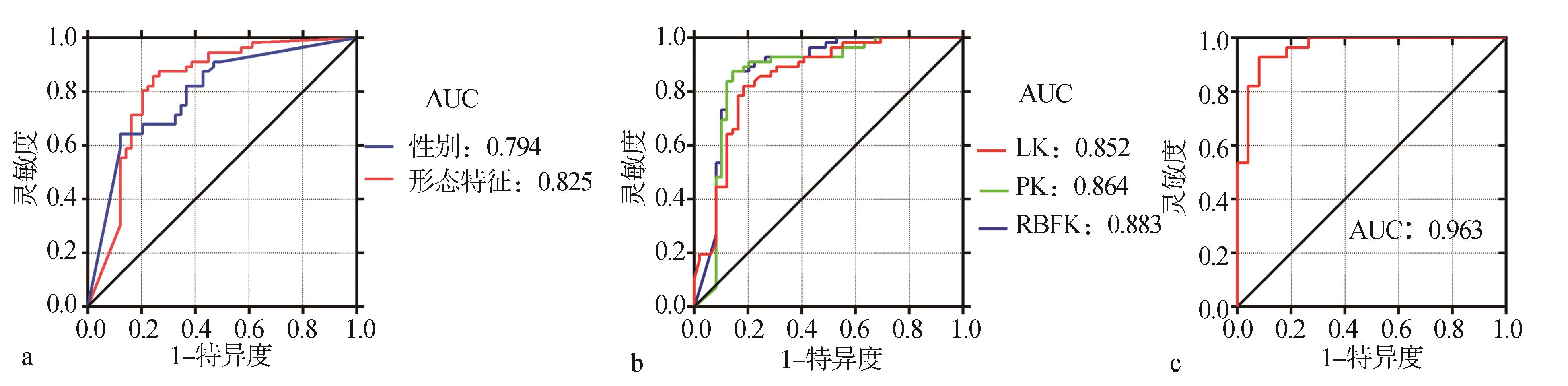
目的 探讨基于CT平扫的形态特征、性别联合放射组学模型对腮腺多形性腺瘤(PA)与腺淋巴瘤(AL)的鉴别应用。 方法 回顾性分析经病理证实的56例PA与49例AL的形态特征,观察分析其形状、边界、囊变、多发以及部位,提取并分析CT平扫图像中肿瘤的6种放射组学特征,包括灰度直方图(HA)、绝对梯度(AG)、灰度共生矩阵(GLCM)、自回归模型(AR)、灰度游程矩阵(GLRLM)和小波变换(WT),对两组间有统计学意义的放射组学特征参数进行筛选,分别以径向基函数核(RBFK)、多项式核(PK)和线性核(LK)对筛选后的放射组学特征建立支持向量机(SVM)分类模型并联合性别及形态特征建立联合模型,运用受试者工作特征曲线(ROC)评价诊断效能。 结果 最终从287个放射组学特征参数中筛出12个特征建立分类模型,以RBFK为核的分类模型诊断效能最高,对应的灵敏度、特异度、准确率及曲线下面积(AUC)分别为90.2%、82.5%、89.6%及0.883;PA以女性多见,AL以男性多见;与PA相比,AL更易多发及囊变(P<0.05);而2组间边界是否清楚、形状是否规则以及肿瘤的部位无明显差异 (P>0.05)。放射组学特征联合性别及形态特征 (多发与囊变) 建立以 RBFK为核的联合模型的灵敏度、特异度、准确率及 AUC 分别为 95.1%、87.6%、92.8%及 0.963。 结论 基于性别及CT形态特征联合放射组学特征建立的联合模型能够在术前对PA与AL进行有效鉴别。
中图分类号:
| 1 | Assili S, Fathi Kazerooni A, Aghaghazvini L, et al. Dynamic contrast magnetic resonance imaging (DCE-MRI) and diffusion weighted MR imaging (DWI) for differentiation between benign and malignant salivary gland tumors[J]. J Biomed Phys Eng, 2015, 5(4): 157-168. |
| 2 | Maahs GS, Oppermann Pde O, Maahs LG, et al. Parotid gland tumors: a retrospective study of 154 patients[J]. Braz J Otorhinolaryngol, 2015, 81(3): 301-306. |
| 3 | Ziółkowska M, Bień S, Okła S, et al. Epidemiological and clinical characteristics of 705 salivary glands neoplasms[J]. Otolaryngol Pol, 2013, 67(3): 154-163. |
| 4 | Comoglu S, Ozturk E, Celik M, et al. Comprehensive analysis of parotid mass: a retrospective study of 369 cases[J]. Auris Nasus Larynx, 2018, 45(2): 320-327. |
| 5 | Noel L, Medford S, Islam S, et al. Epidemiology of salivary gland tumours in an Eastern Caribbean nation: a retrospective study[J]. Ann Med Surg, 2018, 36: 148-151. |
| 6 | 雷晓雯, 程敬亮, 冉云彩. ADC全域直方图分析对腮腺多形性腺瘤和腺淋巴瘤的鉴别诊断价值[J]. 放射学实践, 2020, 35(8): 1005-1008. |
| Lei XW, Cheng JL, Ran YC. The value of ADC global histogram analysis in the differential diagnosis of pleomorphic adenoma and lymphoma of the parotid gland[J]. Pract Radiol, 2020, 35 (8): 1005-1008. | |
| 7 | Qian W, Xu XQ, Zhu LN, et al. Preliminary study of using diffusion kurtosis imaging for characteri-zing parotid gland tumors[J]. Acta Radiol, 2019, 60(7): 887-894. |
| 8 | Valstar MH, Mast H, Ten Hove I, et al. Malignant transformation of salivary gland pleomorphic adenoma: proof of principle[J]. J Pathol Clin Res, 2021, 7(5): 432-437. |
| 9 | Rooker SA, van Abel KM, Yin LX, et al. Risk factors for subsequent recurrence after surgical treatment of recurrent pleomorphic adenoma of the parotid gland[J]. Head Neck, 2021, 43(4): 1088-1096. |
| 10 | Gao M, Hao Y, Huang MX, et al. Salivary gland tumours in a northern Chinese population: a 50-year retrospective study of 7 190 cases[J]. Int J Oral Ma-xillofac Surg, 2017, 46(3): 343-349. |
| 11 | 李炳雨, 唐祖南, 胡耒豪, 等. 腮腺微小肿瘤的临床病理研究[J]. 北京大学学报(医学版), 2022, 54(2): 335-339. |
| Li BY, Tang ZN, Hu LH, et al. Clinicopathological study of parotid gland small tumors[J] J Peking Uni (Med Ed), 2022, 54(2): 335-339. | |
| 12 | 金朋, 蒋翠萍, 陈松旺, 等. 腮腺多形性腺瘤和沃辛瘤的超声特征对比分析[J]. 肿瘤影像学, 2022, 31(2): 174-180. |
| Jin P, Jiang CP, Chen SW, et al. Comparative analysis of ultrasound features between pleomorphic adenoma of the parotid gland and Vosin’s tumor[J]. Tumor Imag, 2022, 31(2): 174-180. | |
| 13 | 于冬洋, 单奔, 柳勇, 等. 3.0T磁共振DWI及动态增强扫描在腮腺常见肿瘤中的诊断价值[J]. 中华全科医师杂志, 2018(4): 303-306. |
| Yu DY, Shan B, Liu Y, et al. The diagnostic value of 3.0T magnetic resonance DWI and dynamic contrast-enhanced scanning in common parotid gland tumors[J]. Chin J General Pract, 2018(4): 303-306. | |
| 14 | 夏飞飞, 秦文娟, 冯佳, 等. 超声灰度直方图对多形性腺瘤与腺淋巴瘤鉴别诊断效能的初步研究[J]. 国际口腔医学杂志, 2022, 49(1): 60-65. |
| Xia FF, Qin WJ, Feng J, et al. Preliminary study on the differential diagnostic efficacy of ultrasound grayscale histogram in pleomorphic adenoma and adenolymphoma[J]. Int J Stomatol, 2022, 49(1): 60-65. | |
| 15 | Psychogios G. Ultrasonography techniques in the preoperative diagnosis of parotid gland tumors-an updated review of the literature[J]. Med Ultrason, 2021, 23(1): 122-123. |
| 16 | 张丹丹, 尤超, 顾雅佳. 基于动态增强 MRI 影像组学参数的观察者一致性研究[J]. 肿瘤影像学, 2021, 30(4): 252-256. |
| Zhang DD, You C, Gu YJ. Observer consistency study based on dynamic enhanced MRI imaging omics parameters[J]. Tumor Imag, 2021, 30(4): 252-256. | |
| 17 | 余先超, 孙宇凤, 李鹏, 等. 影像组学在腮腺多形性腺瘤与腺淋巴瘤鉴别诊断中的应用[J]. 现代肿瘤医学, 2021, 29(5): 837-840. |
| Yu XC, Sun YF, Li P, et al. The application of ima-ging omics in the differential diagnosis of pleomorphic adenoma and lymphoma of the parotid gland[J]. Modern Oncol Med, 2021, 29(5): 837-840. | |
| 18 | Geistlinger L, Oh S, Ramos M, et al. Multiomic analysis of subtype evolution and heterogeneity in high-grade serous ovarian carcinoma[J]. Cancer Res, 2020, 80(20): 4335-4345. |
| 19 | Castellano G, Bonilha L, Li LM, et al. Texture analysis of medical images[J]. Clin Radiol, 2004, 59(12): 1061-1069. |
| 20 | Pressney I, Khoo M, Endozo R, et al. Pilot study to differentiate lipoma from atypical lipomatous tumour/well-differentiated liposarcoma using MR radiomics-based texture analysis[J]. Skeletal Radiol, 2020, 49(11): 1719-1729. |
| 21 | Yuan R, Shi SY, Chen JH, et al. Radiomics in RayPlus: a web-based tool for texture analysis in medical images[J]. J Digit Imaging, 2019, 32(2): 269-275. |
| 22 | Wang S, Meng M, Zhang X, et al. Texture analysis of diffusion weighted imaging for the evaluation of glioma heterogeneity based on different regions of interest[J]. Oncol Lett, 2018, 15(5): 7297-7304. |
| 23 | Suh HB, Choi YS, Bae S, et al. Primary central nervous system lymphoma and atypical glioblastoma: differentiation using radiomics approach[J]. Eur Radiol, 2018, 28(9): 3832-3839. |
| 24 | Jung YJ, Han MR, Ha EJ, et al. Differentiation of salivary gland tumors through tumor heterogeneity: a comparison between pleomorphic adenoma and Warthin tumor using CT texture analysis[J]. Neuroradiology, 2020, 62(11): 1451-1458. |
| 25 | Goyal A, Razik A, Kandasamy D, et al. Role of MR texture analysis in histological subtyping and gra-ding of renal cell carcinoma: a preliminary study[J]. Abdom Radiol (NY), 2019, 44(10): 3336-3349. |
| 26 | Huang SY, Franc BL, Harnish RJ, et al. Exploration of PET and MRI radiomic features for decoding breast cancer phenotypes and prognosis[J]. NPJ Breast Cancer, 2018, 4: 24. |
| 27 | Guo Y, Kong QC, Zhu YQ, et al. Whole-lesion histogram analysis of the apparent diffusion coefficient: evaluation of the correlation with subtypes of mucinous breast carcinoma[J]. J Magn Reson Ima-ging, 2018, 47(2): 391-400. |
| 28 | 王友红, 柳勇, 韩婷婷, 等. 基于MR对比增强T1WI纹理分析鉴别泪腺淋巴瘤与泪腺炎性假瘤[J]. 中国医学影像技术, 2022, 38(6): 837-841. |
| Wang YH, Liu Y, Han TT, et al. Differentiation of lacrimal gland lymphoma and lacrimal gland inflammatory pseudotumor based on contrast-enhanced T1WI texture analysis using MR imaging[J]. Chin Med Imag Technol, 2022, 38(6): 837-841. | |
| 29 | Park M, Kim J, Choi YS, et al. Application of dynamic contrast-enhanced MRI parameters for diffe-rentiating squamous cell carcinoma and malignant lymphoma of the oropharynx[J]. AJR Am J Roentgenol, 2016, 206(2): 401-407. |
| 30 | 王安然. 基于CT影像组学的腮腺良恶性肿瘤分类方法研究[D]. 重庆: 重庆医科大学, 2021. |
| Wang AR. Research on the classification method of benign and malignant parotid gland tumors based on CT imaging omics[D]. Chongqing: Chongqing Me-dical University, 2021. | |
| 31 | Mahato S, Goyal N, Ram D, et al. Detection of depression and scaling of severity using six channel EEG data[J]. J Med Syst, 2020, 44(7): 118. |
| 32 | Cai J, Liu H, Yuan H, et al. A radiomics study to predict invasive pulmonary adenocarcinoma appearing as pure ground-glass nodules[J]. Clin Radiol, 2021, 76(2): 143-151. |
| 33 | Gabelloni M, Faggioni L, Attanasio S, et al. Can magnetic resonance radiomics analysis discriminate parotid gland tumors? A pilot study[J]. Diagnostics (Basel), 2020, 10(11): 900. |
| 34 | Hussain L, Awan IA, Aziz W, et al. Detecting congestive heart failure by extracting multimodal features and employing machine learning techniques[J]. Biomed Res Int, 2020, 2020: 4281243. |
| 35 | Shao S, Mao N, Liu WJ, et al. Epithelial salivary gland tumors: utility of radiomics analysis based on diffusion-weighted imaging for differentiation of benign from malignant tumors[J]. J Xray Sci Technol, 2020, 28(4): 799-808. |
| 36 | 吴梦苇, 魏培英, 邵畅, 等. MRI多参数联合对腮腺腺淋巴瘤与多形性腺瘤的鉴别诊断价值[J]. 中国临床医学影像杂志, 2022, 33(3): 162-165, 171. |
| Wu MW, Wei PY, Shao C, et al. The value of MRI multi parameter combination in the differential diagnosis of parotid gland lymphoma and pleomorphic adenoma[J]. Chin J Clin Med Imag, 2022, 33(3): 162-165, 171. | |
| 37 | 王琴, 黄东琼, 于冬洋, 等. 放射组学模型鉴别腮腺多形性腺瘤与沃辛瘤[J]. 肿瘤影像学, 2021, 30(6): 499-503. |
| Wang Q, Huang DQ, Yu DY, et al. Radioomics mo-del for distinguishing pleomorphic adenoma of the parotid gland from Vosin’s tumor[J]. Tumor Imag, 2021, 30(6): 499-503. | |
| 38 | 林杨, 吴丽芳, 陈懿. 腮腺混合瘤与腺淋巴瘤的磁共振成像表现对比分析[J]. 肿瘤影像学, 2020, 29(3): 319-323. |
| Lin Y, Wu LF, Chen Y. Comparative analysis of magnetic resonance imaging manifestations between parotid mixed tumor and adenolymphoma[J]. Tumor Imag, 2020, 29(3): 319-323. | |
| 39 | 史灵雪. 多参数MR影像组学特征对腮腺多形性腺瘤及腺淋巴瘤诊断价值的研究[D]. 吉林: 吉林大学, 2021. |
| Shi LX. Study on the diagnostic value of multi parameter MR imaging omics features for parotid pleomorphic adenoma and adenolymphoma[D]. Jilin: Jilin University, 2021. | |
| 40 | 郭作梁, 陈晓华, 马兴灿, 等. 腮腺多形性腺瘤与腺淋巴瘤的CT影像特征及对比分析[J]. 心电图杂志(电子版), 2019, 8(1): 28-29. |
| Guo ZL, Chen XH, Ma XC, et al. CT imaging characteristics and comparative analysis of pleomorphic adenoma and lymphoma of the parotid gland[J]. J Electrocardiogram (Electro Ed), 2019, 8(1): 28-29. |
| [1] | 王罗丹,范红. 蝶鞍的形态学特点及其与错 畸形的关系[J]. 国际口腔医学杂志, 2023, 50(6): 653-660. 畸形的关系[J]. 国际口腔医学杂志, 2023, 50(6): 653-660. |
| [2] | 杨雨楠,刘鹏,王虎,游梦. 上颌窦黏膜增厚的锥形束CT影像分析[J]. 国际口腔医学杂志, 2023, 50(3): 302-307. |
| [3] | 延泽宁,孙睿,李菲,张建森. 双能量CT在颈部淋巴结病变评估中的研究进展[J]. 国际口腔医学杂志, 2023, 50(3): 335-340. |
| [4] | 吴文智,冯达兴,陈垂壮,周丽鹃. 海口地区下颌第一恒磨牙近中中央根管发生率及相关因素[J]. 国际口腔医学杂志, 2022, 49(4): 420-425. |
| [5] | 翟晓静,曹石,辛文龙,曹珊,张皓. 伴有广泛角化囊肿的多形性腺瘤1例[J]. 国际口腔医学杂志, 2022, 49(3): 328-331. |
| [6] | 叶泽林,刘璐,龙虎,游梦. 弯曲前牙的影像评价及治疗的研究进展[J]. 国际口腔医学杂志, 2022, 49(2): 173-181. |
| [7] | 夏飞飞,秦文娟,冯佳,周旭阳,孙二灿,黎昌学. 超声灰度直方图对多形性腺瘤与腺淋巴瘤鉴别诊断效能的初步研究[J]. 国际口腔医学杂志, 2022, 49(1): 60-65. |
| [8] | 田浩楠,林敏,谢丛蔓,任嫒姝. 上颌腭侧阻生尖牙与寰椎后桥相关性的锥形束CT研究[J]. 国际口腔医学杂志, 2021, 48(5): 536-540. |
| [9] | 施丹妮,杨鑫,吴建勇. 锥形束CT三维头影测量参考坐标系的研究进展[J]. 国际口腔医学杂志, 2021, 48(4): 398-404. |
| [10] | 丁张帆,郭陟永,苗诚,李春洁,宣鸣,王晓毅,张壮. 基于锥形束CT的三维可视化技术在颌骨囊性病变手术中的应用[J]. 国际口腔医学杂志, 2021, 48(2): 180-186. |
| [11] | 王奔,许喆桢,韦曦. 数字化微创技术在牙髓根尖周病学中的应用与进展[J]. 国际口腔医学杂志, 2021, 48(1): 110-118. |
| [12] | 唐蓓,赵文俊,王虎,郑广宁,游梦. 根管超填导致下牙槽神经损伤2例[J]. 国际口腔医学杂志, 2020, 47(3): 293-296. |
| [13] | 章婷婷,胡常红,彭燕,周文翘,张慧聪,刘蝶. 300例不同年龄段有牙颌人群上唇软组织侧貌的锥形束CT三维测量分析[J]. 国际口腔医学杂志, 2020, 47(2): 182-188. |
| [14] | 王春林,刘从华,宋思吟,周丽淑,林丽佳. 运用锥形束CT诊断上下颌横向发育不调的研究进展[J]. 国际口腔医学杂志, 2020, 47(1): 121-124. |
| [15] | 黎祺, 黄少宏. 岭南地区广府民系人群下颌第二恒磨牙牙根和根管形态的锥形束CT研究[J]. 国际口腔医学杂志, 2019, 46(6): 640-649. |
|