国际口腔医学杂志 ›› 2021, Vol. 48 ›› Issue (5): 497-505.doi: 10.7518/gjkq.2021097
• 专家笔谈 • 下一篇
王剑( )
)
Wang Jian()
摘要:
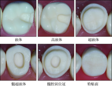
全冠修复是后牙牙体缺损最常见的修复方式之一,然而全冠修复由于备牙量大,对患牙抗折性存在潜在影响。随着粘接技术和微创修复的发展,嵌体和高嵌体可以大大减少牙体预备量,有效保存残留牙体组织,已逐渐成为后牙牙体缺损修复的主流形式,得到越来越广泛的应用。本文就嵌体和高嵌体修复的材料、适应证、修复效果、预备要点及粘接等各方面作一讨论,以期为临床工作提供参考。
中图分类号:
| [1] |
Vagropoulou GI, Klifopoulou GL, Vlahou SG, et al. Complications and survival rates of inlays and onlays vs complete coverage restorations: a systematic review and analysis of studies[J]. J Oral Rehabil, 2018, 45(11):903-920.
doi: 10.1111/joor.12695 pmid: 30019391 |
| [2] |
Batson ER, Cooper LF, Duqum I, et al. Clinical outcomes of three different crown systems with CAD/CAM technology[J]. J Prosthet Dent, 2014, 112(4):770-777.
doi: 10.1016/j.prosdent.2014.05.002 pmid: 24980739 |
| [3] |
Morimoto S, Rebello de Sampaio FB, Braga MM, et al. Survival rate of resin and ceramic inlays, onlays, and overlays: a systematic review and meta-analysis[J]. J Dent Res, 2016, 95(9):985-994.
doi: 10.1177/0022034516652848 pmid: 27287305 |
| [4] | 赵铱民. 口腔修复学[M]. 7版. 北京: 人民卫生出版社, 2014. |
| Zhao YM. Prosthodontics[M]. 7th ed. Beijing: Peo-ple’s Medical Publishing House, 2014. | |
| [5] |
Bindl A, Mörmann WH. Clinical evaluation of adhesively placed Cerec endo-crowns after 2 years: preliminary results[J]. J Adhes Dent, 1999, 1(3):255-265.
pmid: 11725673 |
| [6] | 田宇, 张亚庆, 吕海鹏, 等. 后牙椅旁CAD/CAM修复洞型分类的思考[J]. 牙体牙髓牙周病学杂志, 2016, 26(10):581-588, 611. |
| Tian Y, Zhang YQ, Lü HP, et al. Cavity classification for CAD/CAM ceramic restorations in posterior tee-th[J]. Chin J Conserv Dent, 2016, 26(10):581-588, 611. | |
| [7] |
Angeletaki F, Gkogkos A, Papazoglou E, et al. Direct versus indirect inlay/onlay composite restorations in posterior teeth. A systematic review and meta-analysis[J]. J Dent, 2016, 53:12-21.
doi: 10.1016/j.jdent.2016.07.011 pmid: 27452342 |
| [8] |
Fan J, Xu Y, Si L, et al. Long-term clinical performance of composite resin or ceramic inlays, onlays, and overlays: a systematic review and meta-analysis[J]. Oper Dent, 2021, 46(1):25-44.
doi: 10.2341/19-107-LIT pmid: 33882133 |
| [9] | Bustamante-Hernández N, Montiel-Company JM, Bellot-Arcís C, et al. Clinical behavior of ceramic, hybrid and composite onlays. A systematic review and meta-analysis[J]. Int J Environ Res Public Heal-th, 2020, 17(20):7582. |
| [10] |
Alakkad L, Kostagianni A, Finkelman M, et al. Bia-xial flexural strength of various CAD-CAM glass-ceramic materials[J]. Am J Dent, 2021, 34(2):91-96.
pmid: 33940666 |
| [11] |
González-Serrano C, Phark JH, Fuentes MV, et al. Effect of a single-component ceramic conditioner on shear bond strength of precoated brackets to diffe-rent CAD/CAM materials[J]. Clin Oral Investig, 2021, 25(4):1953-1965.
doi: 10.1007/s00784-020-03504-0 |
| [12] |
Su Y, Xin M, Chen XD, et al. Effect of CAD-CAM ceramic materials on the color match of veneer restorations[J]. J Prosthet Dent, 2021. doi: 10.1016/j.pro-sdent.2021.04.029.
doi: 10.1016/j.pro-sdent.2021.04.029 |
| [13] |
Sagsoz O, Yildiz M, Hojjat Ghahramanzadeh ASL, et al. In vitro fracture strength and hardness of different computer-aided design/computer-aided manufacturing inlays[J]. Niger J Clin Pract, 2018, 21(3):380-387.
doi: 10.4103/njcp.njcp_58_17 pmid: 29519990 |
| [14] |
Mynampati P, Babu MR, Saraswathi DD, et al. Comparison of fracture resistance and failure pattern of endodontically treated premolars with different esthetic onlay systems: an in vitro study[J]. J Conserv Dent, 2015, 18(2):140-143.
doi: 10.4103/0972-0707.153072 |
| [15] |
Belli R, Petschelt A, Hofner B, et al. Fracture rates and lifetime estimations of CAD/CAM all-ceramic restorations[J]. J Dent Res, 2016, 95(1):67-73.
doi: 10.1177/0022034515608187 pmid: 26428908 |
| [16] |
Lauvahutanon S, Takahashi H, Shiozawa M, et al. Mechanical properties of composite resin blocks for CAD/CAM[J]. Dent Mater J, 2014, 33(5):705-710.
pmid: 25273052 |
| [17] |
Awada A, Nathanson D. Mechanical properties of resin-ceramic CAD/CAM restorative materials[J]. J Prosthet Dent, 2015, 114(4):587-593.
doi: 10.1016/j.prosdent.2015.04.016 |
| [18] |
Bottino MA, Campos F, Ramos NC, et al. Inlays ma-de from a hybrid material: adaptation and bond streng-ths[J]. Oper Dent, 2015, 40(3):E83-E91.
doi: 10.2341/13-343-L |
| [19] |
Reeh ES, Messer HH, Douglas WH. Reduction in tooth stiffness as a result of endodontic and resto-rative procedures[J]. J Endod, 1989, 15(11):512-516.
doi: 10.1016/S0099-2399(89)80191-8 |
| [20] |
Chen YN, Chen D, Ding H, et al. Fatigue behavior of endodontically treated maxillary premolars with MOD defects under different minimally invasive restorations[J]. Clin Oral Investig, 2021. doi: 10.1007/s00784-021-03991-9.
doi: 10.1007/s00784-021-03991-9 |
| [21] |
Iaculli F, Rengo C, Lodato V, et al. Fracture resistance of endodontically-treated maxillary premolars restored with different type of posts and direct composite reconstructions: a systematic review and meta-analysis of in vitro studies[J]. Dent Mater, 2021. doi: 10.1016/j.dental.2021.06.007.
doi: 10.1016/j.dental.2021.06.007 |
| [22] |
De Matos LMR, Oliveira LP, Silva AM, et al. Resistance to fracture of endodontically treated teeth: influence of the post systems and cements[J]. Dent Res J (Isfahan), 2020, 17(6):417-423.
doi: 10.4103/1735-3327.302888 |
| [23] | Edelhoff D, Sorensen JA. Tooth structure removal associated with various preparation designs for posterior teeth[J]. Int J Periodontics Restorative Dent, 2002, 22(3):241-249. |
| [24] |
Yu H, Zhao Y, Li J, et al. Minimal invasive microscopic tooth preparation in esthetic restoration: a specialist consensus[J]. Int J Oral Sci, 2019, 11(3):31.
doi: 10.1038/s41368-019-0057-y |
| [25] |
Hofsteenge JW, van den Heijkant IA, Cune MS, et al. Influence of preparation design and restorative material on fatigue and fracture strength of restored ma-xillary premolars[J]. Oper Dent, 2021. doi: 10.2341/20-032-L.
doi: 10.2341/20-032-L |
| [26] |
Kassis C, Khoury P, Mehanna CZ, et al. Effect of inlays, onlays and endocrown cavity design preparation on fracture resistance and fracture mode of en-dodontically treated teeth: an in vitro study[J]. J Pro-sthodont, 2020. doi: 10.1111/jopr.13294.
doi: 10.1111/jopr.13294 |
| [27] |
Dejak B, Młotkowski A. A comparison of mvM stre-ss of inlays, onlays and endocrowns made from va-rious materials and their bonding with molars in a computer simulation of mastication-FEA[J]. Dent Mater, 2020, 36(7):854-864.
doi: 10.1016/j.dental.2020.04.007 |
| [28] | Costa VCD, Machado AC, Soares PV, et al. Influen-ce of material and loading location on stress distribution of inlays[J]. Am J Dent, 2021, 34(3):171-176. |
| [29] |
Kim SY, Kim BS, Kim H, et al. Occlusal stress distribution and remaining crack propagation of a cra-cked tooth treated with different materials and designs: 3D finite element analysis[J]. Dent Mater, 2021, 37(4):731-740.
doi: 10.1016/j.dental.2021.01.020 |
| [30] |
Mamoun J. Post and core build-ups in crown and bri-dge abutments: bio-mechanical advantages and disadvantages[J]. J Adv Prosthodont, 2017, 9(3):232-237.
doi: 10.4047/jap.2017.9.3.232 pmid: 28680556 |
| [31] |
Sedrez-Porto JA, Rosa WL, da Silva AF, et al. Endocrown restorations: a systematic review and meta-analysis[J]. J Dent, 2016, 52:8-14.
doi: 10.1016/j.jdent.2016.07.005 pmid: 27421989 |
| [32] |
Elashmawy Y, Elshahawy W, Seddik M, et al. In-fluence of fatigue loading on fracture resistance of endodontically treated teeth restored with endocrowns[J]. J Prosthodont Res, 2021, 65(1):78-85.
doi: 10.2186/jpr.JPOR_2019_485 |
| [33] |
Belleflamme MM, Geerts SO, Louwette MM, et al. No post-no core approach to restore severely dama-ged posterior teeth: an up to 10-year retrospective study of documented endocrown cases[J]. J Dent, 2017, 63:1-7.
doi: S0300-5712(17)30093-3 pmid: 28456557 |
| [34] | Al-Dabbagh RA. Survival and success of endocro-wns: a systematic review and meta-analysis[J]. J Pro-sthet Dent 2021, 125(3): 415.e1-415.e9. |
| [35] |
Papia E, Habib W, Larsson C. The influence of different designs, materials and cements on the success and survival rate of endocrowns. A systematic review[J]. Eur J Prosthodont Restor Dent, 2020, 28(3):100-111.
doi: 10.1922/EJPRD_1992Papia12 pmid: 32645260 |
| [36] |
Ruggiero MM, Soares Gomes R, Pedroso Bergamo ET, et al. Resin-matrix ceramics for occlusal veneers: effect of thickness on reliability and stress distribution[J]. Dent Mater, 2021, 37(3):e131-e139.
doi: 10.1016/j.dental.2020.11.002 pmid: 33276957 |
| [37] |
Huang XQ, Hong NR, Zou LY, et al. Estimation of stress distribution and risk of failure for maxillary premolar restored by occlusal veneer with different CAD/CAM materials and preparation designs[J]. Clin Oral Investig, 2020, 24(9):3157-3167.
doi: 10.1007/s00784-019-03190-7 |
| [38] |
Bitter K, Meyer-Lueckel H, Fotiadis N, et al. In-fluence of endodontic treatment, post insertion, and ceramic restoration on the fracture resistance of ma-xillary premolars[J]. Int Endod J, 2010, 43(6):469-477.
doi: 10.1111/j.1365-2591.2010.01701.x pmid: 20536574 |
| [39] |
Couegnat G, Fok SL, Cooper JE, et al. Structural optimization of dental restorations using the principle of adaptive growth[J]. Dent Mater, 2006, 22(1):3-12.
doi: 10.1016/j.dental.2005.04.003 |
| [40] |
Guess PC, Schultheis S, Wolkewitz M, et al. Influen-ce of preparation design and ceramic thicknesses on fracture resistance and failure modes of premolar partial coverage restorations[J]. J Prosthet Dent, 2013, 110(4):264-273.
doi: 10.1016/S0022-3913(13)60374-1 |
| [41] | Fages M, Bennasar B. The endocrown: a different ty-pe of all-ceramic reconstruction for molars[J]. J Can Dent Assoc, 2013, 79:d140. |
| [42] |
Sasse M, Krummel A, Klosa K, et al. Influence of restoration thickness and dental bonding surface on the fracture resistance of full-coverage occlusal veneers made from lithium disilicate ceramic[J]. Dent Mater, 2015, 31(8):907-915.
doi: 10.1016/j.dental.2015.04.017 |
| [43] |
Johnson AC, Versluis A, Tantbirojn D, et al. Fracture strength of CAD/CAM composite and compo-site-ceramic occlusal veneers[J]. J Prosthodont Res, 2014, 58(2):107-114.
doi: 10.1016/j.jpor.2014.01.001 pmid: 24636368 |
| [44] | Mante FK, Ozer F, Walter R, et al. The current state of adhesive dentistry: a guide for clinical practice[J]. Compend Contin Educ Dent, 2013, 34(Spec 9):2-8. |
| [45] |
Mohammadi N, Kahnamoii MA, Yeganeh PK, et al. Effect of fiber post and cusp coverage on fracture resistance of endodontically treated maxillary premolars directly restored with composite resin[J]. J Endod, 2009, 35(10):1428-1432.
doi: 10.1016/j.joen.2009.07.010 pmid: 19801245 |
| [1] | 吴礼安. 部分断冠粘接术在儿童恒前牙复杂冠根折中的初步应用[J]. 国际口腔医学杂志, 2023, 50(6): 623-631. |
| [2] | 薛晶, 杨静. 基于循证实践的Ⅱ类洞复合树脂修复的操作要点[J]. 国际口腔医学杂志, 2023, 50(4): 375-387. |
| [3] | 丁景瑜,田子璐,王惠敏,朱轩言,杨宇斌,朱松. 即刻牙本质封闭的研究进展[J]. 国际口腔医学杂志, 2022, 49(1): 121-124. |
| [4] | 刘昱晨,田敏,牛丽娜,方明. 粘接固定桥存留率的影响因素及提高对策[J]. 国际口腔医学杂志, 2021, 48(5): 585-591. |
| [5] | 黎敏,华成舸,蒋丽. 提高氧化锆陶瓷粘接性能新技术的研究进展[J]. 国际口腔医学杂志, 2021, 48(4): 485-490. |
| [6] | 钮晔,曾芸婷,曾悦翔,张泽宇,肖立伟. 数字化技术在直丝弓托槽间接粘接中的应用[J]. 国际口腔医学杂志, 2021, 48(4): 491-496. |
| [7] | 沈冬妮,施莹,傅柏平. 后牙牙合贴面修复的研究进展[J]. 国际口腔医学杂志, 2021, 48(3): 287-291. |
| [8] | 刘敏,张宽收,刘青梅. 激光蚀刻牙体组织在直接粘接技术中的研究进展[J]. 国际口腔医学杂志, 2021, 48(3): 292-296. |
| [9] | 刘恩言,李明云. 茶多酚类化合物在牙本质粘接中应用的研究进展[J]. 国际口腔医学杂志, 2020, 47(6): 732-738. |
| [10] | 韩雨亭,吴燕茹. 应用龈壁提升术修复牙体缺损的研究进展[J]. 国际口腔医学杂志, 2019, 46(3): 349-355. |
| [11] | 秦娇娇,焦珊,王成坤. Er:YAG和Nd:YAG激光对牙本质与瓷修复体粘接面粘接强度影响的研究进展[J]. 国际口腔医学杂志, 2019, 46(3): 361-366. |
| [12] | 侯晔坡,高杰. Er:YAG激光照射对牙科陶瓷材料粘接影响的研究进展[J]. 国际口腔医学杂志, 2019, 46(1): 68-72. |
| [13] | 谭欣,于海洋. 模拟髓腔压力在牙本质粘接强度研究中的应用[J]. 国际口腔医学杂志, 2018, 45(6): 723-727. |
| [14] | 黄璐,钱捷. 三维有限元在嵌体修复中的研究进展[J]. 国际口腔医学杂志, 2018, 45(6): 728-733. |
| [15] | 田斌, 李雨轩, 余娜. 超声器械在肩台预备中的应用现状和研究进展[J]. 国际口腔医学杂志, 2018, 45(1): 97-99. |
|