国际口腔医学杂志 ›› 2023, Vol. 50 ›› Issue (6): 623-631.doi: 10.7518/gjkq.2023099
• 专家笔谈 • 下一篇
吴礼安
Wu Li’an
摘要:
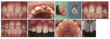
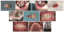
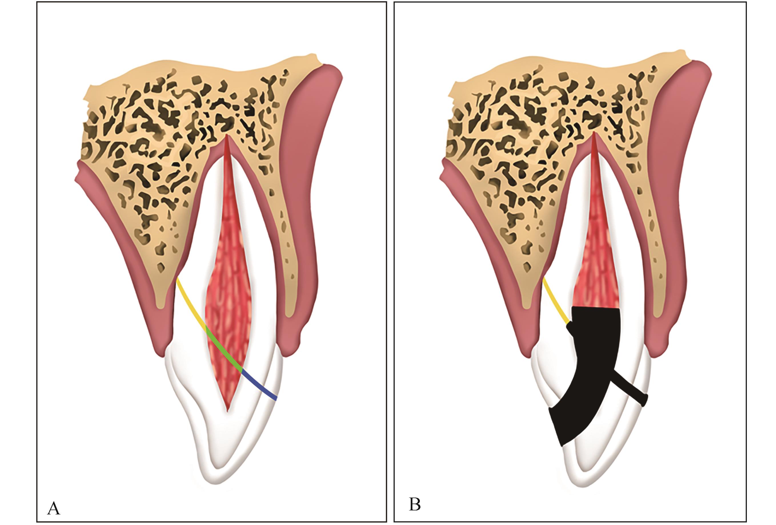
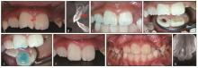
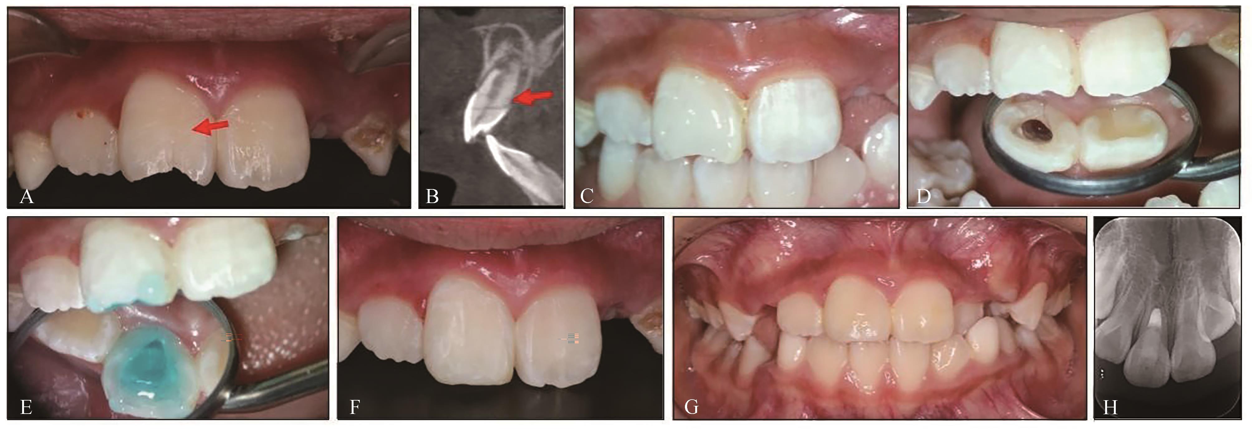
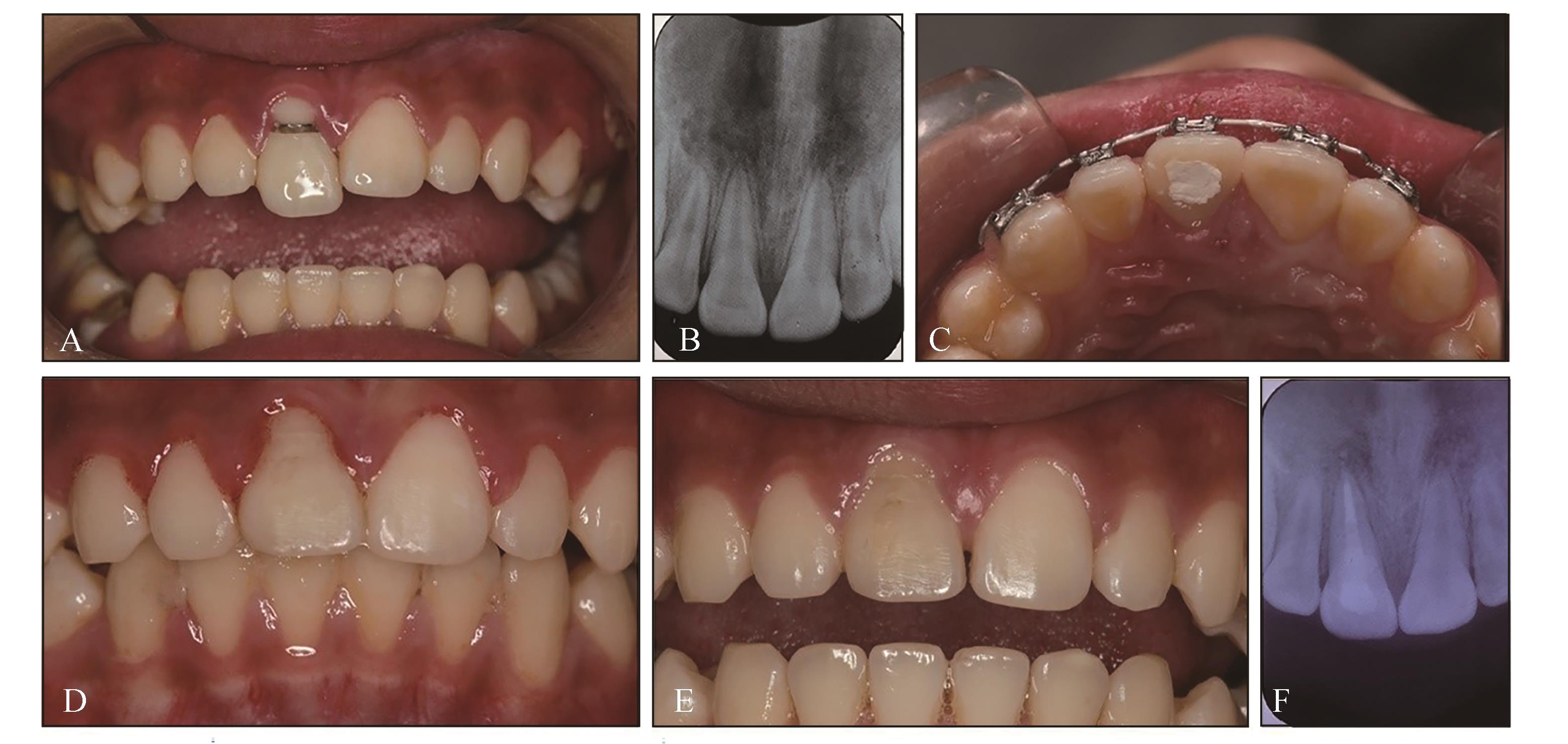
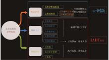
复杂冠根折是牙外伤中较为严重的一种类型,涉及牙体、牙髓和牙周组织损伤,临床上常用治疗方法包括正畸牵引、外科牵引、意向再植和断冠粘接术等。这些方法能够比较有效地保存患牙,但需要先拔出冠部断端,暴露龈下断面,然后再行粘接修复,可能会导致外伤牙冠根比不协调或牙周问题,给远期疗效带来挑战。近期,笔者尝试冠部断端非拔出技术联合部分断冠粘接术,即保留冠部断端不予拔出,仅沿龈上折裂线和龈下折裂线(髓腔侧)进行粘接,而龈下折裂线(牙周侧)不做处理,旨在简化操作,最大限度地减少对牙周组织的二次损伤,降低牙周并发症的可能,短期内取得了较满意的效果,但粘接强度和远期疗效尚有待观察与评估。
中图分类号:
| 1 | Andreasen JO. Etiology and pathogenesis of traumatic dental injuries. A clinical study of 1 298 cases[J]. Scand J Dent Res, 1970, 78(4): 329-342. |
| 2 | Tsilingaridis G, Malmgren B, Andreasen JO, et al. Intrusive luxation of 60 permanent incisors: a retrospective study of treatment and outcome[J]. Dent Traumatol, 2012, 28(6): 416-422. |
| 3 | Castro JC, Poi WR, Manfrin TM, et al. Analysis of the crown fractures and crown-root fractures due to dental trauma assisted by the Integrated Clinic from 1992 to 2002[J]. Dent Traumatol, 2005, 21(3): 121-126. |
| 4 | Oz IA, Haytaç MC, Toroglu MS. Multidisciplinary approach to the rehabilitation of a crown-root fracture with original fragment for immediate esthetics: a case report with 4-year follow-up[J]. Dent Traumatol, 2006, 22(1): 48-52. |
| 5 | Chung MP, Wang SS, Chen CP, et al. Management of crown-root fracture tooth by intra-alveolar transplantation with 180-degree rotation and suture fixation[J]. Oral Surg Oral Med Oral Pathol Oral Radiol Endod, 2010, 109(2): e126-e130. |
| 6 | Diangelis AJ, Andreasen JO, Ebeleseder KA, et al. International Association of Dental Traumatology guidelines for the management of traumatic dental injuries: 1. fractures and luxations of permanent teeth[J]. Dent Traumatol, 2012, 28(1): 2-12. |
| 7 | Bourguignon C, Cohenca N, Lauridsen E, et al. International Association of Dental Traumatology guidelines for the management of traumatic dental injuries: 1. fractures and luxations[J]. Dent Traumatol, 2020, 36(4): 314-330. |
| 8 | Cohen ES. Atlas of cosmetic and reconstructive pe-riodontal surgery[J]. BC Decker Inc Hamilton, 2007, 3: 239-269. |
| 9 | Lee JH, Yoon SM. Surgical extrusion of multiple teeth with crown-root fractures: a case report with 18-months follow up[J]. Dent Traumatol, 2015, 31(2): 150-155. |
| 10 | Murali RV, Rajashekhar L, Rajalingam S. Extrusion of fractured anterior tooth-an invisible[J]. Indian J Multidiscip Dent, 2011, 1(2): 96-99. |
| 11 | Kim SH, Tramontina V, Passanezi E. A new approach using the surgical extrusion procedure as an alternative for the reestablishment of biologic width[J]. Int J Periodontics Restorative Dent, 2004, 24(1): 39-45. |
| 12 | Malmgren O, Malmgren B, Frykholm A. Rapid or-thodontic extrusion of crown root and cervical root fractured teeth[J]. Endod Dent Traumatol, 1991, 7(2): 49-54. |
| 13 | Lee H, Lim CW, Hong HP, et al. Efficacy of the APACHE II score at ICU discharge in predicting post-ICU mortality and ICU readmission in critically ill surgical patients[J]. Anaesth Intensive Care, 2015, 43(2): 175-186. |
| 14 | Elkhadem A, Mickan S, Richards D. Adverse events of surgical extrusion in treatment for crown-root and cervical root fractures: a systematic review of case series/reports[J]. Dent Traumatol, 2014, 30(1): 1-14. |
| 15 | 廖汶晓, 马心笛, 洪志薇, 等. 前牙复杂冠根折外科手术冠向复位治疗的早期效果[J]. 国际口腔医学杂志, 2021, 48(5): 541-548. |
| Liao WX, Ma XD, Hong ZW, et al. Clinical effect of surgical extrusion in the treatment of complicated crown-root fracture of anterior teeth[J]. Int J Stomatol, 2021, 48(5): 541-548. | |
| 16 | 李思逸, 房瑞贞, 袁林天, 等. 180°旋转意向再植术治疗复杂冠根折1例[J]. 牙体牙髓牙周病学杂志, 2016, 26(4): 259-260. |
| Li SY, Fang RZ, Yuan LT, et al. A case of complex crown and root fracture treated by 180° rotation intention replantation[J]. Chin J Conserv Dent, 2016, 26(4): 259-260. | |
| 17 | Tsukiboshi M. Autotransplantation of teeth: requirements for predictable success[J]. Dent Traumatol, 2002, 18(4): 157-180. |
| 18 | Andreasen JO. Periodontal healing after replantation and autotransplantation of incisors in monkeys[J]. Int J Oral Surg, 1981, 10(1): 54-61. |
| 19 | Khandelwal P, Srinivasan S, Arul B, et al. Fragment reattachment after complicated crown-root fractures of anterior teeth: a systematic review[J]. Dent Traumatol, 2021, 37(1): 37-52. |
| 20 | Macedo GV, Diaz PI, De O Fernandes CA, et al. Reattachment of anterior teeth fragments: a conservative approach[J]. J Esthet Restor Dent, 2008, 20(1): 5-20. |
| 21 | 葛立宏. 儿童口腔医学[M]. 5版. 北京: 人民卫生出版社, 2020. |
| Ge LH. Pediatric dentistry[M]. 5th ed. Beijing: People’s Medical Publishing House, 2020. | |
| 22 | Islam MA, Wakia T, Alam MS, et al. Management of a subgingivally fractured central incisor by re-attachment using a fiber post[J]. Update Dent Coll J, 2014, 3(1): 37-40. |
| 23 | Chosack A, Eidelman E. Rehabilitating of a fractured incisor using the patient’s natural crown: a case report[J]. J Dent Child, 1994, 71: 19-21. |
| 24 | Tamilselvam S, Divyanand MJ, Neelakantan P. Biocompatibility of a conventional glass ionomer, ceramic reinforced glass ionomer, giomer and resin composite to fibroblasts: in vitro study[J]. J Clin Pediatr Dent, 2013, 37(4): 403-406. |
| 25 | Selimović-Dragaš M, Huseinbegović A, Kobašlija S, et al. A comparison of the in vitro cytotoxicity of conventional and resin modified glass ionomer cements[J]. Bosn J Basic Med Sci, 2012, 12(4): 273-278. |
| 26 | de Souza Costa CA, Hebling J, Garcia-Godoy F, et al. In vitro cytotoxicity of five glass-ionomer cements[J]. Biomaterials, 2003, 24(21): 3853-3858. |
| 27 | Lang O, Kohidai L, Kohidai Z, et al. Cell physiolo-gical effects of glass ionomer cements on fibroblast cells[J]. Toxicol In Vitro, 2019, 61: 104627. |
| 28 | Frauscher KE, Ilie N. Degree of conversion of nano-hybrid resin-based composites with novel and conventional matrix formulation[J]. Clin Oral Investig, 2013, 17(2): 635-642. |
| 29 | Marigo L, Spagnuolo G, Malara F, et al. Relation between conversion degree and cytotoxicity of a flowable bulk-fill and three conventional flowable resin-composites[J]. Eur Rev Med Pharmacol Sci, 2015, 19(23): 4469-4480. |
| 30 | van Landuyt KL, Nawrot T, Geebelen B, et al. How much do resin-based dental materials release? A meta-analytical approach[J]. Dent Mater, 2011, 27(8): 723-747. |
| 31 | Cândea Ciurea A, Şurlin P, Stratul ŞI, et al. Evaluation of the biocompatibility of resin composite-based dental materials with gingival mesenchymal stromal cells[J]. Microsc Res Tech, 2019, 82(10): 1768-1778. |
| 32 | Kurt A, Altintas SH, Kiziltas MV, et al. Evaluation of residual monomer release and toxicity of self-adhesive resin cements[J]. Dent Mater J, 2018, 37(1): 40-48. |
| 33 | Marczuk-Kolada G, Łuczaj-Cepowicz E, Pawińska M, et al. Evaluation of the cytotoxicity of selected conventional glass ionomer cements on human gingival fibroblasts[J]. Adv Clin Exp Med, 2017, 26(7): 1041-1045. |
| 34 | Alkan A, Keskiner I, Yuzbasioglu E. Connective tissue grafting on resin ionomer in localized gingival recession[J]. J Periodontol, 2006, 77(8): 1446-1451. |
| 35 | Dablanca-Blanco AB, Blanco-Carrión J, Martín-Biedma B, et al. Management of large class Ⅱ lesions in molars: how to restore and when to perform surgical crown lengthening[J]. Restor Dent Endod, 2017, 42(3): 240-252. |
| 36 | 马健, 邵强, 于毅. 不同龈下楔状缺损充填材料对牙龈卟啉单胞菌生物膜形成的影响[J]. 上海口腔医学, 2020, 29(4): 375-379. |
| Ma J, Shao Q, Yu Y. The effect of commonly used subgingival wedge-shaped defect filling materials on the formation of Porphyromonas gingivalis biofilm[J]. Shanghai J Stomatol, 2020, 29(4): 375-379. | |
| 37 | 高磊, 孙书恺, 周子凌, 等. 原位断端再接技术在儿童年轻恒前牙复杂冠根折中的应用二例[J]. 中华口腔医学杂志, 2021, 56(9): 892-896. |
| Gao L, Sun SK, Zhou ZL, et al. Two cases of application of in situ termination and reattachment technique in complex crown root fractures of young permanent anterior teeth in children[J]. Chin J Stomatol, 2021, 56(9): 892-896. |
| [1] | 高若凡,夏斌. 基于慢性疾病管理理念的重度低龄儿童龋管理方法[J]. 国际口腔医学杂志, 2023, 50(3): 341-346. |
| [2] | 任海霞,刘颍凤,梁慧敏,李家勇,温春琴,王春梅. 虚拟现实技术在儿童深龋治疗中对牙科畏惧的干预效果研究[J]. 国际口腔医学杂志, 2022, 49(5): 529-536. |
| [3] | 贺红. 伴扁桃体肥大Ⅲ类错𬌗畸形儿童早期矫治的临床诊疗策略[J]. 国际口腔医学杂志, 2022, 49(3): 249-254. |
| [4] | 朱锦怡,樊琪,周媛,邹静,黄睿洁. 唾液蛋白作为低龄儿童龋预测标志物的研究进展[J]. 国际口腔医学杂志, 2022, 49(2): 212-219. |
| [5] | 廖汶晓,马心笛,洪志薇,吴昕彧,邢云娣,刘经纬,陈蕾. 前牙复杂冠根折外科手术冠向复位治疗的早期效果[J]. 国际口腔医学杂志, 2021, 48(5): 541-548. |
| [6] | 邓晓宇,张蕴涵,邹静. 低龄儿童龋的早期生物学管理[J]. 国际口腔医学杂志, 2020, 47(5): 581-588. |
| [7] | 邓晓宇,仁青色格,霍媛媛,崔晨,亓文婷,韩轩,黄睿洁,周媛,朱林,邹静,旦增念扎. 西藏自治区城区和牧区儿童患龋情况与相关因素分析[J]. 国际口腔医学杂志, 2020, 47(4): 383-390. |
| [8] | 于林彤,宋光泰. 铒激光在儿童口腔医学中的应用[J]. 国际口腔医学杂志, 2020, 47(3): 351-355. |
| [9] | 王晓波,林世耀,李霞. 母亲与儿童龋病关系的研究进展[J]. 国际口腔医学杂志, 2019, 46(4): 469-474. |
| [10] | 赵金,赖光云,汪俊. 全身麻醉下儿童口腔疾病治疗家长接受度现况的研究进展[J]. 国际口腔医学杂志, 2018, 45(6): 739-744. |
| [11] | 丁杰, 宋光泰. 微创技术在儿童龋病治疗中的应用[J]. 国际口腔医学杂志, 2018, 45(4): 473-479. |
| [12] | 游文喆, 夏斌. 精神/智力残疾儿童的口腔健康促进和维护[J]. 国际口腔医学杂志, 2018, 45(4): 492-496. |
| [13] | 刘晓华, 王恩博. 儿童埋伏多生牙拔除术锥形束计算机断层扫描影像学定位的应用进展[J]. 国际口腔医学杂志, 2018, 45(3): 295-300. |
| [14] | 蔡东霖, 卢锐. 儿童复发性阿弗他溃疡病因学的研究进展[J]. 国际口腔医学杂志, 2018, 45(2): 145-149. |
| [15] | 高雪彬, 张琦, 李晶, 毕也, 杨华, 黄洋. 低龄儿童行窝沟封闭术时酸蚀剂选择的临床研究[J]. 国际口腔医学杂志, 2017, 44(4): 433-436. |
|