国际口腔医学杂志 ›› 2021, Vol. 48 ›› Issue (6): 644-655.doi: 10.7518/gjkq.2021104
何蓉( ),刘学军(),周宇琨
He Rong(),Liu Xuejun(),Zhou Yukun
摘要:
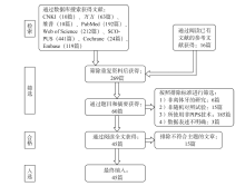
目的 评价掺铒钇铝石榴石(Er: YAG)激光光子引导的光声流(PIPS)效应在根管荡洗中的作用效果。方法 计算机检索中国知网、万方数据知识服务平台、维普中文科技期刊数据库、PubMed、Embase、Cochrane Library、Web of Science、SCOPUS数据库,纳入有关PIPS根管荡洗作用的随机对照试验,检索时间从2010年1月至2020年7月,所选文献按照纳入和排除标准进行筛选,根据Cochrane系统评价的方法进行风险评估,根据《系统综述和荟萃分析优先报告的条目:PRISMA声明》确定的项目进行评价。结果 最终共纳入45篇文章,通过归类描述和系统评价,这些文章体现了PIPS技术在灭菌、去除玷污层及牙本质碎屑、去除根管内氢氧化钙、增加牙本质小管渗透性等方面有显著的成效,但是仍有部分实验结果未能体现PIPS技术的优势。目前,PIPS技术的研究中存在实验对象(离体牙根管长度和弯曲度的差异)以及实验过程和方法未标准化、研究方法存在差异等问题,缺少根管微裂、根尖溢出冲洗液和碎屑、标准参数、不同根管荡洗方法作用效果的比较、随机对照的临床试验等方面的相关研究。结论 大多数实验结果表明PIPS在灭菌、去除玷污层及牙本质碎屑、去除根管内氢氧化钙、增加牙本质小管渗透性等方面有显著的成效,但仍有实验结果存在争议,需要更多长期、大样本、高质量、设计严谨的实验来验证这些结论。
中图分类号:
| [1] | 刘正. 感染根管的细菌学研究[J]. 国外医学·口腔医学分册, 1982, 9(6):336-339. |
| Liu Z. Bacteriological study of infected root canals[J]. Foreign Med Sci (Stomatol), 1982, 9(6):336-339. | |
| [2] |
Santos AL, Siqueira JF, Rôças IN, et al. Comparing the bacterial diversity of acute and chronic dental root canal infections[J]. PLoS One, 2011, 6(11):e28088.
doi: 10.1371/journal.pone.0028088 |
| [3] | 周学东. 成人根管系统形态与根管治疗难度评估[J]. 中国实用口腔科杂志, 2008, 1(1):5-9. |
| Zhou XD. Morphology of the adult root canal system and assessment of the difficulty of root canal treat-ment[J]. Chin J Pract Stomatol, 2008, 1(1):5-9. | |
| [4] |
Arnold M, Ricucci D, Siqueira JF. Infection in a complex network of apical ramifications as the cause of persistent apical periodontitis: a case report[J]. J Endod, 2013, 39(9):1179-1184.
doi: 10.1016/j.joen.2013.04.036 |
| [5] | Takahashi K, Machida T, Kimura Y, et al. The morphological study of root canal walls with Er: YAG laser[J]. J Jpn Endod Assoc, 1996, 17(2):197-203. |
| [6] |
Roper MJ, White JM, Goodis HE, et al. Two-dimensional changes and surface characteristics from an erbium laser used for root canal preparation[J]. Lasers Surg Med, 2010, 42(5):379-383.
doi: 10.1002/lsm.20918 pmid: 20583251 |
| [7] |
Ozses Ozkaya B, Gulsahi K, Ungor M, et al. A comparison of Er: YAG laser with photon-initiated photoacoustic streaming, Nd: YAG laser, and conventional irrigation on the eradication of root dentinal tubule infection by Enterococcus faecalis biofilms: a scanning electron microscopy study[J]. Scanning, 2017, 2017:6215482.
doi: 10.1155/2017/6215482 pmid: 29279728 |
| [8] |
Suk M, Bago I, Katić M, et al. The efficacy of photon-initiated photoacoustic streaming in the removal of calcium silicate-based filling remnants from the root canal after rotary retreatment[J]. Lasers Med Sci, 2017, 32(9):2055-2062.
doi: 10.1007/s10103-017-2325-4 |
| [9] |
Swimberghe RCD, Coenye T, De Moor RJG, et al. Biofilm model systems for root canal disinfection: a literature review[J]. Int Endod J, 2019, 52(5):604-628.
doi: 10.1111/iej.13050 pmid: 30488449 |
| [10] |
Golob BS, Olivi G, Vrabec M, et al. Efficacy of photon-induced photoacoustic streaming in the reduction of Enterococcus faecalis within the root canal: different settings and different sodium hypochlorite concentrations[J]. J Endod, 2017, 43(10):1730-1735.
doi: 10.1016/j.joen.2017.05.019 |
| [11] |
Al Shahrani M, DiVito E, Hughes CV , et al. Enhanced removal of Enterococcus faecalis biofilms in the root canal using sodium hypochlorite plus photon-induced photoacoustic streaming: an in vitro study[J]. Photomed Laser Surg, 2014, 32(5):260-266.
doi: 10.1089/pho.2014.3714 |
| [12] |
Balić M, Lucić R, Mehadžić K, et al. The efficacy of photon-initiated photoacoustic streaming and so-nic-activated irrigation combined with QMiX solution or sodium hypochlorite against intracanal E. faecalis biofilm[J]. Lasers Med Sci, 2016, 31(2):335-342.
doi: 10.1007/s10103-015-1864-9 |
| [13] |
Pedullà E, Genovese C, Campagna E, et al. Decontamination efficacy of photon-initiated photoacoustic streaming (PIPS) of irrigants using low-energy laser settings: an ex vivo study[J]. Int Endod J, 2012, 45(9):865-870.
doi: 10.1111/j.1365-2591.2012.02044.x pmid: 22486805 |
| [14] |
Olivi G, DiVito E, Peters O, et al. Disinfection efficacy of photon-induced photoacoustic streaming on root canals infected with Enterococcus faecalis: an ex vivo study[J]. J Am Dent Assoc, 2014, 145(8):843-848.
doi: 10.14219/jada.2014.46 |
| [15] |
Zhu X, Yin X, Chang JW, et al. Comparison of the antibacterial effect and smear layer removal using photon-initiated photoacoustic streaming aided irrigation versus a conventional irrigation in single-rooted canals: an in vitro study[J]. Photomed Laser Surg, 2013, 31(8):371-377.
doi: 10.1089/pho.2013.3515 |
| [16] |
Wang XL, Cheng XG, Liu X, et al. Bactericidal effect of various laser irradiation systems on Enterococcus faecalis biofilms in dentinal tubules: a confocal laser scanning microscopy study[J]. Photomed Laser Surg, 2018, 36(9):472-479.
doi: 10.1089/pho.2017.4430 |
| [17] |
Peters OA, Bardsley S, Fong J, et al. Disinfection of root canals with photon-initiated photoacoustic strea-ming[J]. J Endod, 2011, 37(7):1008-1012.
doi: 10.1016/j.joen.2011.03.016 pmid: 21689561 |
| [18] | 田甜甜. PIPS-Er: YAG激光在不同根尖终末工作宽度时对根管内粪肠球菌杀灭效果的研究[D]. 西安: 第四军医大学, 2016. |
| Tian TT. Bactericidal effect of PIPS-Er: YAG laser with different apical terminal working width on Enterococcus faecalis in experimentally infected root canals[D]. Xi’an: The Fourth Military Medical University, 2016. | |
| [19] |
Akyuz Ekim SN, Erdemir A. Comparison of diffe-rent irrigation activation techniques on smear layer removal: an in vitro study[J]. Microsc Res Tech, 2015, 78(3):230-239.
doi: 10.1002/jemt.v78.3 |
| [20] |
Doğanay Yıldız E, Dinçer B, Fidan ME. Effect of different laser-assisted irrigation activation techni-ques on apical debris extrusion[J]. Acta Odontol Scand, 2020, 78(5):332-336.
doi: 10.1080/00016357.2020.1717603 pmid: 31986947 |
| [21] |
Korkut E, Torlak E, Gezgin O, et al. Antibacterial and smear layer removal efficacy of Er: YAG laser irradiation by photon-induced photoacoustic strea-ming in primary molar root canals: a preliminary study[J]. Photomed Laser Surg, 2018, 36(9):480-486.
doi: 10.1089/pho.2017.4369 pmid: 29905503 |
| [22] |
Keles A, Kamalak A, Keskin C, et al. The efficacy of laser, ultrasound and self-adjustable file in remo-ving smear layer debris from oval root canals follo-wing retreatment: a scanning electron microscopy study[J]. Aust Endod J, 2016, 42(3):104-111.
doi: 10.1111/aej.12145 pmid: 26786709 |
| [23] |
Sippus J, Gutknecht N. Deep disinfection and tubular smear layer removal with Er: YAG using photon-induced photoacoustic streaming (PIPS) contra laser-activated irrigation (LAI) technics[J]. Lasers Dent Sci, 2019, 3(1):37-42.
doi: 10.1007/s41547-018-0050-3 |
| [24] | 孙宁佳, 郭威. Er: YAG激光和超声荡洗对根管内壁形态及微渗漏的比较研究[J]. 牙体牙髓牙周病学杂志, 2017, 27(12):689-697, 712. |
| Sun NJ, Guo W. Comparison of root canal morpho-logy and microleakage after cleaning with Er: YAG laser and ultrasonic irrigation[J]. Chin J Conserv Dent, 2017, 27(12):689-697, 712. | |
| [25] | 刘敏, 彭彬. 两种功率PIPS-Er: YAG激光对根管内玷污层去除效果的比较研究[J]. 口腔医学研究, 2018, 34(10):1067-1071. |
| Liu M, Peng B. Comparison of photon-initiated photoacoustic streaming with two kinds of power settings on removal of smear layer[J]. J Oral Sci Res, 2018, 34(10):1067-1071. | |
| [26] |
Passalidou S, Calberson F, de Bruyne M , et al. Debris removal from the mesial root canal system of mandibular molars with laser-activated irrigation[J]. J Endod, 2018, 44(11):1697-1701.
doi: S0099-2399(18)30434-5 pmid: 30241679 |
| [27] |
Todea C, Mocuta D, Manescu A, et al. Laser use in endodontic for increase the adhesion of root canal filling. A synchrotron radiation micro tomography study[J]. Rev Chim, 2018, 69(8):2144-2149.
doi: 10.37358/RC.18.8.6489 |
| [28] |
Verstraeten J, Jacquet W, De Moor RJG, et al. Hard tissue debris removal from the mesial root canal system of mandibular molars with ultrasonically and laser-activated irrigation: a micro-computed tomography study[J]. Lasers Med Sci, 2017, 32(9):1965-1970.
doi: 10.1007/s10103-017-2297-4 |
| [29] |
Lloyd A, Navarrete G, Marchesan MA, et al. Removal of calcium hydroxide from Weine TypeⅡsystems using photon-induced photoacoustic strea-ming, passive ultrasonic, and needle irrigation: a microcomputed tomography study[J]. J Appl Oral Sci, 2016, 24(6):543-548.
doi: S1678-77572016000600543 pmid: 28076457 |
| [30] |
Sathe S, Hegde V, Jain P, et al. Effectiveness of Er: YAG (PIPS) and Nd: YAG activation on final irri-gants for smear layer removal-SEM observation[J]. J Dent Lasers, 2014, 8(1):8.
doi: 10.4103/0976-2868.134110 |
| [31] |
Ozbay Y, Erdemir A. Effect of several laser systems on removal of smear layer with a variety of irrigation solutions[J]. Microsc Res Tech, 2018, 81(10):1214-1222.
doi: 10.1002/jemt.v81.10 |
| [32] | Luca R, Carmen Todea MD, Bălăbuc C, et al. Alternative techniques in root canal debridement[C]//Proc SPIE 8925, Fifth International Conference on lasers in medicine: biotechnologies integrated in daily medicine. Berlin: Springer, 2014: 89250F1-89250F7. |
| [33] |
Nasher R, Franzen R, Gutknecht N. The effectiveness of the Erbium: Yttrium aluminum garnet PIPS technique in comparison to different chemical solutions in removing the endodontic smear layer-an in vitro profilometric study[J]. Lasers Med Sci, 2016, 31(9):1871-1882.
doi: 10.1007/s10103-016-2063-z |
| [34] |
Akcay M, Arslan H, Durmus N, et al. Dentinal tubule penetration of AH Plus, iRoot SP, MTA fillapex, and guttaflow bioseal root canal sealers after different final irrigation procedures: a confocal microscopic study[J]. Lasers Surg Med, 2016, 48(1):70-76.
doi: 10.1002/lsm.22446 |
| [35] |
Turkel E, Onay EO, Ungor M. Comparison of three final irrigation activation techniques: effects on canal cleanness, smear layer removal, and dentinal tubule penetration of two root canal sealers[J]. Photomed Laser Surg, 2017, 35(12):672-681.
doi: 10.1089/pho.2016.4234 pmid: 28437194 |
| [36] |
DiVito E, Peters OA, Olivi G. Effectiveness of the Erbium: YAG laser and new design radial and stripped tips in removing the smear layer after root canal instrumentation[J]. Lasers Med Sci, 2012, 27(2):273-280.
doi: 10.1007/s10103-010-0858-x |
| [37] |
Kamaci A, Aydin B, Erdilek N. The effect of ultrasonically activated irrigation and laser based root canal irrigation methods on debris removal[J]. Int J Artif Organs, 2017. doi: 10.5301/ijao.5000646.
doi: 10.5301/ijao.5000646 |
| [38] |
Lloyd A, Uhles JP, Clement DJ, et al. Elimination of intracanal tissue and debris through a novel laser-activated system assessed using high-resolution micro-computed tomography: a pilot study[J]. J Endod, 2014, 40(4):584-587.
doi: 10.1016/j.joen.2013.10.040 pmid: 24666917 |
| [39] |
Deleu E, Meire MA, De Moor RJ. Efficacy of laser-based irrigant activation methods in removing debris from simulated root canal irregularities[J]. Lasers Med Sci, 2015, 30(2):831-835.
doi: 10.1007/s10103-013-1442-y |
| [40] |
Parčina I, Amižić, Miletić I, et al. Influence of laser activated irrigation with two erbium lasers on bond strength of inidividually formed fiber reinforced composite posts to root canal dentin[J]. Acta Stomatol Croat, 2016, 50(4):321-328.
doi: 10.15644/asc50/4/5 pmid: 28275279 |
| [41] |
Akcay M, Arslan H, Mese M, et al. Effect of photon-initiated photoacoustic streaming, passive ultraso-nic, and sonic irrigation techniques on dentinal tubule penetration of irrigation solution: a confocal microscopic study[J]. Clin Oral Investig, 2017, 21(7):2205-2212.
doi: 10.1007/s00784-016-2013-y |
| [42] |
Miletić I, Chieffi N, Rengo C, et al. Effect of photon induced photoacoustic streaming (PIPS) on bond strength to dentine of two root canal filling materials[J]. Lasers Surg Med, 2016, 48(10):951-954.
doi: 10.1002/lsm.22536 pmid: 27254395 |
| [43] |
Vidas J, Snjaric D, Braut A, et al. Comparison of apical irrigant solution extrusion among conventio-nal and laser-activated endodontic irrigation[J]. Lasers Med Sci, 2020, 35(1):205-211.
doi: 10.1007/s10103-019-02846-w |
| [44] |
Arslan D, Kustarci A. Efficacy of photon-initiated photoacoustic streaming on apically extruded debris with different preparation systems in curved canals[J]. Int Endod J, 2018, 51(Suppl 1):e65-e72.
doi: 10.1111/iej.2018.51.issue-S1 |
| [45] |
Guneser MB, Arslan D, Usumez A. Tissue dissolution ability of sodium hypochlorite activated by photon-initiated photoacoustic streaming technique[J]. J Endod, 2015, 41(5):729-732.
doi: 10.1016/j.joen.2015.01.014 pmid: 25728817 |
| [46] |
Kamalak A, Uzun I, Arslan H, et al. Fracture resistance of endodontically retreated roots after retreatment using self-adjusting file, passive ultrasonic irrigation, photon-induced photoacoustic streaming, or laser[J]. Photomed Laser Surg, 2016, 34(10):467-472.
pmid: 27598303 |
| [47] |
Dagher J, El Feghali R, Parker S, et al. Postoperative quality of life following conventional endodontic intracanal irrigation compared with laser-activa-ted irrigation: a randomized clinical study[J]. Photobiomodul Photomed Laser Surg, 2019, 37(4):248-253.
doi: 10.1089/photob.2018.4558 |
| [48] |
Azim AA, Aksel H, Zhuang TT, et al. Efficacy of 4 irrigation protocols in killing bacteria colonized in dentinal tubules examined by a novel confocal laser scanning microscope analysis[J]. J Endod, 2016, 42(6):928-934.
doi: 10.1016/j.joen.2016.03.009 |
| [49] |
Ayranci LB, Arslan H, Akcay M, et al. Effectiveness of laser-assisted irrigation and passive ultrasonic irrigation techniques on smear layer removal in middle and apical thirds[J]. Scanning, 2016, 38(2):121-127.
doi: 10.1002/sca.21247 pmid: 26183211 |
| [50] |
Jezeršek M, Jereb T, Lukač N, et al. Evaluation of apical extrusion during novel Er: YAG laser-activa-ted irrigation modality[J]. Photobiomodul Photomed Laser Surg, 2019, 37(9):544-550.
doi: 10.1089/photob.2018.4608 pmid: 31335265 |
| [51] | Jezeršek M, Lukač N, Lukač M, et al. Measurement of pressures generated in root canal during Er: YAG laser-activated irrigation[J]. Photobiomodul Photo-med Laser Surg, 2020, 38(10):625-631. |
| [52] | 何新宇, 彭丽莉, 王众, 等. 光子引导的光声流效应在根管治疗术中的研究进展[J]. 牙体牙髓牙周病学杂志, 2018, 28(11):673-676, 679. |
| He XY, Peng LL, Wang Z, et al. Disinfection of root canals with photon-initiated photoacoustic streaming: a review[J]. Chin J Conserv Dent, 2018, 28(11):673-676, 679. | |
| [53] | De Moor RJG, Meire M. High-power lasers in en-dodontics-fiber placement for laser-enhanced en-dodontics: in the canal or at the orifice[J]. J Laser Health Acad, 2014, 2014(1):20-28. |
| [54] | 谷苗, 陈兴兴, 林媛, 等. 自制根管润滑剂与不同浓度次氯酸钠液组合对粪肠球菌感染根管壁的清洁效果[J]. 牙体牙髓牙周病学杂志, 2009, 19(5):275-279. |
| Gu M, Chen XX, Lin Y, et al. Cleaning efficacy of a self-made lubricant combined with different concen-trations of NaClO on root canal walls infected by Enterococcus faecalis[J]. Chin J Conserv Dent, 2009, 19(5):275-279. | |
| [55] |
da Silva LA, Nelson-Filho P, Faria G, et al. Bacterial profile in primary teeth with necrotic pulp and periapical lesions[J]. Braz Dent J, 2006, 17(2):144-148.
doi: 10.1590/S0103-64402006000200012 |
| [56] |
Dönmez Özkan H, Kaval ME, Özkan G, et al. Efficacy of two different nickel-titanium rotary systems in retreatment procedure with or without laser-activated irrigation: an in vitro study[J]. Photobiomodul Photomed Laser Surg, 2019, 37(8):495-499.
doi: 10.1089/photob.2019.4637 |
| [57] |
Mamootil K, Messer HH. Penetration of dentinal tubules by endodontic sealer cements in extracted teeth and in vivo[J]. Int Endod J, 2007, 40(11):873-881.
pmid: 17764458 |
| [58] |
Lin ZM, Ling JQ, Fang JY, et al. Physicochemical properties, sealing ability, bond strength and cytoto-xicity of a new dimethacrylate-based root canal sealer[J]. J Formos Med Assoc, 2010, 109(11):819-827.
doi: 10.1016/S0929-6646(10)60127-1 |
| [59] |
Abbaszadeh HA, Peyvandi AA, Peyvandi AA, et al. Er: YAG laser and cyclosporine-a effect on cell cycle regulation of human gingival fibroblast cells[J]. J Lasers Med Sci, 2017, 8(3):143-149.
doi: 10.15171/jlms.2017.26 |
| [60] | Lukac N, Muc BT, Jezersek M, et al. Photoacoustic endodontics using the novel SWEEPS Er: YAG laser modality[J]. J Laser Health Acad, 2017, 2047(1):1-7. |
| [1] | 吴思佳,舒畅,王洋,王媛,邓淑丽,王慧明. 根管内感染控制对年轻恒牙牙髓再生治疗的影响及研究进展[J]. 国际口腔医学杂志, 2023, 50(4): 388-394. |
| [2] | 高宇天,苏勤. 酸性氧化电位水在根管治疗中的研究与应用[J]. 国际口腔医学杂志, 2023, 50(4): 401-406. |
| [3] | 汪牡丹,宋东哲,黄定明. 开髓洞型对患牙根管治疗术后抗折性能影响的研究进展[J]. 国际口腔医学杂志, 2023, 50(2): 186-194. |
| [4] | 王璐璇,侯本祥. 根管内氢氧化钙残留对根管治疗的影响[J]. 国际口腔医学杂志, 2022, 49(3): 367-372. |
| [5] | 杨加震,张颖,刘育含,李帆,曾飞,李修珍,马玉莹,杨芳. 口腔诊疗环境细菌群落的时间变化趋势研究[J]. 国际口腔医学杂志, 2022, 49(2): 132-137. |
| [6] | 戢晓,景钫淇,李雅,薛晶. 根管预备顺序的数据模拟优化研究[J]. 国际口腔医学杂志, 2022, 49(1): 37-47. |
| [7] | 邢桂琪,郭林溪,苏勤. 根管治疗后疾病的综合评估和治疗决策[J]. 国际口腔医学杂志, 2021, 48(5): 579-584. |
| [8] | 彭玮琪,高原,徐欣. 髓腔通路设计的微创理念及其研究进展[J]. 国际口腔医学杂志, 2021, 48(4): 433-438. |
| [9] | 李米雪子,张琛. 椅旁计算机辅助设计/计算机辅助制作髓腔固位冠修复根管治疗后磨牙的临床考量[J]. 国际口腔医学杂志, 2021, 48(3): 274-279. |
| [10] | 李诗佳,陈秋宇,邹静,黄睿洁. 尼古丁对口腔细菌单独或混合培养时菌群数目调控的研究[J]. 国际口腔医学杂志, 2021, 48(3): 305-311. |
| [11] | 易祖木,王昕宇,伍颖颖. 糖尿病患者口腔细菌多样性的变化[J]. 国际口腔医学杂志, 2020, 47(5): 522-529. |
| [12] | 谭凯璇,李帆,张利娟,李姗姗,卢洁,张颖,杨芳. 根管再治疗并发皮下气肿1例[J]. 国际口腔医学杂志, 2020, 47(5): 563-566. |
| [13] | 唐蓓,赵文俊,王虎,郑广宁,游梦. 根管超填导致下牙槽神经损伤2例[J]. 国际口腔医学杂志, 2020, 47(3): 293-296. |
| [14] | 王蕊,盖阔,刘梦齐,蒋丽. 原子力显微镜在细菌黏附力学研究中的应用[J]. 国际口腔医学杂志, 2019, 46(6): 687-692. |
| [15] | 许庆安,樊明文. 非器械根管治疗与多声波超洁净系统[J]. 国际口腔医学杂志, 2019, 46(5): 522-525. |
|