国际口腔医学杂志 ›› 2019, Vol. 46 ›› Issue (6): 640-649.doi: 10.7518/gjkq.2019105
黎祺1,2,黄少宏1( )
)
Li Qi1,2,Huang Shaohong1()
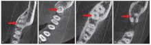
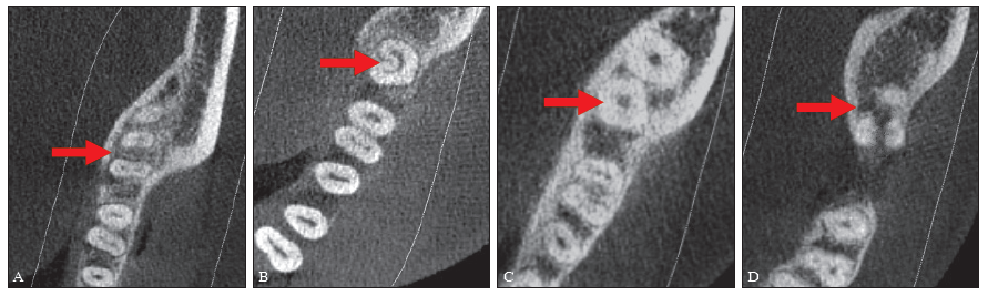
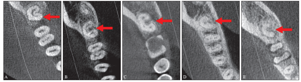
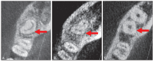
摘要: 目的 研究岭南地区广府民系人群下颌第二恒磨牙的牙根和根管形态。 方法 收集广府民系患者815份锥形束CT(CBCT)影像学资料共1 279颗下颌第二恒磨牙,观察和统计牙根和根管形态,并与近年的国内外同类研究进行比较和分析。 结果 下颌第二恒磨牙双根型占57.16%、C形根型占32.84%、锥形融合根型占9.77%、三根型占0.23%,其中双根型检出率男性高于女性(P<0.05),C形根型检出率女性高于男性(P<0.05),女性中不同年龄段的双根型和C形根型检出率差异均有统计学意义(P<0.01);双根型的近中根的根管形态主要为Ⅳ型(2-2)38.58%、Ⅱ型(2-1)28.73%和Ⅰ型(1-1)22.98%,其中Ⅰ型(1-1)根管检出率女性明显高于男性(P<0.01),Ⅳ型(2-2)根管检出率男性明显高于女性(P<0.01),双根型的远中根以Ⅰ型(1-1)根管为主,占81.67%,近中根和远中根各个根管形态检出率的差异有统计学意义(P<0.01);C形根管检出率女性高于男性(P<0.05),各种根管形态检出率在根管不同部位的差异有统计学意义(P<0.01);锥形融合根型的C形根管型62.40%、多根管型20.00%、单根管型17.60%,其中多根管型检出率男性明显高于女性(P<0.01);双侧牙根形态对称68.10%,双侧牙根和根管形态完全对称62.93%。 结论 岭南地区广府民系人群下颌第二恒磨牙牙根及根管形态复杂多样,与国内外其他地区和种族有一定差异性。
中图分类号:
| [1] | 黄少宏 . 广东省口腔健康30年趋势研究[M]. 广州: 广东科技出版社, 2019: 25-64. |
| Huang SH. A 30-year trend study on oral health in Guangdong province[M]. Guangzhou: Guangdong Science and Technology Press, 2019: 25-64. | |
| [2] | von Zuben M, Martins JNR, Berti L , et al. Worldwide prevalence of mandibular second molar C- shaped morphologies evaluated by cone-beam computed tomography[J]. J Endod, 2017,43(9):1442-1447. |
| [3] | Kashyap RR, Beedubail SP, Kini R , et al. Assessment of the number of root canals in the maxillary and mandibular molars: a radiographic study using cone beam computed tomography[J]. J Conserv Dent, 2017,20(5):288-291. |
| [4] | Patel S, Durack C, Abella F , et al. Cone beam computed tomography in endodontics—a review[J]. Int Endod J, 2015,48(1):3-15. |
| [5] | Hillson S. Tooth development in human evolution and bioarchaeology[M]. London: Cambridge Uni-versity Press, 2014: 1-307. |
| [6] | 广东省统计局, 国家统计局广东调查总队. 广东统计年鉴[M]. 北京: 中国统计出版社, 2018: 24-25. |
| Guangdong Statistical Bureau, Guangdong Investigation Team of the National Bureau of Statistics. Guangdong statistical yearbook[M]. Beijing: China Statistical Press, 2018: 24-25. | |
| [7] | 黄淑娉 . 广东族群与区域文化研究[M]. 广州: 广东高等教育出版社, 1999: 50-75. |
| Huang SP. Study on ethnic groups and regional culture in Guangdong[M]. Guangzhou: Guangdong Higher Education Press, 1999: 50-75. | |
| [8] | 翟丹然 . 专家论证广府文化发祥地[N]. 南方日报, 2012-02-11(9). |
| Zhai DR . Experts demonstrate the birthplace of Guangfu culture[N]. Nanfang Daily, 2012-02-11(9). | |
| [9] | Vertucci FJ . Root canal anatomy of the human per-manent teeth[J]. Oral Surg Oral Med Oral Pathol, 1984,58(5):589-599. |
| [10] | Melton DC, Krell KV, Fuller MW . Anatomical and histological features of C-shaped canals in mandibular second molars[J]. J Endod, 1991,17(8):384-388. |
| [11] | 吴汝康, 吴新智, 张振标 . 人体测量方法[M]. 北京: 科学出版社, 1984: 12-13. |
| Wu RK, Wu XZ, Zhang ZB. Anthropometric mea-surement[M]. Beijing: Science Press, 1984: 12-13. | |
| [12] | Pérez-Heredia M, Ferrer-Luque CM, Bravo M , et al. Cone-beam computed tomographic study of root anatomy and canal configuration of molars in a Spanish population[J]. J Endod, 2017,43(9):1511-1516. |
| [13] | Madani ZS, Mehraban N, Moudi E , et al. Root and canal morphology of mandibular molars in a selected Iranian population using cone-beam computed tomo-graphy[J]. Iran Endod J, 2017,12(2):143-148. |
| [14] | Pawar AM, Pawar M, Kfir A , et al. Root canal morphology and variations in mandibular second molar teeth of an Indian population: an in vivo cone-beam computed tomography analysis[J]. Clin Oral Investig, 2017,21(9):2801-2809. |
| [15] | Kim SY, Kim BS, Kim Y . Mandibular second molar root canal morphology and variants in a Korean subpopulation[J]. Int Endod J, 2016,49(2):136-144. |
| [16] | 谢霓, 王九龙, 朱友家 . 下颌第二磨牙牙根及根管形态学研究[J]. 临床口腔医学杂志, 2015,31(7):415-417. |
| Xie N, Wang JL, Zhu YJ . The research on mandibular second molars morphology[J]. J Clin Stomatol, 2015,31(7):415-417. | |
| [17] | 程倩, 黄巍, 王昊 . 下颌第二恒磨牙牙根及根管形态的锥形束CT研究[J]. 北京口腔医学, 2017,25(5):284-286. |
| Cheng Q, Huang W, Wang H . Evaluation of the root and canal morphology of mandibular second permanent molars using cone-beam computed tomography[J]. Beijing J Stomatol, 2017,25(5):284-286. | |
| [18] | Martins JNR, Marques D, Mata A , et al. Root and root canal morphology of the permanent dentition in a Caucasian population: a cone-beam computed tomography study[J]. Int Endod J, 2017,50(11):1013-1026. |
| [19] | Torres A, Jacobs R, Lambrechts P , et al. Characterization of mandibular molar root and canal morphology using cone beam computed tomography and its variability in Belgian and Chilean population samples[J]. Imaging Sci Dent, 2015,45(2):95-101. |
| [20] | Pan JYY, Parolia A, Chuah SR , et al. Root canal morphology of permanent teeth in a Malaysian sub-population using cone-beam computed tomogra-phy[J]. BMC Oral Health, 2019,19(1):14. |
| [21] | 张博森, 吴家媛 . 下颌磨牙近中中根管研究进展[J]. 口腔疾病防治, 2018,26(4):258-262. |
| Zhang BS, Wu JY . Research progress on the middle mesial root canal in the mandibular molar[J]. J Prev Treat Stomatol Dis, 2018,26(4):258-262. | |
| [22] | Tahmasbi M, Jalali P, Nair MK , et al. Prevalence of middle mesial canals and isthmi in the mesial root of mandibular molars: an in vivo cone-beam computed tomographic study[J]. J Endod, 2017,43(7):1080-1083. |
| [23] | 史志芸, 胡楠, 石校伟 , 等. 应用锥形束CT对187例患者下颌第二磨牙根管形态的研究[J]. 解放军医学院学报, 2018,39(5):437-440. |
| Shi ZY, Hu N, Shi XW , et al. Root and canal morphology of mandibular second molars: a cone-beam computed tomography analysis[J]. Acad J Chin PLA Med Sch, 2018,39(5):437-440. | |
| [24] | 殷铭, 王俊, 刘青梅 . C形根管系统的研究进展[J]. 全科口腔医学杂志, 2016,3(18):14-18. |
| Yin M, Wang J, Liu QM . The research situation of C-shaped canal system[J]. General J Stomatol, 2016,3(18):14-18. | |
| [25] | Janani M, Rahimi S, Jafari F , et al. Anatomic features of C-shaped mandibular second molars in a selected Iranian population using CBCT[J]. Iran Endod J, 2018,13(1):120-125. |
| [26] | Wadhwani S, Singh MP, Agarwal M , et al. Prevalence of C-shaped canals in mandibular second and third molars in a central India population: a cone beam computed tomography analysis[J]. J Conserv Dent, 2017,20(5):351-354. |
| [27] | Vaz de Azevedo KR, Lopes CB, Andrade RHTLR , et al. C-shaped canals in first and second mandibular molars from Brazilian individuals: a prevalence study using cone-beam computed tomography[J]. PLoS One, 2019,14(2):e0211948. |
| [28] | Alfawaz H, Alqedairi A, Alkhayyal AK , et al. Prevalence of C-shaped canal system in mandibular first and second molars in a Saudi population assessed via cone beam computed tomography: a retrospective study[J]. Clin Oral Investig, 2019,23(1):107-112. |
| [29] | 刘佼佼, 王晨, 杨勇 . 辽宁地区汉族人下颌第二恒磨牙牙根和根管数目的锥形束CT研究[J]. 口腔颌面修复学杂志, 2013,14(6):337-340. |
| Liu JJ, Wang C, Yang Y . Use of cone-beam computed tomography to evaluate the number of roots and canals of mandibular second permanent molars in Han people of Liaoning province[J]. J Chin Prosthodont, 2013,14(6):337-340. | |
| [30] | 周涛, 赵长铭, 柴治国 , 等. 下颌第二恒磨牙牙根及根管系统的显微CT观察[J]. 牙体牙髓牙周病学杂志, 2014,24(8):469-472, 480. |
| Zhou T, Zhao CM, Chai ZG , et al. A micro-CT analysis of the root and root canal systems of permanent mandibular second molars[J]. Chin J Conserv Dent, 2014,24(8):469-472, 480. | |
| [31] | Wang Y, Guo J, Yang HB , et al. Incidence of C- shaped root canal systems in mandibular second molars in the native Chinese population by analysis of clinical methods[J]. Int J Oral Sci, 2012,4(3):161-165. |
| [32] | 梁学萍, 张洋洋, 孙玉亮 , 等. 锥体束CT对维吾尔族成人下颌第二磨牙C形根管的研究[J]. 口腔医学研究, 2016,32(3):294-296. |
| Liang XP, Zhang YY, Sun YL , et al. Evaluation of C-shaped root canals in mandibular second molars in Uyghur adults by cone-beam computed tomography[J]. J Oral Sci Res, 2016,32(3):294-296. | |
| [33] | 李斌, 王娟, 杨静 , 等. 宁夏回族人群下颌第二磨牙C形根管发生率及形态变化的研究[J]. 宁夏医学杂志, 2018,40(4):314-316. |
| Li B, Wang J, Yang J , et al. Study the incidence and morphological changes of the mandibular second molars with C-shaped canal among Hui nationality in Ningxia[J]. Ningxia Med J, 2018,40(4):314-316. | |
| [34] | Zheng Q, Zhang L, Zhou X , et al. C-shaped root canal system in mandibular second molars in a Chinese population evaluated by cone-beam computed tomography[J]. Int Endod J, 2011,44(9):857-862. |
| [35] | 邓岚, 朱友家 . 下颌磨牙融合根的根管系统研究[J]. 临床口腔医学杂志, 2015,31(2):71-73. |
| Deng L, Zhu YJ . Study of pupl canal system about mandibular molars’ fusional roots[J]. J Clin Stomatol, 2015,31(2):71-73. | |
| [36] | 陈筑苏, 盘荣剑, 李晓敏 . 应用锥形束CT研究下颌第二磨牙根管数目和构型[J]. 广东牙病防治, 2013,21(10):521-523. |
| Chen ZS, Pan RJ, Li XM . Cone-beam CT study of the morphology of canals in mandibular second molars[J]. J Dent Prev Treat, 2013,21(10):521-523. | |
| [37] | Parashar SR, Kowsky RD, Natanasabapathy V . Mandibular second molar exhibiting a unique “Y-” and “J-” “shaped” root canal anatomy diagnosed using cone-beam computed tomographic scanning: a case report[J]. J Conserv Dent, 2017,20(1):50-53. |
| [38] | 张治勇, 孙洁 . 牙科数字成像系统、锥形束CT及透明牙对离体第一恒磨牙根管系统诊断价值的对比性研究[J]. 华西口腔医学杂志, 2013,31(5):441-447. |
| Zhang ZY, Sun J . Comparative study of the diagnostic values of radio visio graphy, cone-beam computed tomography, and transparent teeth in the in vitro diagnosis of the first molar root canal system[J]. West China J Stomatol, 2013,31(5):441-447. | |
| [39] | Felsypremila G, Vinothkumar TS, Kandaswamy D . Anatomic symmetry of root and root canal morphology of posterior teeth in Indian subpopulation using cone beam computed tomography: a retrospective study[J]. Eur J Dent, 2015,9(4):500-507. |
| [40] | Kim HS, Jung D, Lee H , et al. C-shaped root canals of mandibular second molars in a Korean population: a CBCT analysis[J]. Restor Dent Endod, 2018,43(4):e42. |
| [41] | 刘武, 邢松, 张银运 . 中国直立人牙齿特征变异及其演化意义[J]. 人类学学报, 2015,34(4):425-441. |
| Liu W, Xing S, Zhang YY . Dental morphological variation and evolutionary implications of Homo erectus in China[J]. Acta Anthropologica Sinica, 2015,34(4):425-441. |
| [1] | 黄昕,许晓杰,张荣华,赵媛. 牙髓钙化及其治疗方法的研究进展[J]. 国际口腔医学杂志, 2024, 51(1): 82-90. |
| [2] | 王京楠,邓淑丽. 牙根发育异常疾病概述[J]. 国际口腔医学杂志, 2023, 50(6): 639-645. |
| [3] | 赵苑汐,苏勤. 根管再治疗中根管充填物去除辅助技术的应用与发展[J]. 国际口腔医学杂志, 2023, 50(5): 581-586. |
| [4] | 高宇天,苏勤. 酸性氧化电位水在根管治疗中的研究与应用[J]. 国际口腔医学杂志, 2023, 50(4): 401-406. |
| [5] | 杨雨楠,刘鹏,王虎,游梦. 上颌窦黏膜增厚的锥形束CT影像分析[J]. 国际口腔医学杂志, 2023, 50(3): 302-307. |
| [6] | 汪牡丹,宋东哲,黄定明. 开髓洞型对患牙根管治疗术后抗折性能影响的研究进展[J]. 国际口腔医学杂志, 2023, 50(2): 186-194. |
| [7] | 霍帜远,岳林,邹晓英. 声波根管冲洗的研究进展[J]. 国际口腔医学杂志, 2023, 50(1): 91-99. |
| [8] | 曾杨林,谭学莲,宋东哲,黄定明. 牙根内吸收临床诊治方法的研究进展[J]. 国际口腔医学杂志, 2022, 49(5): 561-568. |
| [9] | 李转转,格根塔娜. 牙髓血运重建术根管冲洗消毒药物的研究进展[J]. 国际口腔医学杂志, 2022, 49(5): 569-577. |
| [10] | 颜愈佳,邹玲. 生物陶瓷类根管封闭剂的研究进展[J]. 国际口腔医学杂志, 2022, 49(5): 578-585. |
| [11] | 吴文智,冯达兴,陈垂壮,周丽鹃. 海口地区下颌第一恒磨牙近中中央根管发生率及相关因素[J]. 国际口腔医学杂志, 2022, 49(4): 420-425. |
| [12] | 朱嘉妮,苏勤. 难治性根尖周炎根管内及根尖外菌群的研究现状[J]. 国际口腔医学杂志, 2022, 49(3): 283-289. |
| [13] | 王璐璇,侯本祥. 根管内氢氧化钙残留对根管治疗的影响[J]. 国际口腔医学杂志, 2022, 49(3): 367-372. |
| [14] | 叶泽林,刘璐,龙虎,游梦. 弯曲前牙的影像评价及治疗的研究进展[J]. 国际口腔医学杂志, 2022, 49(2): 173-181. |
| [15] | 戢晓,景钫淇,李雅,薛晶. 根管预备顺序的数据模拟优化研究[J]. 国际口腔医学杂志, 2022, 49(1): 37-47. |
|