国际口腔医学杂志 ›› 2024, Vol. 51 ›› Issue (1): 82-90.doi: 10.7518/gjkq.2024008
黄昕( ),许晓杰,张荣华,赵媛()
Huang Xin(),Xu Xiaojie,Zhang Ronghua,Zhao Yuan()
摘要:
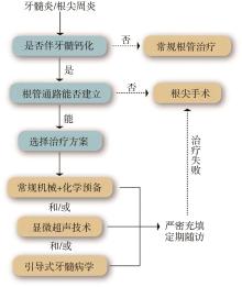
牙髓钙化是牙髓组织的一种增龄性变化或病理性反应,当牙髓病或根尖周病伴有牙髓钙化时,根管治疗是其最主要的治疗方式。临床治疗中,钙化根管往往因其疏通困难,难以建立准确的髓腔通路,易发生根管侧穿、台阶形成和器械分离等并发症,导致治疗失败率高,给临床医生带来巨大的困难。近年来,随着显微超声技术及引导式牙髓病学在钙化根管治疗中的应用及推广,钙化根管治疗的成功率大幅提升。本文通过文献回顾,对牙髓钙化的病因、诊断及治疗策略等方面进行详细阐述,以期为临床医生在钙化根管的治疗策略和方案的选择上提供帮助。
中图分类号:
| 1 | Carvalho TS, Lussi A. Age-related morphological, histological and functional changes in teeth[J]. J Oral Rehabil, 2017, 44(4): 291-298. |
| 2 | Jannati R, Afshari M, Moosazadeh M, et al. Pre-valence of pulp stones: a systematic review and meta-analysis[J]. J Evid Based Med, 2019, 12(2): 133-139. |
| 3 | 于世凤. 口腔组织病理学[M]. 7版. 北京: 人民卫生出版社, 2012: 181-192. |
| Yu SF. Oral histopathology[M]. 7th ed. Beijing: Peo-ple’s Medical Publishing House, 2012: 181-192. | |
| 4 | Mello-Moura AC, Bonini GA, Zardetto CG, et al. Pulp calcification in traumatized primary teeth: prevalence and associated factors[J]. J Clin Pediatr Dent, 2011, 35(4): 383-387. |
| 5 | Malhotra N, Mala K. Calcific metamorphosis. Li-terature review and clinical strategies[J]. Dent Update, 2013, 40(1): 48-50, 53-54, 57-58. |
| 6 | 史瑞棠, 侯本祥. 牙髓钙化的病因、诊断和治疗策略[J]. 中华口腔医学杂志, 2022, 57(3): 220-226. |
| Shi RT, Hou BX. Causes, diagnosis and treatment strategies for dental pulp calcification[J]. Chin J Stomatol, 2022, 57(3): 220-226. | |
| 7 | Tang L, Sun TQ, Gao XJ, et al. Tooth anatomy risk factors influencing root canal working length accessibility[J]. Int J Oral Sci, 2011, 3(3): 135-140. |
| 8 | Yang YM, Guo B, Guo LY, et al. CBCT-aided microscopic and ultrasonic treatment for upper or middle thirds calcified root canals[J]. Biomed Res Int, 2016, 2016: 4793146. |
| 9 | Aslantas EE, Buzoglu HD, Karapinar SP, et al. Age-related changes in the alkaline phosphatase activity of healthy and inflamed human dental pulp[J]. J Endod, 2016, 42(1): 131-134. |
| 10 | Gulsahi A, Cebeci AI, Özden S. A radiographic assessment of the prevalence of pulp stones in a group of Turkish dental patients[J]. Int Endod J, 2009, 42(8): 735-739. |
| 11 | 张文萍, 陈瑞扬. 牙髓钙化形成的原因探讨[J]. 临床口腔医学杂志, 2008, 24(5): 272-274. |
| Zhang WP, Chen RY. Analysis of the causes of the pulp calcification formation[J]. Clin J Stomatol, 2008, 24(5): 272-274. | |
| 12 | McCabe PS, Dummer PM. Pulp canal obliteration: an endodontic diagnosis and treatment challenge[J]. Int Endod J, 2012, 45(2): 177-197. |
| 13 | Andreasen FM, Kahler B. Pulpal response after acute dental injury in the permanent dentition: clinical implications-a review[J]. J Endod, 2015, 41(3): 299-308. |
| 14 | Huang LG, Chen G. A histological and radiographic study of pulpal calcification in periodontally in-volved teeth in a Taiwanese population[J]. J Dent Sci, 2016, 11(4): 405-410. |
| 15 | Nissrin B, Basma R, Majid S. Association between periodontitis and pulp calcifications: radiological study[J]. Int J Dent, 2022, 2022: 9599554. |
| 16 | Sabeti M, Tayeed H, Kurtzman G, et al. Histo-pathological investigation of dental pulp reactions related to periodontitis[J]. Eur Endod J, 2021, 6(2): 164-169. |
| 17 | Youssef AR, Emara R, Taher MM, et al. Effects of mineral trioxide aggregate, calcium hydroxide, biodentine and emdogain on osteogenesis, odon-togenesis, angiogenesis and cell viability of dental pulp stem cells[J]. BMC Oral Health, 2019, 19(1): 133. |
| 18 | Okabe T, Sakamoto M, Takeuchi H, et al. Effects of pH on mineralization ability of human dental pulp cells[J]. J Endod, 2006, 32(3): 198-201. |
| 19 | Almutairi W, Al-Dahman Y, Alnassar F, et al. In-tracanal calcification following regenerative endo-dontic treatment: a systematic review and meta-analysis[J]. Clin Oral Investig, 2022, 26(4): 3333-3342. |
| 20 | Zaen El-Din AM, Hamama HH, Abo El-Elaa MA, et al. The effect of four materials on direct pulp capping: an animal study[J]. Aust Endod J, 2020, 46(2): 249-256. |
| 21 | Tassoker M, Magat G, Sener S. A comparative study of cone-beam computed tomography and digital panoramic radiography for detecting pulp stones[J]. Imaging Sci Dent, 2018, 48(3): 201-212. |
| 22 | Fleig S, Attin T, Jungbluth H. Narrowing of the radicular pulp space in coronally restored teeth[J]. Clin Oral Investig, 2017, 21(4): 1251-1257. |
| 23 | da Silva EJNL, Prado MC, Queiroz PM, et al. Assessing pulp stones by cone-beam computed tomography[J]. Clin Oral Investig, 2017, 21(7): 2327-2333. |
| 24 | Vitali FC, Cardoso IV, Mello FW, et al. Association between orthodontic force and dental pulp changes: a systematic review of clinical and radiographic outcomes[J]. J Endod, 2022, 48(3): 298-311. |
| 25 | Srivastava KC, Shrivastava D, Nagarajappa AK, et al. Assessing the prevalence and association of pulp stones with cardiovascular diseases and diabetes mellitus in the Saudi Arabian population-a CBCT based study[J]. Int J Environ Res Public Health, 2020, 17(24): 9293. |
| 26 | Alsamahi S, Milne TM, Hussaini H, et al. Type 2 diabetes and the clinically normal pulp: an in vitro study[J]. Int Endodontic J, 2022, 55(6): 660-671. |
| 27 | Inagaki Y, Yoshida K, Ohba H, et al. High glucose levels increase osteopontin production and patho-logic calcification in rat dental pulp tissues[J]. J Endod, 2010, 36(6): 1014-1020. |
| 28 | Nakajima Y, Inagaki Y, Hiroshima Y, et al. Ad-vanced glycation end-products enhance calcifica-tion in cultured rat dental pulp cells[J]. J Endod, 2013, 39(7): 873-878. |
| 29 | Sugiyama K, Miura J, Shimizu M, et al. Effects of advanced glycation end products on dental pulp calcification[J]. Oral Dis, 2022, 28(3): 745-755. |
| 30 | Nayak M, Kumar J, Prasad LK. A radiographic correlation between systemic disorders and pulp stones[J]. Indian J Dent Res, 2010, 21(3): 369-373. |
| 31 | Ezoddini-Ardakani F, Nemayandeh SM, Sadrbafghi SM, et al. Diagnostic value of dental pulp stones in the early diagnosis of ischemic heart diseases[J]. Health, 2015, 7(3): 336-345. |
| 32 | Babu J, Swarnalatha C, Rao P, et al. Pulp stones as risk predictors for coronary artery disease[J]. Int J Prev Med, 2020, 11(1): 7. |
| 33 | Pettiette MT, Zhong S, Moretti AJ, et al. Potential correlation between statins and pulp chamber calcification[J]. J Endod, 2013, 39(9): 1119-1123. |
| 34 | American Academy of Pediatric Dentistry. Guide-line on dental management of heritable dental de-velopmental anomalies[J]. Pediatr Dent, 2013, 35(5): E179-E184. |
| 35 | Amler MH. Pulp calcification as a possible factor in trigeminal neuralgia: a case report[J]. N Y State Dent J, 1986, 52(1): 32-33. |
| 36 | Patel S. New dimensions in endodontic imaging: part 2. cone beam computed tomography[J]. Int Endod J, 2009, 42(6): 463-475. |
| 37 | McClammy TV. Endodontic applications of cone beam computed tomography[J]. Dent Clin North Am, 2014, 58(3): 545-559. |
| 38 | 徐琼, 刘建伟, 凌均棨. 手术显微镜配合超声技术处理钙化根管的临床疗效[J]. 中华口腔医学研究杂志(电子版), 2011, 5(1): 52-57. |
| Xu Q, Liu JW, Ling JQ. Clinical effect of calcified root canals with ultrasonic instruments under dental operating microscope[J]. Chin J Stomatol Res (Electr Ed), 2011, 5(1): 52-57. | |
| 39 | 赵隽隽, 周卓君, 韩俊力, 等. Mtwo 镍钛器械在钙化阻塞根管治疗中的应用[J]. 上海口腔医学, 2016, 25(2): 204. |
| Zhao JJ, Zhou ZJ, Han JL, et al. Clinical mana-gement of calcified root canal with Mtwo NiTi files[J]. Shanghai J Stomatol, 2016, 25(2): 204. | |
| 40 | Sayin TC, Serper A, Cehreli ZC, et al. Calcium loss from root canal dentin following EDTA, EGTA, EDTAC, and tetracycline-HCl treatment with or without subsequent NaOCl irrigation[J]. J Endod, 2007, 33(5): 581-584. |
| 41 | Sato T, Fujimaki R, Suzuki J, et al. Bactericidal effect of a novel alkaline EDTA root canal cleaning solution[J]. Eur J Dent, 2021, 15(3): 546-550. |
| 42 | Chu T, Ni XF, Zhu YH. EDTA combined with C-pilot files and microultrasound for root canal calcification: dredging effect and safety analysis[J]. Comput Math Methods Med, 2022, 2022: 1911448. |
| 43 | Kumar R. Report of a rare case: a maxillary first molar with seven canals confirmed with cone-beam computed tomography[J]. Iran Endod J, 2014, 9(2): 153-157. |
| 44 | Lim A, le Clerc J. Endodontic treatment of a hypertaurodontic mandibular left second molar in a patient with many taurodonts combined with multi-ple pulp stones[J]. Aust Endod J, 2019, 45(3): 414-419. |
| 45 | Robinson JP, Macedo RG, Verhaagen B, et al. Cleaning lateral morphological features of the root canal: the role of streaming and cavitation[J]. Int Endod J, 2018, 51(): e55-e64. |
| 46 | Ashwinkumar V, Nandini S, Velmurugan N. Endo-dontic management of three-canalled mandibular lateral incisor using dental operating microscope[J]. J Dent (Tehran), 2014, 11(4): 490-494. |
| 47 | 毛学理, 凌均棨, 林正梅, 等. 锥形束CT结合显微超声技术处理钙化根管的效果评价[J]. 中华口腔医学研究杂志(电子版), 2011, 5(6): 594-599. |
| Mao XL, Ling JQ, Lin ZM, et al. Evaluation of cone beam computed tomography combining with dental microscopy and ultrasonic instrument in the treatment of canal calcification[J]. Chin J Stomatol Res (Electr Ed), 2011, 5(6): 594-599. | |
| 48 | Aydemir S, Cimilli H, Mumcu G, et al. Crack formation on resected root surfaces subjected to conventional, ultrasonic, and laser root-end cavity preparation[J]. Photomed Laser Surg, 2014, 32(6): 351-355. |
| 49 | Krastl G, Zehnder MS, Connert T, et al. Guided endodontics: a novel treatment approach for teeth with pulp canal calcification and apical pathology[J]. Dent Traumatol, 2016, 32(3): 240-246. |
| 50 | Torres A, Shaheen E, Lambrechts P, et al. Micro-guided endodontics: a case report of a maxillary lateral incisor with pulp canal obliteration and apical periodontitis[J]. Int Endod J, 2019, 52(4): 540-549. |
| 51 | Connert T, Krug R, Eggmann F, et al. Guided endodontics versus conventional access cavity preparation: a comparative study on substance loss using 3-dimensional-printed teeth[J]. J Endod, 2019, 45(3): 327-331. |
| 52 | Todd R, Resnick S, Zicarelli T, et al. Template-guided endodontic access[J]. J Am Dent Assoc, 2021, 152(1): 65-70. |
| 53 | Kaur G, Venkatesh KV, Sihivahanan D. Micro-guided endodontics: a case report of conservative approach for the management of calcified maxillary lateral incisors[J]. Saudi Endod J, 2021, 11(2): 266. |
| 54 | Shi XL, Zhao SY, Wang WD, et al. Novel na-vigation technique for the endodontic treatment of a molar with pulp canal calcification and apical pathology[J]. Aust Endod J, 2018, 44(1): 66-70. |
| 55 | Dianat O, Gupta S, Price JB, et al. Guided endo-dontic access in a maxillary molar using a dynamic navigation system[J]. J Endod, 2021, 47(4): 658-662. |
| 56 | Zehnder MS, Connert T, Weiger R, et al. Guided endodontics: accuracy of a novel method for guided access cavity preparation and root canal location[J]. Int Endod J, 2016, 49(10): 966-972. |
| 57 | Connert T, Zehnder MS, Weiger R, et al. Micro-guided endodontics: accuracy of a minia-turized technique for apically extended access cavity pre-paration in anterior teeth[J]. J Endod, 2017, 43(5): 787-790. |
| 58 | Peña-Bengoa F, Valenzuela M, Flores MJ, et al. Effectiveness of guided endodontics in locating calcified root canals: a systematic review[J]. Clin Oral Investig, 2023, 27(5): 2359-2374. |
| 59 | Torres A, Boelen GJ, Lambrechts P, et al. Dynamic navigation: a laboratory study on the accuracy and potential use of guided root canal treatment[J]. Int Endod J, 2021, 54(9): 1659-1667. |
| 60 | Jain SD, Carrico CK, Bermanis I. 3-dimensional accuracy of dynamic navigation technology in lo-cating calcified canals[J]. J Endod, 2020, 46(6): 839-845. |
| 61 | Gambarini G, Galli M, Stefanelli LV, et al. Endo-dontic microsurgery using dynamic navigation sys-tem: a case report[J]. J Endod, 2019, 45(11): 1397-1402.e6. |
| 62 | Giacomino CM, Ray JJ, Wealleans JA. Targeted endodontic microsurgery: a novel approach to anato-mically challenging scenarios using 3-dimensional-printed guides and trephine burs-a report of 3 cases[J]. J Endod, 2018, 44(4): 671-677. |
| 63 | Fu WT, Chen C, Bian Z, et al. Endodontic micro-surgery of posterior teeth with the assistance of dynamic navigation technology: a report of three cases[J]. J Endod, 2022, 48(7): 943-950. |
| 64 | Peng L, Zhao J, Wang ZH, et al. Accuracy of root-end resection using a digital guide in endodontic surgery: an in vitro study[J]. J Dent Sci, 2021, 16(1): 45-50. |
| [1] | 卢妍,吴宾,闫卉. 4种根管预备系统根尖推出物和冠方带出物的比较研究[J]. 国际口腔医学杂志, 2019, 46(5): 503-508. |
| [2] | 吴杉杉, 张茹, 侯本祥. 钙化根管的诊断与治疗[J]. 国际口腔医学杂志, 2017, 44(3): 279-283. |
| [3] | 赵旭,李玉娇,刘莉,赵洪岩,张志民. 根管初步预备器械的研究现状[J]. 国际口腔医学杂志, 2017, 44(1): 75-78. |
| [4] | 朱玉婷 刘江峰 黄江勇 徐妍 李艳利 李晓星 陈秉勋. 控制记忆型镍钛锉HyFlex CM根管预备的临床效果分析[J]. 国际口腔医学杂志, 2014, 41(5): 521-525. |
| [5] | 赵朋朋 秦宗长. 根管治疗与根管壁微裂[J]. 国际口腔医学杂志, 2014, 41(4): 478-482. |
| [6] | 刘昭慧 凌均棨. 三维自调节根管预备锉[J]. 国际口腔医学杂志, 2014, 41(3): 320-323. |
| [7] | 蔡雪 王晓燕. 根管超声冲洗效果的影响因素[J]. 国际口腔医学杂志, 2014, 41(1): 72-76. |
| [8] | 毛姣姣综述 林正梅审校. 根管冲洗方法的研究进展[J]. 国际口腔医学杂志, 2012, 39(5): 635-638. |
| [9] | 张晶1综述 刘丽2审校. 根管治疗与根折的关系[J]. 国际口腔医学杂志, 2012, 39(4): 501-505. |
| [10] | 李姝慧 邱志远. 妊娠期牙髓炎一次性根管预备的疗效观察[J]. 国际口腔医学杂志, 2012, 39(1): 43-45. |
| [11] | 杨利杰综述 吴红崑审校. 根管成形评价方法的研究进展[J]. 国际口腔医学杂志, 2010, 37(5): 562-565. |
| [12] | 郑庆华综述 李文, 黄定明审校. 恒牙根管弯曲的解剖学特征[J]. 国际口腔医学杂志, 2010, 37(3): 317-317~319. |
| [13] | 张健综述 葛久禹, 孙卫斌审校. Mtwo 镍钛器械的研究进展[J]. 国际口腔医学杂志, 2009, 36(5): 607-609. |
| [14] | 邵美瑛, 郑广宁, 胡涛. 牛牙症根管治疗1 例和文献综述[J]. 国际口腔医学杂志, 2009, 36(3): 288-290. |
| [15] | 李艳, 赵今, 孙玉亮, 牛巧丽. 机用镍钛器械ProTaper 用于再治疗根管预备效果的临床评价[J]. 国际口腔医学杂志, 2009, 36(3): 267-269. |
|