国际口腔医学杂志 ›› 2019, Vol. 46 ›› Issue (4): 387-392.doi: 10.7518/gjkq.2019070
颜丹1,张锡忠2,王建国2( )
)
Yan Dan1,Zhang Xizhong2,Wang Jianguo2()
摘要:
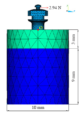
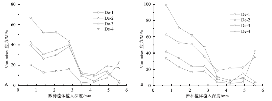
目的 分析螺纹深度变化对微种植体及颌骨上的应力分布和位移的影响,为支抗微种植体的临床选择和优化设计提供理论参照。方法 运用Pro/E软件建立不同螺纹深度的微种植体和下颌骨三维有限元模型,用Hypermesh软件对该模型进行网格划分,并在微种植体顶部施加与颌骨面平行的正畸力2.94 N,用ANSYS软件进行三维模拟计算。结果 微种植体-骨界面应力峰值集中在骨密质内,当螺纹深度为0.3 mm时,微种植体颈部及其相邻颌骨面的Von-mises应力峰值最小,为18.12 MPa;螺纹深度为0.4 mm时,应力峰值最大,为60.28 MPa。结论 螺纹深度影响微种植体和颌骨的应力分布,在本实验条件下,螺纹深度为0.3 mm时,微种植体-骨界面、微种植体上的应力和位移分布最优。
中图分类号:
| 5 | 参考文献 |
| [1] | 邵雯婷, 王学金 . 正畸微种植体支抗稳定性研究方法现状[J]. 口腔医学研究, 2016,32(10):1106-1109. |
| Shao WT, Wang XJ . Status of research methods on the stability of micro-implants as orthodontic anchorage[J]. J Oral Sci Res, 2016,32(10):1106-1109. | |
| [2] | Favero L, Giagnorio C, Cocilovo F . Comparative analysis of anchorage systems for micro implant orthodontics[J]. Prog Orthod, 2010,11(2):105-117. |
| [3] | 林东, 林珊, 陈江 , 等. 不同螺纹深度微小种植体的生物力学三维有限元研究[J]. 福建医科大学学报, 2009,43(5):381-383. |
| Lin D, Lin S, Chen J , et al. A three-dimensional finite element analysis on the influence of different depth of thread on biomechanical properties of micro-im-plant-bone interface[J]. J Fujian Med Univ, 2009,43(5):381-383. | |
| [4] | 颜丹, 王建国, 张锡忠 . 不同骨内段直径对支抗微种植体和颌骨表面影响的三维有限元分析[J]. 天津医药, 2013,41(1):22-25, 97. |
| Yan D, Wang JG, Zhang XZ . Three-dimensional finite element analysis for the influence of micro-implant endosteal diameter on micro-implant and surface of mandibular[J]. Tianjin Med J, 2013,41(1):22-25, 97. | |
| [5] | Tada S, Stegaroiu R, Kitamura E , et al. Influence of implant design and bone quality on stress/strain distribution in bone around implants: a 3-dimensional finite element analysis[J]. Int J Oral Maxillofac Implants, 2003,18(3):357-368. |
| [6] | Duaibis R, Kusnoto B, Natarajan R , et al. Factors affecting stresses in cortical bone around miniscrew implants: a three-dimensional finite element study[J]. Angle Orthod, 2012,82(5):875-880. |
| [7] | Sebbar M, Bourzgui F, Badre L , et al. Anchorage miniscrews: a histologic study of peri-implant soft tissue[J]. Int Orthod, 2012,10(1):85-95. |
| [8] | Abuhussein H, Pagni G, Rebaudi A , et al. The effect of thread pattern upon implant osseointegration[J]. Clin Oral Implants Res, 2010,21(2):129-136. |
| [9] |
Chowdhary R, Halldin A, Jimbo R , et al. Evaluation of stress pattern generated through various thread designs of dental implants loaded in a condition of immediately after placement and on osseointegration: an FEA study[J]. Implant Dent, 2013,22(1):91-96.
doi: 10.1097/ID.0b013e31827daf55 |
| [10] | Mischkowski RA, Kneuertz P, Florvaag B , et al. Bio-mechanical comparison of four different miniscrew types for skeletal anchorage in the mandibulo-maxil-lary area[J]. Int J Oral Maxillofac Surg, 2008,37(10):948-954. |
| [11] | 周栾慧, 杨四维, 黄跃 , 等. 植入角度及深度对种植体支抗稳定性影响的三维有限元研究[J]. 口腔医学, 2010,30(8):488-490. |
| Zhou LH, Yang SW, Huang Y , et al. 3D-FE study of implant anchorage stability under effects of inserting angulation and depth[J]. Stomatology, 2010,30(8):488-490. | |
| [12] | Lin TS, Tsai FD, Chen CY , et al. Factorial analysis of variables affecting bone stress adjacent to the orthodontic anchorage mini-implant with finite ele-ment analysis[J]. Am J Orthod Dentofacial Orthop, 2013,143(2):182-189. |
| [13] | 曾婷艳, 黄生高 . 种植体支抗稳定性的三维有限元分析[J]. 国际口腔医学杂志, 2018,45(1):112-118. |
| Zeng TY, Huang SG . A three-dimensional finite ele-ment analysis for stability of mini-implant anchorage[J]. Int J Stomatol, 2018,45(1):112-118. |
| [1] | 孙晓倩, 张军. 机械力环境影响头颈癌生物学行为及作用机制的研究进展[J]. 国际口腔医学杂志, 2023, 50(4): 414-418. |
| [2] | 黄依欢,李委航,马典,陈瑾,钱捷,李旭东. IPS e.maxCAD和Lava Ultimate在 贴面修复中的有限元分析[J]. 国际口腔医学杂志, 2023, 50(4): 423-432. 贴面修复中的有限元分析[J]. 国际口腔医学杂志, 2023, 50(4): 423-432. |
| [3] | 方苓力,谭玺,叶雨丝,黄兰,何瑶. 颞下颌关节退行性变早期髁突软骨细胞行为改变的实验研究[J]. 国际口腔医学杂志, 2021, 48(4): 417-425. |
| [4] | 孟秀萍,侯建华,李怡然,孙梦瑶. 龈壁提升术材料选择及边缘设计的研究进展[J]. 国际口腔医学杂志, 2021, 48(3): 280-286. |
| [5] | 季梦真,漆美瑶,杜珂芯,全淑琪,张煜强,郑庆华. 开髓洞型对全冠修复后隐裂牙抗力影响的三维有限元研究[J]. 国际口腔医学杂志, 2021, 48(1): 41-49. |
| [6] | 李静雅,税钰森,郭永文. 循环牵张应力影响人牙周膜细胞成骨分化机制的研究进展[J]. 国际口腔医学杂志, 2020, 47(6): 652-660. |
| [7] | 陈昕,毛渤淳,鲁雨晴,董博,朱卓立,岳莉,于海洋. 钴铬合金和聚醚醚酮用于可摘局部义齿支架的三维有限元分析[J]. 国际口腔医学杂志, 2019, 46(5): 526-531. |
| [8] | 黄璐,钱捷. 三维有限元在嵌体修复中的研究进展[J]. 国际口腔医学杂志, 2018, 45(6): 728-733. |
| [9] | 曾婷艳, 黄生高. 种植体支抗稳定性的三维有限元分析[J]. 国际口腔医学杂志, 2018, 45(1): 112-118. |
| [10] | 张晓, 邓青完, 杜琼, 谢静. 主桩辅桩联合修复对前磨牙应力的有限元分析[J]. 国际口腔医学杂志, 2017, 44(5): 559-565. |
| [11] | 曹国庆, 王林霞, 杜莉平. 有限元法在桩核冠修复研究中的应用[J]. 国际口腔医学杂志, 2017, 44(2): 209-213. |
| [12] | 赵静子1 张文君2 张晓东2. 上颌后牙区正畸微种植体支抗植入安全区的测量研究[J]. 国际口腔医学杂志, 2015, 42(6): 659-663. |
| [13] | 颜丹,张锡忠,王增全,王建国,关泽建. 螺距对支抗微种植体—骨界面影响的三维有限元分析[J]. 国际口腔医学杂志, 2015, 42(5): 557-561. |
| [14] | 关卿 金涛 顾永春 杨犇 倪龙兴. 3种根管预备器械在弯曲根管中扭转负载下的三维有限元分析[J]. 国际口腔医学杂志, 2015, 42(3): 269-272. |
| [15] | 张强1 李英2. Ⅱ类骨质中平台转换种植体植入深度对周围骨应力影响的有限元分析[J]. 国际口腔医学杂志, 2014, 41(1): 31-35. |
|