国际口腔医学杂志 ›› 2019, Vol. 46 ›› Issue (2): 209-212.doi: 10.7518/gjkq.2019002
曹焜,李家锋( ),孙玉华,鲍强,卢秋宁,唐巍
Kun Cao,Jiafeng Li(),Yuhua Sun,Qiang Bao,Qiuning Lu,Wei Tang
摘要:
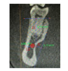
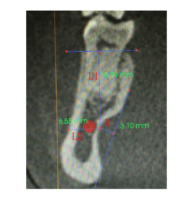
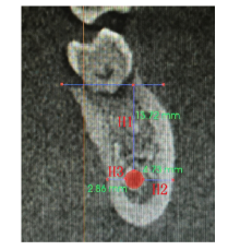
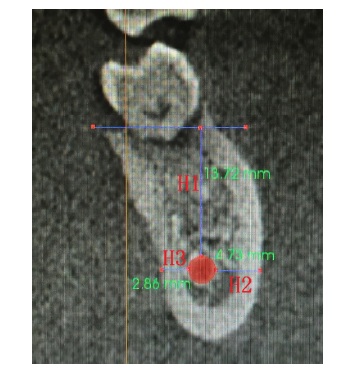
目的 通过锥形束CT(CBCT)测量分析下颌骨下颌下窝的形态,为牙种植术提供一定的参考。方法 对100例CBCT影像中下颌下窝骨倒凹的分布规律进行观察分析,测量下颌下窝深度、骨倒凹最深点至牙槽嵴顶和颊侧骨壁的距离,分析其与下颌管的毗邻关系。结果 下颌下窝位于下颌磨牙区,大部分下颌下窝骨倒凹最深点低于下颌管上缘;第一磨牙区下颌下窝骨倒凹深度为(1.2±0.73)mm,第二磨牙区为(1.7±0.54)mm,性别、左右及是否有牙的差异无统计学意义。第一磨牙区下颌下窝最深点至牙槽嵴顶的垂直距离在有牙和无牙时分别为(17.2±2.16)mm和(13.1±2.71)mm,至颊侧骨壁的水平距离为(10.1±2.07)mm,第二磨牙区下颌下窝最深点至牙槽嵴顶的垂直距离在有牙和无牙时分别为(15.3±2.43)mm和(9.6±2.56)mm,至颊侧骨壁的水平距离为(11.3±1.53)mm。结论 在下颌磨牙区行种植手术时,应综合考虑下颌下窝和下颌管对手术的影响,CBCT可以实现对下颌下窝的精确测量,能够清晰显示下颌下窝与下颌管的位置关系,为下颌磨牙区种植手术提供重要信息。
中图分类号:
| [1] |
Chan HL, Benavides E, Yeh CY , et al. Risk assess-ment of lingual plate perforation in posterior mandi-bular region: a virtual implant placement study using cone-beam computed tomography[J]. J Periodontol, 2011,82(1):129-135.
doi: 10.1902/jop.2010.100313 pmid: 20653440 |
| [2] |
Parnia F, Fard EM, Mahboub F , et al. Tomographic volume evaluation of submandibular fossa in patients requiring dental implants[J]. Oral Surg Oral Med Oral Pathol Oral Radiol Endod, 2010,109(1):e32-e36.
doi: 10.1016/j.tripleo.2009.08.035 pmid: 20123366 |
| [3] |
Yang J, Cavalcanti MG, Ruprecht A , et al. 2-D and 3-D reconstructions of spiral computed tomography in localization of the inferior alveolar canal for den-tal implants[J]. Oral Surg Oral Med Oral Pathol Oral Radiol Endod, 1999,87(3):369-374.
doi: 10.1016/S1079-2104(99)70226-X pmid: 10102603 |
| [4] | 韩科 . 种植义齿[M]. 北京: 人民军医出版社, 2007: 180. |
| Han K. Implant denture[M]. Beijing: People’s Mili-tary Medical Press, 2007: 180. | |
| [5] |
李立峰, 平飞云 . 成人下颌骨舌下腺窝形态的分析研究[J]. 实用口腔医学杂志, 2012,28(5):589-591.
doi: 10.3969/j.issn.1001-3733.2012.05.12 |
|
Li LF, Ping YF . The morphological analysis of sub-lingual gland fossa on in-vitro adult mandibles[J]. J Pract Stomatol, 2012,28(5):589-591.
doi: 10.3969/j.issn.1001-3733.2012.05.12 |
|
| [6] |
Scarfe WC, Farman AG, Sukovic P . Clinical applica-tions of cone-beam computed tomography in dental practice[J]. J Can Dent Assoc, 2006,72(1):75-80.
doi: 10.1016/j.prosdent.2005.11.018 pmid: 16480609 |
| [7] |
Benninger B, Peterson A, Cook V . Assessing validity of actual tooth height and width from cone beam images of cadavers with subsequent dissection to aid oral surgery[J]. J Oral Maxillofac Surg, 2012,70(2):302-306.
doi: 10.1016/j.joms.2011.03.014 pmid: 21684648 |
| [8] |
Sumer AP, Zengin AZ, Uzun C , et al. Evaluation of submandibular fossa using computed tomography and panoramic radiography[J]. Oral Radiol, 2015,31(1):23-27.
doi: 10.1007/s11282-014-0175-3 |
| [9] |
Yildiz S, Bayar GR, Guvenc I , et al. Tomographic evaluation on bone morphology in posterior mandi-bular region for safe placement of dental implant[J]. Surg Radiol Anat, 2015,37(2):167-173.
doi: 10.1007/s00276-014-1351-1 pmid: 25078676 |
| [10] |
Park CW, Park KB, Park KP , et al. Bucco-lingual implant path in the posterior mandible[J]. Korean J Phys Anthropol. 1998,11(1):73-81.
doi: 10.11637/kjpa.1998.11.1.73 |
| [11] |
Kim SE, Kim JS, Kim JD . Comparison of the buccolingual inclination in alveolar bone and tooth using dental CBCT[J]. Korean J Oral Maxillofac Radiol, 2008,38(1):17-22.
doi: 10.1016/j.irfa.2015.08.001 |
| [12] |
Güler AU, Sumer M, Sumer P , et al. The evaluation of vertical heights of maxillary and mandibular bones and the location of anatomic landmarks in panoramic radiographs of edentulous patients for implant dentistry[J]. J Oral Rehabil, 2005,32(10):741-746.
doi: 10.1111/j.1365-2842.2005.01499.x pmid: 16159352 |
| [13] |
de Souza LA, Souza Picorelli Assis NM, Ribeiro RA , et al. Assessment of mandibular posterior regional landmarks using cone-beam computed tomography in dental implant surgery[J]. Ann Anat, 2016,205:53-59.
doi: 10.1016/j.aanat.2016.01.006 pmid: 26851559 |
| [1] | 杨雨楠,刘鹏,王虎,游梦. 上颌窦黏膜增厚的锥形束CT影像分析[J]. 国际口腔医学杂志, 2023, 50(3): 302-307. |
| [2] | 陆倩,夏海斌,王敏. 种植体磨光整形术治疗种植体周围炎的研究进展[J]. 国际口腔医学杂志, 2023, 50(2): 152-158. |
| [3] | 吴文智,冯达兴,陈垂壮,周丽鹃. 海口地区下颌第一恒磨牙近中中央根管发生率及相关因素[J]. 国际口腔医学杂志, 2022, 49(4): 420-425. |
| [4] | 叶泽林,刘璐,龙虎,游梦. 弯曲前牙的影像评价及治疗的研究进展[J]. 国际口腔医学杂志, 2022, 49(2): 173-181. |
| [5] | 田浩楠,林敏,谢丛蔓,任嫒姝. 上颌腭侧阻生尖牙与寰椎后桥相关性的锥形束CT研究[J]. 国际口腔医学杂志, 2021, 48(5): 536-540. |
| [6] | 施丹妮,杨鑫,吴建勇. 锥形束CT三维头影测量参考坐标系的研究进展[J]. 国际口腔医学杂志, 2021, 48(4): 398-404. |
| [7] | 丁张帆,郭陟永,苗诚,李春洁,宣鸣,王晓毅,张壮. 基于锥形束CT的三维可视化技术在颌骨囊性病变手术中的应用[J]. 国际口腔医学杂志, 2021, 48(2): 180-186. |
| [8] | 王奔,许喆桢,韦曦. 数字化微创技术在牙髓根尖周病学中的应用与进展[J]. 国际口腔医学杂志, 2021, 48(1): 110-118. |
| [9] | 唐蓓,赵文俊,王虎,郑广宁,游梦. 根管超填导致下牙槽神经损伤2例[J]. 国际口腔医学杂志, 2020, 47(3): 293-296. |
| [10] | 章婷婷,胡常红,彭燕,周文翘,张慧聪,刘蝶. 300例不同年龄段有牙颌人群上唇软组织侧貌的锥形束CT三维测量分析[J]. 国际口腔医学杂志, 2020, 47(2): 182-188. |
| [11] | 王春林,刘从华,宋思吟,周丽淑,林丽佳. 运用锥形束CT诊断上下颌横向发育不调的研究进展[J]. 国际口腔医学杂志, 2020, 47(1): 121-124. |
| [12] | 黎祺, 黄少宏. 岭南地区广府民系人群下颌第二恒磨牙牙根和根管形态的锥形束CT研究[J]. 国际口腔医学杂志, 2019, 46(6): 640-649. |
| [13] | 于婉琦,周延民,赵静辉. 口腔种植体新材料的研究现状[J]. 国际口腔医学杂志, 2019, 46(4): 488-496. |
| [14] | 孟怡彤,张晓东. 成人个别正常颌上气道不同软件三维测量的比较研究[J]. 国际口腔医学杂志, 2018, 45(6): 690-694. |
| [15] | 徐迅, 黄建生, 甘泽坤, 罗震. 上颌第一磨牙区腭侧骨板的锥形束CT测量结果及其临床意义[J]. 国际口腔医学杂志, 2017, 44(6): 686-690. |
|