Int J Stomatol ›› 2024, Vol. 51 ›› Issue (2): 241-248.doi: 10.7518/gjkq.2024011
• Reviews • Previous Articles
Jiaping Si( ),Lü Lin,Sijie Wang,Yu Zhou,Xiaoyan Chen()
CLC Number:
| 1 | Yu X, Zhang H, Sun LY, et al. Prevalence of malocclusion and occlusal traits in the early mixed dentition in Shanghai, China[J]. PeerJ, 2019, 7: e6630. |
| 2 |
程锦, 刘冬梅, 曾照斌, 等. 辽宁地区成人深覆 患者颌面部硬组织形态特征的CBCT研究[J]. 实用口腔医学杂志, 2017, 33(3): 364-367. 患者颌面部硬组织形态特征的CBCT研究[J]. 实用口腔医学杂志, 2017, 33(3): 364-367.
|
| Chen J, Liu DM, Zeng ZB, et al. A CBCT study of the hard tissue morphology of the maxillofacial region in adult patients with deep overbite in Liaoning area[J]. J Pract Stomatol, 2017, 33(3): 364-367. | |
| 3 | Huang GJ, Bates SB, Ehlert AA, et al. Stability of deep-bite correction: a systematic review[J]. J World Fed Orthod, 2012, 1(3): e89-e96. |
| 4 |
陆史俊, 王林, 王震东. 前牙压低技术在深覆 患者矫治中的应用进展[J]. 国际口腔医学杂志, 2011, 38(6): 674-676, 680. 患者矫治中的应用进展[J]. 国际口腔医学杂志, 2011, 38(6): 674-676, 680.
|
| Lu SJ, Wang L, Wang ZD. Progress of anterior teeth intrusion in deep overbite treatment[J]. Int J Stomatol, 2011, 38(6): 674-676, 680. | |
| 5 | Rozzi M, Mucedero M, Pezzuto C, et al. Long-term stability of curve of Spee levelled with continuous archwires in subjects with different vertical patterns: a retrospective study[J]. Eur J Orthod, 2019, 41(3): 286-293. |
| 6 | Ghafari JG, Macari AT, Haddad RV. Deep bite: treatment options and challenges[J]. Semin Orthod, 2013, 19(4): 253-266. |
| 7 | Deguchi T, Murakami T, Kuroda S, et al. Comparison of the intrusion effects on the maxillary incisors between implant anchorage and J-hook headgear[J]. Am J Orthod Dentofacial Orthop, 2008, 133(5): 654-660. |
| 8 |
邓晓姝, 黄宁, 陈绪道, 等. 压低辅弓有效改正成人前牙深覆 的正畸治疗[J]. 国际口腔医学杂志, 2011, 38(4): 395-398. 的正畸治疗[J]. 国际口腔医学杂志, 2011, 38(4): 395-398.
|
| Deng XS, Huang N, Chen XD, et al. Intrusive auxi-liary arch can correct the anterior deep bite in adult patients effectively[J]. Int J Stomatol, 2011, 38(4): 395-398. | |
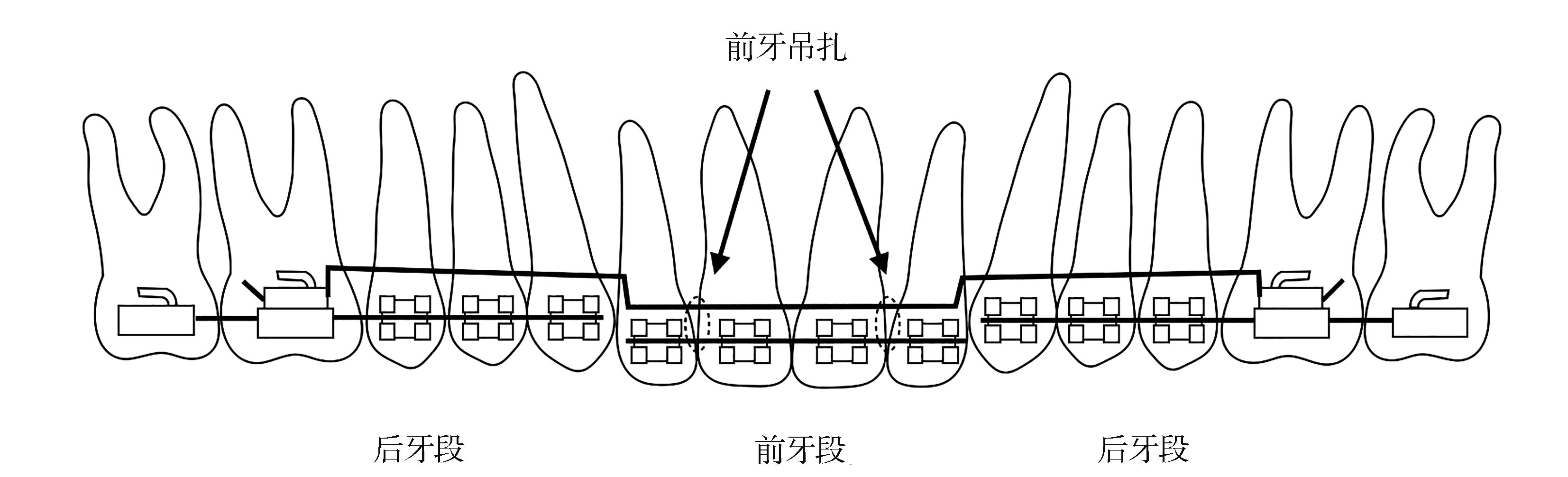
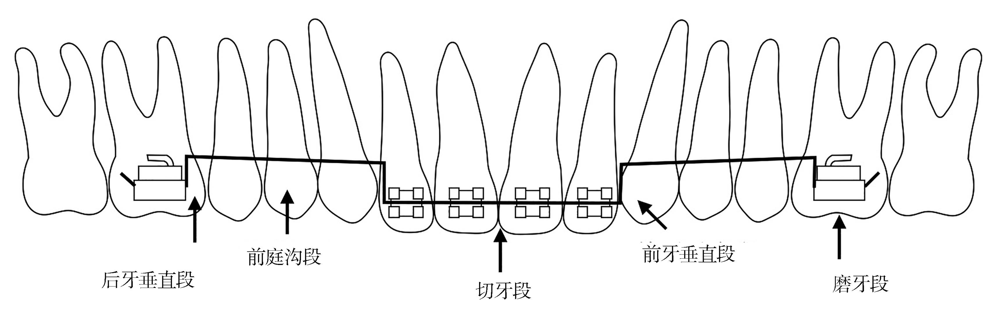
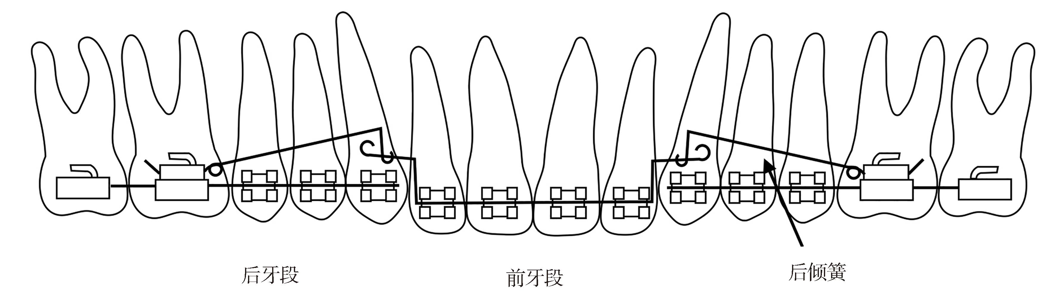
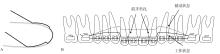
| 9 | Braun S, Marcotte MR. Rationale of the segmented approach to orthodontic treatment[J]. Am J Orthod Dentofacial Orthop, 1995, 108(1): 1-8. |
| 10 | Burstone CR. Deep overbite correction by intrusion[J]. Am J Orthod, 1977, 72(1): 1-22. |
| 11 | Ricketts RM. Bioprogressive therapy as an answer to orthodontic needs Part Ⅰ[J]. Am J Orthod, 1976, 70(3): 241-268. |
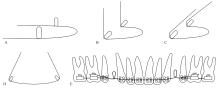
| 12 | Davidovitch M, Rebellato J. Two-couple orthodontic appliance systems utility arches: a two-couple intrusion arch[J]. Semin Orthod, 1995, 1(1): 25-30. |
| 13 | Andrews LF. The six keys to normal occlusion[J]. Am J Orthod, 1972, 62(3): 296-309. |
| 14 | Shroff B, Lindauer SJ, Burstone CJ, et al. Segmen-ted approach to simultaneous intrusion and space closure: biomechanics of the three-piece base arch appliance[J]. Am J Orthod Dentofacial Orthop, 1995, 107(2): 136-143. |
| 15 | Nanda R, Marzban R, Kuhlberg A. The connecticut intrusion arch[J]. J Clin Orthod, 1998, 32(12): 708-715. |
| 16 | Bagri K, Kannan S, Singh AK, et al. Orthodontic management of deep bite in adult: a review[J]. TMU J Dent, 2018, 5(2): 23-26. |
| 17 | Kalra V. Simultaneous intrusion and retraction of the anterior teeth[J]. J Clin Orthod, 1998, 32(9): 535-540. |
| 18 | Raje Batham P, Diana Pereira Kalia U, Ramchan-dani B. An innovative use of the K-SIR arch[J]. IP Indian J Orthod Dentofac Res, 2020, 4(3): 161-163. |
| 19 | Shakti P, Ani GS, Peter E, et al. Maxillary incisor intrusion using two conventional intrusion arches and mini implants: a prospective study[J]. J Contemp Dent Pract, 2021, 22(8): 907-913. |
| 20 | Goel P, Tandon R, Agrawal KK. A comparative study of different intrusion methods and their effect on maxillary incisors[J]. J Oral Biol Craniofac Res, 2014, 4(3): 186-191. |
| 21 | Sifakakis I, Pandis N, Makou M, et al. A comparative assessment of the forces and moments genera-ted with various maxillary incisor intrusion biomechanics[J]. Eur J Orthod, 2010, 32(2): 159-164. |
| 22 | Sharma S, Vora S, Pandey V. Clinical evaluation of efficacy of CIA and CNA intrusion arches[J]. J Clin Diagn Res, 2015, 9(9): ZC29-ZC33. |
| 23 | Schwertner A, Almeida RR, Gonini A Jr, et al. Photoelastic analysis of stress generated by Connecticut Intrusion Arch (CIA)[J]. Dental Press J Orthod, 2017, 22(1): 57-64. |
| 24 | Costopoulos G, Nanda R. An evaluation of root resorption incident to orthodontic intrusion[J]. Am J Orthod Dentofacial Orthop, 1996, 109(5): 543-548. |
| 25 | de Almeida MR, Marçal ASB, Fernandes TMF, et al. A comparative study of the effect of the intrusion arch and straight wire mechanics on incisor root resorption: a randomized, controlled trial[J]. Angle Orthod, 2018, 88(1): 20-26. |
| 26 | McFadden WM, Engstrom C, Engstrom H, et al. A study of the relationship between incisor intrusion and root shortening[J]. Am J Orthod Dentofacial Orthop, 1989, 96(5): 390-396. |
| 27 | Dermaut LR, De Munck A. Apical root resorption of upper incisors caused by intrusive tooth movement: a radiographic study[J]. Am J Orthod Dentofacial Orthop, 1986, 90(4): 321-326. |
| 28 | Matsumoto K, Sherrill-Mix S, Boucher N, et al. A cone-beam computed tomographic evaluation of alveolar bone dimensional changes and the periodontal limits of mandibular incisor advancement in ske-letal Class Ⅱ patients[J]. Angle Orthod, 2020, 90(3): 330-338. |
| 29 | Guo R, Zhang L, Hu M, et al. Alveolar bone chan-ges in maxillary and mandibular anterior teeth during orthodontic treatment: a systematic review and meta-analysis[J]. Orthod Craniofac Res, 2021, 24(2): 165-179. |
| 30 | Han JY, Jung GU. Labial and lingual/palatal bone thickness of maxillary and mandibular anteriors in human cadavers in Koreans[J]. J Periodontal Implant Sci, 2011, 41(2): 60-66. |
| 31 | Kaied IB, Tanielian RH. Comparative radiographic evaluation of the alveolar bone support changes after incisal intrusion[J]. Orthodontics (Chic), 2012, 13(1): 60-71. |
| 32 | Atik E, Gorucu-Coskuner H, Akarsu-Guven B, et al. Evaluation of changes in the maxillary alveolar bone after incisor intrusion[J]. Korean J Orthod, 2018, 48(6): 367-376. |
| 33 | Erkan M, Pikdoken L, Usumez S. Gingival response to mandibular incisor intrusion[J]. Am J Orthod Dentofacial Orthop, 2007, 132(2): 143.e9-143.e13. |
| 34 | Senışık NE, Türkkahraman H. Treatment effects of intrusion arches and mini-implant systems in deepbite patients[J]. Am J Orthod Dentofacial Orthop, 2012, 141(6): 723-733. |
| 35 | Verma P, Jain RK. Intrusion effects on maxillary anteriors using mini implant anchorage and K-sir loop in subjects with deep overbite-a cohort study[J]. J Clin Diag Res, 2020: 21-25. |
| 36 | Kale Varlık S, Onur Alpakan Ö, Türköz Ç. Deepbite correction with incisor intrusion in adults: a long-term cephalometric study[J]. Am J Orthod Dentofacial Orthop, 2013, 144(3): 414-419. |
| 37 | Dake ML, Sinclair PM. A comparison of the Ric-ketts and Tweed-type arch leveling techniques[J]. Am J Orthod Dentofacial Orthop, 1989, 95(1): 72-78. |