国际口腔医学杂志 ›› 2021, Vol. 48 ›› Issue (5): 536-540.doi: 10.7518/gjkq.2021071
田浩楠( ),林敏,谢丛蔓,任嫒姝()
Tian Haonan(),Lin Min,Xie Congman,Ren Aishu()
摘要:
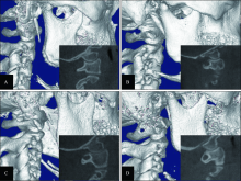
目的 利用锥形束CT(CBCT)研究上颌腭侧阻生尖牙患者寰椎后桥的发生情况及其相关性。方法 选取117例上颌尖牙腭侧阻生患者作为实验组,147例上颌尖牙正常萌出者作为对照组。通过CBCT影像结合Mimics 20.0三维测量软件观察寰椎后桥形态。采用SPSS 21.0软件进行数据统计分析。结果 实验组寰椎后桥发生率为35.04%,对照组寰椎后桥发生率为21.09%,组间寰椎后桥发生率的差异具有统计学意义(P<0.05)。组间单侧与双侧寰椎后桥发生率无明显差异(P>0.05)。实验组男性寰椎后桥发生率明显高于女性(P<0.05),而对照组性别无明显差异(P>0.05)。实验组中寰椎后桥1+2型的发生率高于对照组(P<0.05)。结论 上颌尖牙腭侧阻生患者寰椎后桥的发生率明显高于上颌尖牙正常萌出者,且男性上颌尖牙腭侧阻生患者发生寰椎后桥的概率更大。
中图分类号:
| [1] |
Bishara SE. Impacted maxillary canines: a review[J]. Am J Orthod Dentofacial Orthop, 1992, 101(2):159-171.
doi: 10.1016/0889-5406(92)70008-X |
| [2] |
Becker A, Chaushu S. Etiology of maxillary canine impaction: a review[J]. Am J Orthod Dentofacial Orthop, 2015, 148(4):557-567.
doi: 10.1016/j.ajodo.2015.06.013 |
| [3] |
Brin I, Becker A, Shalhav M. Position of the maxillary permanent canine in relation to anomalous or missing lateral incisors: a population study[J]. Eur J Orthod, 1986, 8(1):12-16.
doi: 10.1093/ejo/8.1.12 |
| [4] |
Peck S, Peck L, Kataja M. The palatally displaced canine as a dental anomaly of genetic origin[J]. Angle Orthod, 1994, 64(4):249-256.
pmid: 7978519 |
| [5] |
Haji Ghadimi M, Amini F, Hamedi S, et al. Associations among sella Turcica bridging, atlas arcuate foramen (ponticulus posticus) development, atlas posterior arch deficiency, and the occurrence of palatally displaced canine impaction[J]. Am J Orthod Dentofacial Orthop, 2017, 151(3):513-520.
doi: 10.1016/j.ajodo.2016.08.024 |
| [6] |
Leonardi R, Barbato E, Vichi M, et al. Skeletal ano-malies and normal variants in patients with palatally displaced canines[J]. Angle Orthod, 2009, 79(4):727-732.
doi: 10.2319/082408-448.1 pmid: 19537879 |
| [7] |
Matsuoka T, Ahlberg PE, Kessaris N, et al. Neural crest origins of the neck and shoulder[J]. Nature, 2005, 436(7049):347-355.
doi: 10.1038/nature03837 |
| [8] |
Miletich I, Sharpe PT. Neural crest contribution to mammalian tooth formation[J]. Birth Defects Res C Embryo Today, 2004, 72(2):200-212.
doi: 10.1002/(ISSN)1542-9768 |
| [9] |
Cederberg RA, Benson BW, Nunn M, et al. Arcuate foramen: prevalence by age, gender, and degree of calcification[J]. Clin Orthod Res, 2000, 3(3):162-167.
doi: 10.1034/j.1600-0544.2000.30309.x |
| [10] |
Tripodi D, Tieri M, Demartis P, et al. Ponticulus posticus: clinical and CBCT analysis in a young Italian population[J]. Eur J Paediatr Dent, 2019, 20(3):219-223.
doi: 10.23804/ejpd.2019.20.03.10 pmid: 31489822 |
| [11] | Bayrakdar IŞ, Miloğlu Ö, Yeşiltepe S, et al. Ponticulus posticus in a cohort of orthodontic children and adolescent patients with different sagittal skeletal ano-malies: a comparative cone beam computed tomography investigation[J]. Folia Morphol (Warsz), 2018, 77(1):65-71. |
| [12] | 刘安琪, 钱玉芬. 上颌尖牙阻生原因及其对周围结构的影响[J]. 上海口腔医学, 2017, 26(6):684-688. |
| Liu AQ, Qian YF. Features and influence of impacted maxillary permanent canine on surrounding structures[J]. Shanghai J Stomatol, 2017, 26(6):684-688. | |
| [13] |
Elliott RE, Tanweer O. The prevalence of the ponticulus posticus (arcuate foramen) and its importance in the Goel-Harms procedure: Meta-analysis and review of the literature[J]. World Neurosurg, 2014, 82(1/2):e335-e343.
doi: 10.1016/j.wneu.2013.09.014 |
| [14] |
Morotomi T, Kawano S, Toyono T, et al. In vitro differentiation of dental epithelial progenitor cells th-rough epithelial-mesenchymal interactions[J]. Arch Oral Biol, 2005, 50(8):695-705.
doi: 10.1016/j.archoralbio.2004.12.006 |
| [15] | 李璐, 陈智, 张露. 牙型发育的研究进展[J]. 武汉大学学报(医学版), 2008, 29(6):831-833. |
| Li L, Chen Z, Zhang L. Research advance in dental patterning[J]. Med J Wuhan Univ, 2008, 29(6):831-833. | |
| [16] | 陈澜月, 赵阳, 郑博文, 等. 腭侧埋伏阻生尖牙与鞍桥的相关性研究[J]. 口腔医学研究, 2017, 33(3):295-297. |
| Chen LY, Zhao Y, Zheng BW, et al. Study on the relevance of palatal impacted canine and sella bri-dges[J]. J Oral Sci Res, 2017, 33(3):295-297. | |
| [17] | 陈澜月, 牛磊, 陈艳娜, 等. 腭侧埋伏阻生尖牙与颈椎骨骼异常的相关性研究[J]. 上海口腔医学, 2017, 26(1):73-75. |
| Chen LY, Niu L, Chen YN, et al. Cervical skeletal abnormalities in patients with palatally displaced canine[J]. Shanghai J Stomatol, 2017, 26(1):73-75. | |
| [18] |
Geist JR, Geist SM, Lin LM. A cone beam CT investigation of ponticulus posticus and lateralis in children and adolescents[J]. Dentomaxillofac Radiol, 2014, 43(5):20130451.
doi: 10.1259/dmfr.20130451 |
| [19] |
Bebnowski D, Hänggi MP, Markic G, et al. Cervical vertebrae anomalies in subjects with ClassⅡmalocclusion assessed by lateral cephalogram and cone beam computed tomography[J]. Eur J Orthod, 2012, 34(2):226-231.
doi: 10.1093/ejo/cjq192 |
| [20] | 王嘉艺, 王珊, 王林. CBCT在口腔正畸学头影测量中的应用与发展[J]. 口腔医学, 2016, 36(11):1047-1050. |
| Wang JY, Wang S, Wang L. Application and deve-lopment of CBCT in the orthodontics cephalometrics[J]. Stomatology, 2016, 36(11):1047-1050. |
| [1] | 杨雨楠,刘鹏,王虎,游梦. 上颌窦黏膜增厚的锥形束CT影像分析[J]. 国际口腔医学杂志, 2023, 50(3): 302-307. |
| [2] | 吴文智,冯达兴,陈垂壮,周丽鹃. 海口地区下颌第一恒磨牙近中中央根管发生率及相关因素[J]. 国际口腔医学杂志, 2022, 49(4): 420-425. |
| [3] | 叶泽林,刘璐,龙虎,游梦. 弯曲前牙的影像评价及治疗的研究进展[J]. 国际口腔医学杂志, 2022, 49(2): 173-181. |
| [4] | 施丹妮,杨鑫,吴建勇. 锥形束CT三维头影测量参考坐标系的研究进展[J]. 国际口腔医学杂志, 2021, 48(4): 398-404. |
| [5] | 丁张帆,郭陟永,苗诚,李春洁,宣鸣,王晓毅,张壮. 基于锥形束CT的三维可视化技术在颌骨囊性病变手术中的应用[J]. 国际口腔医学杂志, 2021, 48(2): 180-186. |
| [6] | 王奔,许喆桢,韦曦. 数字化微创技术在牙髓根尖周病学中的应用与进展[J]. 国际口腔医学杂志, 2021, 48(1): 110-118. |
| [7] | 唐蓓,赵文俊,王虎,郑广宁,游梦. 根管超填导致下牙槽神经损伤2例[J]. 国际口腔医学杂志, 2020, 47(3): 293-296. |
| [8] | 章婷婷,胡常红,彭燕,周文翘,张慧聪,刘蝶. 300例不同年龄段有牙颌人群上唇软组织侧貌的锥形束CT三维测量分析[J]. 国际口腔医学杂志, 2020, 47(2): 182-188. |
| [9] | 王春林,刘从华,宋思吟,周丽淑,林丽佳. 运用锥形束CT诊断上下颌横向发育不调的研究进展[J]. 国际口腔医学杂志, 2020, 47(1): 121-124. |
| [10] | 黎祺, 黄少宏. 岭南地区广府民系人群下颌第二恒磨牙牙根和根管形态的锥形束CT研究[J]. 国际口腔医学杂志, 2019, 46(6): 640-649. |
| [11] | 曹焜,李家锋,孙玉华,鲍强,卢秋宁,唐巍. 下颌下窝的锥形束CT影像分析[J]. 国际口腔医学杂志, 2019, 46(2): 209-212. |
| [12] | 孟怡彤,张晓东. 成人个别正常颌上气道不同软件三维测量的比较研究[J]. 国际口腔医学杂志, 2018, 45(6): 690-694. |
| [13] | 徐迅, 黄建生, 甘泽坤, 罗震. 上颌第一磨牙区腭侧骨板的锥形束CT测量结果及其临床意义[J]. 国际口腔医学杂志, 2017, 44(6): 686-690. |
| [14] | 袁艺航, 张成晓雪, 王扬, 何双双, 宋雪娟, 王虎. 成都正常人群上颌前牙区鼻腭管相关解剖结构的锥形束CT研究[J]. 国际口腔医学杂志, 2017, 44(5): 566-572. |
| [15] | 吴杉杉, 张茹, 侯本祥. 钙化根管的诊断与治疗[J]. 国际口腔医学杂志, 2017, 44(3): 279-283. |
|