国际口腔医学杂志 ›› 2020, Vol. 47 ›› Issue (2): 182-188.doi: 10.7518/gjkq.2020050
章婷婷,胡常红,彭燕,周文翘,张慧聪,刘蝶( )
)
Zhang Tingting,Hu Changhong,Peng Yan,Zhou Wenqiao,Zhang Huicong,Liu Die()
摘要:
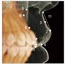
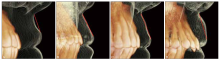
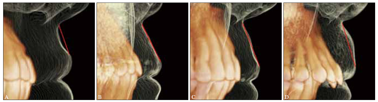
目的 通过锥形束CT(CBCT)进行三维测量,分析不同年龄段有牙颌人群上唇软组织侧貌特征,为无牙颌患者的全口义齿修复提供面部美学的客观参考依据。方法 选择300例有牙颌人群的CBCT影像资料进行研究,按照患者年龄分为青年组(20~39岁)、中年组(40~59岁)、老年组(60岁及以上),对上唇长度、厚度等相关指标进行测量,对上唇皮肤段侧面轮廓进行形态学分析。结果 上唇长度、厚度测量指标结果显示,随着年龄增加,上唇长度逐渐变大,唇红暴露量及唇红厚度逐渐减少,差异有统计学意义(P<0.05)。青年组、中年组和老年组上唇下缘到上中切牙切点的距离分别为(1.962 2±1.107 1)、(0.835 3±1.206 6)、(0.014 1±1.225 6)mm,上唇红部厚度分别为(12.355 8±1.950 3)、(10.634 2±1.782 4)、(9.924 9±1.951 4)mm。归纳上唇侧面轮廓表现为4种形态:凹形、直形、波浪形、凸形,不同年龄段人群的侧面轮廓表现特点有明显差异,青年组凹形比例(68%)最大,中年组凹形和直形比例(80%)最大,而老年组波浪形与凸形比例(67%)最大。结论 为老年无牙颌患者进行全口义齿修复时,应充分考虑与其衰老相关的上唇侧貌特征,人工牙排列应注意上唇下缘到中切牙切缘的距离与患者年龄相符合,上唇丰满度的恢复也应考虑与同年龄有牙颌侧貌轮廓形态表现相符合。
中图分类号:
| [1] | Ellis JS, Thomason JM, McAndrew R . A pilot study examining the effects of enhanced aesthetics on oral health related quality of life and patient’s satisfaction with complete dentures[J]. Eur J Prosthodont Restor Dent, 2010,18(3):116-122. |
| [2] | McCunniff M, Liu W, Dawson D , et al. Patients’ es- thetic expectations and satisfaction with complete dentures[J]. J Prosthet Dent, 2017,118(2):159-165. |
| [3] | Marachlioglou CR, Dos Santos JF, Cunha VP , et al. Expectations and final evaluation of complete den-tures by patients, dentist and dental technician[J]. J Oral Rehabil, 2010,37(7):518-524. |
| [4] | Waliszewski M . Restoring dentate appearance: a literature review for modern complete denture es-thetics[J]. J Prosthet Dent, 2005,93(4):386-394. |
| [5] | Krajicek DD . Guides for natural facial appearance as related to complete denture construction[J]. J Pros-thet Dent, 1969,21(6):654-662. |
| [6] | Patras M, Kourtis S, Sykaras N . Creating natural-looking removable prostheses: combining art and science to imitate nature[J]. J Esthet Restor Dent, 2012,24(3):160-168. |
| [7] | Dai N, Yu XL, Sun YC . Gingival morphology-controlled design of the complete denture baseplate[J]. Int J Numer Method Biomed Eng, 2018,34(2). doi: 10.1002/cnm.2911. |
| [8] | Barão VA, Ogawa ES, Moreno A , et al. Long-term clinical evaluation of the color stability and stainabi-lity of acrylic resin denture teeth[J]. J Prosthet Dent, 2015,113(6):628-635. |
| [9] | Kamashita Y, Kamada Y, Kawahata N , et al. Influence of lip support on the soft-tissue profile of complete denture wearers[J]. J Oral Rehabil, 2006,33(2):102-109. |
| [10] | Gu Y, McNamara JA, Sigler LM , et al. Comparison of craniofacial characteristics of typical Chinese and Caucasian young adults[J]. Eur J Orthod, 2011,33(2):205-211. |
| [11] | Shindoi JM, Matsumoto Y, Sato Y , et al. Soft tissue cephalometric norms for orthognathic and cosmetic surgery[J]. J Oral Maxillofac Surg, 2013,71(1):e24-e30. |
| [12] | Hwang HS, Kim WS, KMcNamaraim JA . Ethnic diffe-rences in the soft tissue profile of Korean and Euro-pean-American adults with normal occlusions and well-balanced faces[J]. Angle Orthod, 2002,72(1):72-80. |
| [13] | Penna V, Stark GB, Eisenhardt SU , et al. The aging lip: a comparative histological analysis of age-related changes in the upper lip complex[J]. Plast Reconstr Surg, 2009,124(2):624-628. |
| [14] | Iblher N, Kloepper J, Penna V , et al. Changes in the aging upper lip: a photomorphometric and MRI-based study (on a quest to find the right rejuvenation ap-proach)[J]. J Plast Reconstr Aesthet Surg, 2008,61(10):1170-1176. |
| [15] | Fujimura T, Haketa K, Hotta M , et al. Loss of skin elasticity precedes to rapid increase of wrinkle levels[J]. J Dermatol Sci, 2007,47(3):233-239. |
| [16] | Drummond S, Capelli J Jr . Incisor display during speech and smile: age and gender correlations[J]. Angle Orthod, 2016,86(4):631-637. |
| [17] | Formby WA, Nanda RS, Currier GF . Longitudinal changes in the adult facial profile[J]. Am J Orthod Dentofacial Orthop, 1994,105(5):464-476. |
| [18] | Vig RG, Brundo GC . The kinetics of anterior tooth display[J]. J Prosthet Dent, 1978,39(5):502-504. |
| [19] | Wulf HC, Sandby-Møller J, Kobayasi T , et al. Skin aging and natural photoprotection[J]. Micron, 2004,35(3):185-191. |
| [20] | Penna V, Stark GB, Voigt M , et al. Classification of the aging lips: a foundation for an integrated appr-oach to perioral rejuvenation[J]. Aesth Plast Surg, 2015,39(1):1-7. |
| [21] | Wohlert AB . Reflex responses of lip muscles in young and older women[J]. J Speech Hear Res, 1996,39(3):578-589. |
| [22] | Shaw RB Jr, Katzel EB, Koltz PF , et al. Facial bone density: effects of aging and impact on facial re-juvenation[J]. Aesthet Surg J, 2012,32(8):937-942. |
| [23] | Shaw RB Jr, Katzel EB, Koltz PF , et al. Aging of the facial skeleton: aesthetic implications and rejuvena-tion strategies[J]. Plast Reconstr Surg, 2011,127(1):374-383. |
| [1] | 汤春波. 无牙颌患者种植治疗修复空间与修复方式的选择策略[J]. 国际口腔医学杂志, 2024, 51(1): 1-9. |
| [2] | 杨雨楠,刘鹏,王虎,游梦. 上颌窦黏膜增厚的锥形束CT影像分析[J]. 国际口腔医学杂志, 2023, 50(3): 302-307. |
| [3] | 吴文智,冯达兴,陈垂壮,周丽鹃. 海口地区下颌第一恒磨牙近中中央根管发生率及相关因素[J]. 国际口腔医学杂志, 2022, 49(4): 420-425. |
| [4] | 雒琪玥,柳叶语,罗依麟,满毅. 以正中关系为中心、面部美学及修复为导向的数字化无牙颌种植修复1例[J]. 国际口腔医学杂志, 2022, 49(4): 426-431. |
| [5] | 叶泽林,刘璐,龙虎,游梦. 弯曲前牙的影像评价及治疗的研究进展[J]. 国际口腔医学杂志, 2022, 49(2): 173-181. |
| [6] | 曾芳,王剑. 全锆冠美学修复效果的影响因素[J]. 国际口腔医学杂志, 2022, 49(2): 233-238. |
| [7] | 田浩楠,林敏,谢丛蔓,任嫒姝. 上颌腭侧阻生尖牙与寰椎后桥相关性的锥形束CT研究[J]. 国际口腔医学杂志, 2021, 48(5): 536-540. |
| [8] | 施丹妮,杨鑫,吴建勇. 锥形束CT三维头影测量参考坐标系的研究进展[J]. 国际口腔医学杂志, 2021, 48(4): 398-404. |
| [9] | 丁张帆,郭陟永,苗诚,李春洁,宣鸣,王晓毅,张壮. 基于锥形束CT的三维可视化技术在颌骨囊性病变手术中的应用[J]. 国际口腔医学杂志, 2021, 48(2): 180-186. |
| [10] | 王奔,许喆桢,韦曦. 数字化微创技术在牙髓根尖周病学中的应用与进展[J]. 国际口腔医学杂志, 2021, 48(1): 110-118. |
| [11] | 唐蓓,赵文俊,王虎,郑广宁,游梦. 根管超填导致下牙槽神经损伤2例[J]. 国际口腔医学杂志, 2020, 47(3): 293-296. |
| [12] | 王剑,张鑫. 种植盾构术修复前牙外伤1例[J]. 国际口腔医学杂志, 2020, 47(1): 10-16. |
| [13] | 王春林,刘从华,宋思吟,周丽淑,林丽佳. 运用锥形束CT诊断上下颌横向发育不调的研究进展[J]. 国际口腔医学杂志, 2020, 47(1): 121-124. |
| [14] | 黎祺, 黄少宏. 岭南地区广府民系人群下颌第二恒磨牙牙根和根管形态的锥形束CT研究[J]. 国际口腔医学杂志, 2019, 46(6): 640-649. |
| [15] | 曹焜,李家锋,孙玉华,鲍强,卢秋宁,唐巍. 下颌下窝的锥形束CT影像分析[J]. 国际口腔医学杂志, 2019, 46(2): 209-212. |
|