国际口腔医学杂志 ›› 2026, Vol. 53 ›› Issue (2): 230-238.doi: 10.7518/gjkq.2026009
• 综述 • 上一篇
洪曼瑶( ),郭米嘉,周婷,邓子龙()
Manyao Hong(),Mijia Guo,Ting Zhou,Zilong Deng()
摘要:
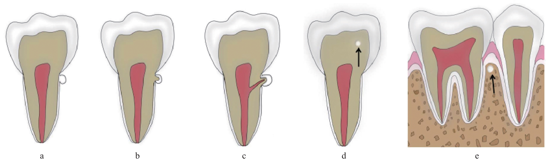
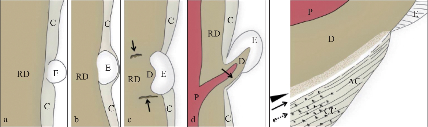
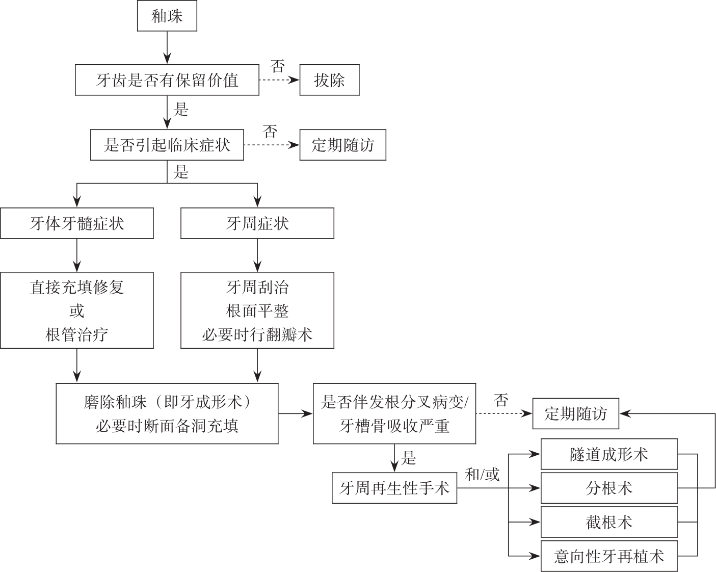
釉珠是最常见的异位牙釉质,发生机制尚不明确。临床中釉珠主要通过影像学检查或引起相关临床症状时被发现。釉珠与牙周组织为半桥粒连接,这种连接减弱了牙周组织防御感染的能力。此外,釉珠的存在使菌斑免受唾液酶及口腔清洁措施的影响,利于菌斑滞留,易导致发生牙体牙髓和牙周疾病。釉珠的早期诊断、评估、处理具有重要的临床意义。本文通过回顾釉珠相关文献,对釉珠的命名、发生率、诊断及鉴别诊断、分类、形态特点、分布特点、理化性质、组织学特征、组织来源与发生机制、临床意义、处理策略等方面进行综述,为釉珠的临床诊治与进一步研究提供参考。
中图分类号:
| [1] | Gaspersic D. Histogenetic aspects of the composition and structure of human ectopic enamel, studied by scanning electron microscopy[J]. Arch Oral Biol, 1992, 37(8): 603-611. |
| [2] | Moskow BS, Canut PM. Studies on root enamel (2). Enamel pearls. A review of their morphology, locali-zation, nomenclature, occurrence, classification, histogenesis and incidence[J]. J Clin Periodontol, 1990, 17(5): 275-281. |
| [3] | Darwazeh A, Hamasha AA. Radiographic evidence of enamel pearls in Jordanian dental patients[J]. Oral Surg Oral Med Oral Pathol Oral Radiol Endod, 2000, 89(2): 255-258. |
| [4] | Akgül N, Caglayan F, Durna N, et al. Evaluation of enamel pearls by cone-beam computed tomography (CBCT)[J]. Med Oral Patol Oral Cir Bucal, 2012, 17(2): e218-e222. |
| [5] | Risnes S. The prevalence, location, and size of enamel pearls on human molars[J]. Scand J Dent Res, 1974, 82(6): 403-412. |
| [6] | Chrcanovic BR, Abreu MHNG, Custódio ALN. Prevalence of enamel pearls in teeth from a human teeth bank[J]. J Oral Sci, 2010, 52(2): 257-260. |
| [7] | Sutalo J, Ciglar I, Njemirovskij V. Incidence of enamel pearls in our population[J]. Acta Stomatol Croat, 1986, 20(2): 123-129. |
| [8] | Grine FE, Holt S, Brink JS, et al. Enamel pearls: their occurrence in recent human populations and earliest manifestation in the modern human lineage[J]. Arch Oral Biol, 2019, 101: 147-155. |
| [9] | Negra ES, Oliveira OL. Morphology and locali-zation of enamel pearls in human teeth (author’s transl)[J]. Arq Cent Estud Fac Odontol UFMG, 1974, 11(2): 195-215. |
| [10] | Loh HS. A local study on enamel pearls[J]. Singapore Dent J, 1980, 5(1): 55-59. |
| [11] | 沙继春, 陈金华, 包美星. 10 000枚离体牙上釉珠发生情况[J]. 口腔医学纵横, 1987, 3(1): 18-20. |
| Sha JC, Chen JH, Bao MX. Prevalence of enamel pearls on 10 000 extracted teeth[J]. J Comprehens Stomatol, 1987, 3(1): 18-20. | |
| [12] | 解危. 离体牙上釉珠的观测与临床意义[J]. 昆明医学院学报, 1991, 3(12): 49-51. |
| Xie W. Observation on enamel pearls in extracted teeth and their clnical significance[J]. Acad J Kunming Med Colle, 1991, 3(12): 49-51. | |
| [13] | Kaminagakura E, Salmon C, Fonseca DC, et al. Prevalence and microscopicfeatures of enamel pearls from permanent human molars[J]. Braz J Oral Sci, 2011, 10(4): 268-271. |
| [14] | Versiani MA, Cristescu RC, Saquy PC, et al. Enamel pearls in permanent dentition: case report and micro-CT evaluation[J]. Dentomaxillofac Radiol, 2013, 42(6): 20120332. |
| [15] | Çolak H, Hamidi MM, Uzgur R, et al. Radiographic evaluation of the prevalence of enamel pearls in a sample adult dental population[J]. Eur Rev Med Pharmacol Sci, 2014, 18(3): 440-444. |
| [16] | Al-Zoubi IA, Patil SR, Alam MK, et al. A radiographic study of prevalence and location of enamel pearls in a Saudi Arabian adolescent population[J]. Pesqui Bras Odontopediatria Clín Integr, 2018, 18(1): 1-6. |
| [17] | Zengin AZ, Sumer AP, Ozturk G, et al. Imaging characteristics of enamel pearls on CBCT and their co-relation with supernumerary tooth[J]. Oral Ra-diol, 2022, 38(3): 370-377. |
| [18] | Cavanha AO. Enamel pearls[J]. Oral Surg Oral Med Oral Pathol, 1965, 19: 373-382. |
| [19] | Kupietzky A, Rozenfarb N. Enamel pearls in the primary dentition: report of two cases[J]. ASDC J Dent Child, 1993, 60(1): 63-66. |
| [20] | Saini T, Ogunleye A, Levering N, et al. Multiple enamel pearls in two siblings detected by volume-tric computed tomography[J]. Dentomaxillofac Radiol, 2008, 37(4): 240-244. |
| [21] | 张祖燕. 口腔颌面医学影像诊断学[M]. 7版. 北京: 人民卫生出版社, 2020: 46-47, 100. |
| Zhang ZY. Stomatological image diagnostics[M]. 7th ed. Beijing: People’s Medical Publishing Hou-se, 2020: 46-47, 100. | |
| [22] | Kaugars GE. Internal enamel pearls: report of case[J]. J Am Dent Assoc, 1983, 107(6): 941-943. |
| [23] | Pardiñas López S, Warren RN, Bromage TG, et al. Treatment of an unusual non-tooth related enamel pearl (EP) and 3 teeth-related EPs with localized periodontal disease without teeth extractions: a case report[J]. Compend Contin Educ Dent, 2015, 36(8): 592-599. |
| [24] | 毛小泉, 蒙亚娇, 李海芳. 根样釉珠1例[J]. 国际口腔医学杂志, 2016, 43(4): 406-408. |
| Mao XQ, Meng YJ, Li HF. A case report of root-like enamel pearl[J]. Int J Stomatol, 2016, 43(4): 406-408. | |
| [25] | Rathva V. Ectopic enamel pearl[J]. Clin Pract, 2012, 2(2): e46. |
| [26] | Risnes S, Segura JJ, Casado A, et al. Enamel pearls and cervical enamel projections on 2 maxillary molars with localized periodontal disease: case report and histologic study[J]. Oral Surg Oral Med Oral Pathol Oral Radiol Endod, 2000, 89(4): 493-497. |
| [27] | Takiguchi R, Funaki T. Scanning electron microscopy of enamel drop[J]. Bull Tokyo Dent Coll, 1977, 18(2): 57-70. |
| [28] | Anderson P, Elliott JC, Bose U, et al. A comparison of the mineral content of enamel and dentine in human premolars and enamel pearls measured by X-ray microtomography[J]. Arch Oral Biol, 1996, 41(3): 281-290. |
| [29] | Gaspersic D. Enamel microhardness and histological features of composite enamel pearls of different size[J]. J Oral Pathol Med, 1995, 24(4): 153-158. |
| [30] | Risnes S. Ectopic tooth enamel. An SEM study of the structure of enamel in enamel pearls[J]. Adv Dent Res, 1989, 3(2): 258-264. |
| [31] | Shiota K, Tamamura S, Okamoto K, et al. Enamel pearls; the incidence and pathology[J]. Jpn J Oral Biol, 1970, 12(3): 185-197. |
| [32] | Schroeder HE, Listgarten MA. Fine structure of the developing epithelial attachment of human teeth[J]. Monogr Dev Biol, 1971, 2: 1-134. |
| [33] | Romeo U, Palaia G, Botti R, et al. Enamel pearls as a predisposing factor to localized periodontitis[J]. Quintessence Int, 2011, 42(1): 69-71. |
| [34] | Goldstein AR. Enamel pearls as contributing factor in periodontal breakdown[J]. J Am Dent Assoc, 1979, 99(2): 210-211. |
| [35] | 周学东. 牙体牙髓病学[M]. 5版. 北京: 人民卫生出版社, 2020: 60-61. |
| Zhou XD. Endodontics[M]. 5th ed. Beijing: People’s Medical Publishing House, 2020: 60-61. | |
| [36] | Shojaeian S, Ghoddusi J, Hajian S. A case report of maxillary second molar with two palatal root canals and a furcal enamel pearl[J]. Iran Endod J, 2013, 8(1): 37-39. |
| [37] | 吴丽. 右上第三磨牙多发釉珠1例[J]. 实用口腔医学杂志, 2013, 29(4): 510. |
| Wu L. The multiple enamel pearls in right maxilla third molar[J]. J Pract Stomatol, 2013, 29(4): 510. | |
| [38] | 张震康, 俞光岩. 实用口腔科学[M]. 北京: 人民卫生出版社, 2009: 37-38. |
| Zhang ZK, Yu GY. Practice of stomatology[M]. Beijing: People’s Medical Publishing House, 2009: 37-38. | |
| [39] | Zenóbio EG, Vieira TR, Bustamante RP, et al. Enamel pearls implications on periodontal disease[J]. Case Rep Dent, 2015, 2015: 236462. |
| [1] | 张琳涵,汤亚玲. 拉曼光谱技术在口腔鳞状细胞癌和口腔潜在恶性疾病诊断和治疗中的应用进展[J]. 国际口腔医学杂志, 2026, 53(1): 107-115. |
| [2] | 张诗铭,石冰,黄汉尧. 唇裂鼻畸形非手术塑形治疗的临床应用[J]. 国际口腔医学杂志, 2026, 53(1): 116-123. |
| [3] | 侯旭彤,李贵民,叶玲,汪成林. 牙体牙髓病的舒适化治疗[J]. 国际口腔医学杂志, 2026, 53(1): 76-83. |
| [4] | 王诗雅,袁国华,邹静. 异位釉质的形成机制及临床诊疗策略[J]. 国际口腔医学杂志, 2025, 52(6): 713-721. |
| [5] | 张书旸, 胡顺佳怡, 戚琳珑, 梁筱瑶, 邓淑丽. 外伤年轻恒牙行牙髓再生治疗的研究进展[J]. 国际口腔医学杂志, 2025, 52(6): 738-747. |
| [6] | 张俭, 白雪, 何小倩, 葛振林. 多模态数据融合技术在口腔正畸领域的应用及研究进展[J]. 国际口腔医学杂志, 2025, 52(6): 764-770. |
| [7] | 陈禹黄,梁星,李然. 牙周炎患者缺失牙修复的临床考量及预后评估[J]. 国际口腔医学杂志, 2025, 52(6): 823-831. |
| [8] | 林超英,张岚,黄定明. 人工智能在根管治疗中的研究进展[J]. 国际口腔医学杂志, 2025, 52(5): 572-578. |
| [9] | 周小洁,侯本祥. 基于深度学习技术诊断龋病方法的研究进展[J]. 国际口腔医学杂志, 2025, 52(5): 579-585. |
| [10] | 蒋小菊,陈婧,苏勤. 牙髓组织应对牙外伤的生物学反应及相关临床诊疗策略[J]. 国际口腔医学杂志, 2025, 52(5): 586-593. |
| [11] | 王晓萌,邝海,何灏逾,李鸿艺,林洁舲,李飞燕. 唇腭裂语音治疗效果及相关因素研究分析[J]. 国际口腔医学杂志, 2025, 52(5): 627-633. |
| [12] | 朱然,严静,孙卫斌,吴文蕾,刘玉. 服用抗血栓药物患者牙周基础治疗期间的出血风险管理[J]. 国际口腔医学杂志, 2025, 52(5): 670-676. |
| [13] | 任家银,游梦. 基于影像学证据的牙骨质—骨结构不良区域的种植考量[J]. 国际口腔医学杂志, 2025, 52(4): 421-427. |
| [14] | 蒋丽,何飞. 再生性牙髓治疗在成熟恒牙中应用的临床研究进展[J]. 国际口腔医学杂志, 2025, 52(4): 449-455. |
| [15] | 齐斌,徐海明,卢志山. 天然植物提取物作为根管冲洗剂的研究进展[J]. 国际口腔医学杂志, 2025, 52(4): 466-472. |
|
||