国际口腔医学杂志 ›› 2018, Vol. 45 ›› Issue (5): 539-545.doi: 10.7518/gjkq.2018.05.008
邓雪阳,潘兰兰,胡婷,李文华,向学熔( )
)
Xueyang Deng,Lanlan Pan,Ting Hu,Wenhua Li,Xuerong. Xiang()
摘要:
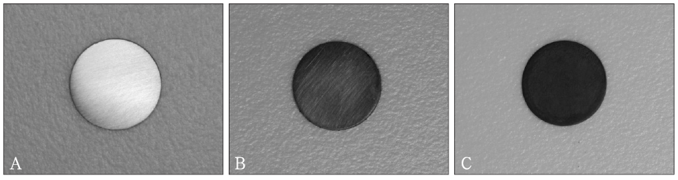
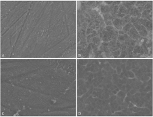
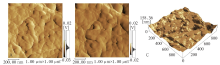
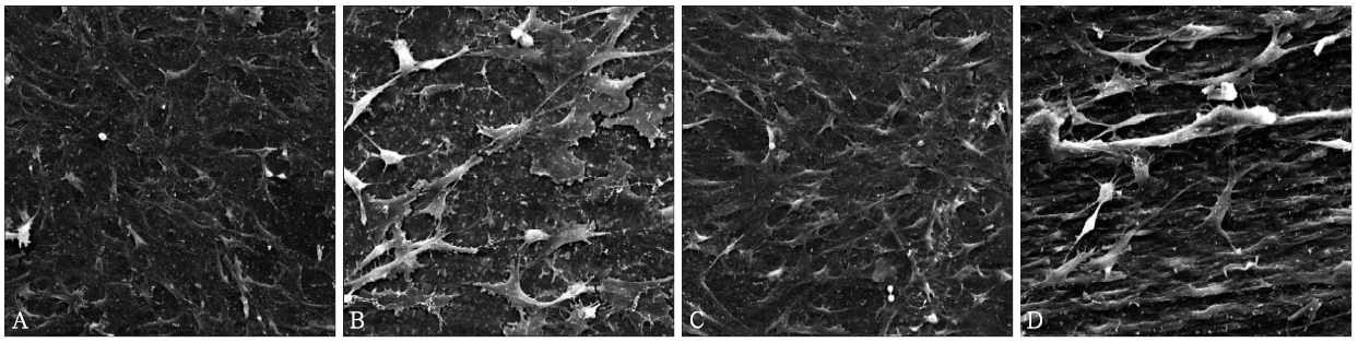
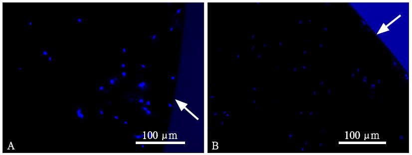
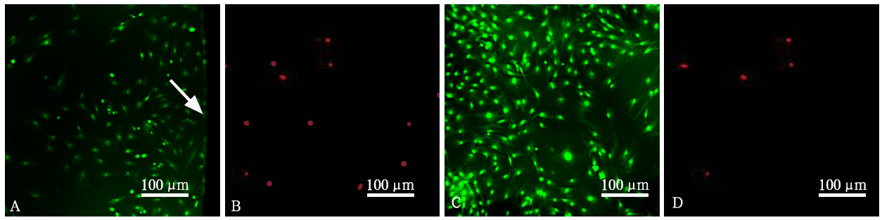
目的 利用氧化石墨烯(GO)制备钛合金表面涂层,初步评价其生物相容性。方法 采用层层自组装的方法,在碱热处理后的钛合金表面形成由GO阳离子(GO-NH3 +)、GO阴离子(GO-COO -)溶液组成的氧化石墨烯涂层。使用接触角测量仪、扫描电子显微镜、原子力显微镜、万能材料试验机初步分析其亲水性、表面形貌、涂层厚度、涂层粘接强度等特征,扫描电子显微镜、荧光显微镜观察骨髓间充质干细胞在其表面的生长情况,贴膜实验观察其抑菌性能。 结果 通过层层自组装方法在碱热处理后的钛合金表面制备出具有较好亲水性和粘接强度的、纳米级形貌的GO涂层。骨髓间充质干细胞在具有GO涂层的钛片的表面易于黏附,而且骨髓间充质干细胞活性保持良好。GO涂层对牙龈卟啉单胞菌的杀菌率为86.34%。结论 GO涂层的钛合金有良好的亲水性、生物相容性及抗菌性。
中图分类号:
| [1] | Brånemark PI, Hansson BO, Adell R , et al. Os-seointegrated implants in the treatment of the eden-tulous jaw. Experience from a 10-year period[J]. Scand J Plast Reconstr Surg Suppl, 1977,16:1-132. |
| [2] |
Wang G, Moya S, Lu Z , et al. Enhancing orthopedic implant bioactivity: refining the nanotopography[J]. Nanomedicine (Lond), 2015,10(8):1327-1341.
doi: 10.2217/nnm.14.216 pmid: 25955126 |
| [3] |
Wu SY, An SS, Hulme J , Current applications of graphene oxide in nanomedicine[J]. Int J Nanome-dicine, 2015,10(Spec Iss):9-24.
doi: 10.2147/IJN.S88285 pmid: 4554423 |
| [4] | Dubey N, Bentini R, Islam I , et al. Graphene: a ve-rsatile carbon-based material for bone tissue en-gineering[J]. Stem Cells Int, 2015,2015:804213. |
| [5] |
Sahni D, Jea A, Mata JA , et al. Biocompatibility of pristine graphene for neuronal interface[J]. J Neuro-surg Pediatr, 2013,11(5):575-583.
doi: 10.3171/2013.1.PEDS12374 pmid: 23473006 |
| [6] |
Shen H, Zhang L, Liu M , et al. Biomedical applica-tions of graphene[J]. Theranostics, 2012,2(3):283-294.
doi: 10.7150/thno.3642 |
| [7] |
Akhavan O, Ghaderi E , Toxicity of graphene and graphene oxide nanowalls against bacteria[J]. ACS Nano, 2010,4(10):5731-5736.
doi: 10.1021/nn101390x pmid: 20925398 |
| [8] | La WG, Jin M, Park S , et al. Delivery of bone mor-phogenetic protein-2 and substance P using graphene oxide for bone regeneration[J]. Int J Nanomedicine, 2014,9(Suppl 1):107-116. |
| [9] |
Grieznis L, Apse P, Blumfelds L , Passive tactile sensibility of teeth and osseointegrated dental im-plants in the maxilla[J]. Stomatologija, 2010,12(3):80-86.
pmid: 21063137 |
| [10] | Jemat A, Ghazali MJ, Razali M , et al. Surface modi-fications and their effects on titanium dental implants[J]. Biomed Res Int, 2015,2015:791725. |
| [11] | Smeets R, Stadlinger B, Schwarz F , et al. Impact of dental implant surface modifications on osseointe-gration[J]. Biomed Res Int, 2016,2016:6285620. |
| [12] |
Gentile P, Carmagnola I, Nardo T , et al. Layer-by-layer assembly for biomedical applications in the last decade[J]. Nanotechnology, 2015,26(42):422001.
doi: 10.1088/0957-4484/26/42/422001 pmid: 26421916 |
| [13] |
Park JS, Cho SM, Kim WJ , et al. Fabrication of gra-phene thin films based on layer-by-layer self-assembly of functionalized graphene nanosheets[J]. ACS Appl Mater Interfaces, 2011,3(2):360-368.
doi: 10.1021/am100977p |
| [14] |
La WG, Park S, Yoon HH , et al. Delivery of a the-rapeutic protein for bone regeneration from a sub-strate coated with graphene oxide[J]. Small, 2013,9(23):4051-4060.
doi: 10.1002/smll.201300571 |
| [15] | 吴昊, 刘洪臣 . 种植体周围炎的易感因素研究现状[J]. 口腔颌面修复学杂志, 2012,13(3):184-187. |
| Wu H, Liu HC . Research status of possible risk fac-tors of peri-implantitis: a review[J]. Chin J Prostho-dont, 2012,13(3):184-187. | |
| [16] |
Persson GR, Renvert S , Cluster of bacteria asso-ciated with peri-implantitis[J]. Clin Implant Dent Relat Res, 2014,16(6):783-793.
doi: 10.1111/cid.12052 pmid: 23527870 |
| [17] |
Liu S, Zeng TH, Hofmann M , et al. Antibacterial activity of graphite, graphite oxide, graphene oxide, and reduced graphene oxide: membrane and oxida-tive stress[J]. ACS Nano, 2011,5(9):6971-6980.
doi: 10.1021/nn202451x |
| [18] |
Tu Y, Lv M, Xiu P , et al. Destructive extraction of phospholipids from Escherichia coli membranes by graphene nanosheets[J]. Nat Nanotechnol, 2013,8(8):594-601.
doi: 10.1038/nnano.2013.125 pmid: 23832191 |
| [1] | 颜愈佳,邹玲. 生物陶瓷类根管封闭剂的研究进展[J]. 国际口腔医学杂志, 2022, 49(5): 578-585. |
| [2] | 张曦丹,孙吉宇,付馨靓,甘雪琦. 介孔硅酸钙纳米材料在牙体牙髓及颅颌面修复领域的研究进展[J]. 国际口腔医学杂志, 2022, 49(4): 476-482. |
| [3] | 王婷, 葛少华. 氧化石墨烯在生物医学领域方面应用的研究进展[J]. 国际口腔医学杂志, 2017, 44(5): 591-595. |
| [4] | 刘奕1,周荣静2,费伟1. 高迁移率族蛋白N2免疫组织化学定位及其活性结构抑菌性能的体外实验研究[J]. 国际口腔医学杂志, 2016, 43(6): 661-665. |
| [5] | 聂蕾. 口腔贱金属合金修复体的生物相容性试验[J]. 国际口腔医学杂志, 2015, 42(1): 79-93. |
| [6] | 吴雨鸿,林居红,张红梅. 三氧化聚合物与波特兰水门汀的理化和生物学性能及其应用[J]. 国际口腔医学杂志, 2014, 41(6): 699-702. |
| [7] | 赵惠1 李潇2 金柱坤2 杨凯2. 影响种植支架材料生物相容性的因素[J]. 国际口腔医学杂志, 2014, 41(3): 341-346. |
| [8] | 李晓宁综述 樊明文 杨雪超审校. 牙髓病治疗新材料BioAggregate的研究进展[J]. 国际口腔医学杂志, 2013, 40(4): 471-472. |
| [9] | 史彦 杨健. 次氯酸钠溶液抗菌体积分数的测定[J]. 国际口腔医学杂志, 2012, 39(5): 590-592. |
| [10] | 王传勇综述 李伟 蒋丽审校. 生物陶瓷表面蛋白吸附的研究进展[J]. 国际口腔医学杂志, 2012, 39(4): 550-553. |
| [11] | 邓霞1 夏熹2. 纳米羟磷灰石-脂肪族聚酯酰胺复合材料对成骨细胞的生物学作用[J]. 国际口腔医学杂志, 2012, 39(1): 33-36. |
| [12] | 周晗 张陶 岳源 乔梦婷 李燕 肖丽英. 葡萄多酚和茶多酚抗变异链球菌活性的比较[J]. 国际口腔医学杂志, 2011, 38(6): 643-648. |
| [13] | 赵耀 刘嘉俊综述 孟玉坤审校. 牙科合金腐蚀行为的研究进展[J]. 国际口腔医学杂志, 2011, 38(3): 339-342. |
| [14] | 吴卫锋 雷丹 姜苏 高雳. 安止痛牙龈膏对牙龈卟啉单胞菌和伴放线嗜血菌及具核梭杆菌的抑菌作用[J]. 国际口腔医学杂志, 2011, 38(2): 138-140. |
| [15] | 叶园园综述 章燕珍审校. 植物精油的药理作用及其在口腔医学中的应用[J]. 国际口腔医学杂志, 2011, 38(2): 185-187. |
|