国际口腔医学杂志 ›› 2022, Vol. 49 ›› Issue (1): 100-108.doi: 10.7518/gjkq.2022006
刘力嘉1( ),毛婧1,龙欢1,蒲亚龙1,王军2()
Liu Lijia1(),Mao Jing1,Long Huan1,Pu Yalong1,Wang Jun2()
摘要:
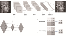
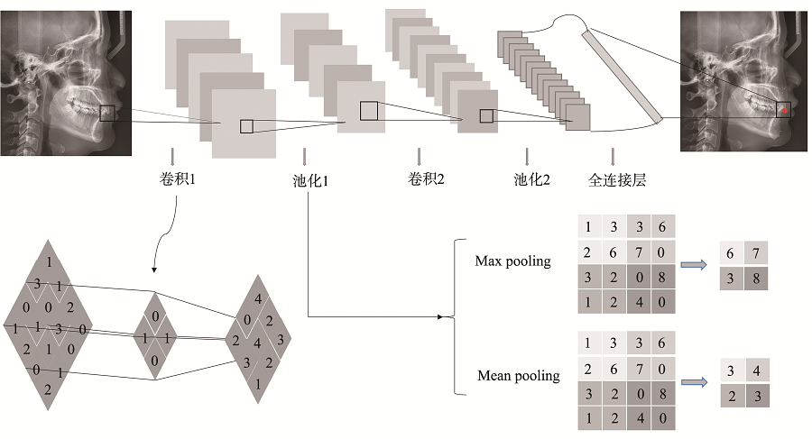
头影测量是正畸、正颌诊疗过程中不可或缺的分析手段。随着计算机辅助技术的发展,头影测量自动定点已经在二维头影测量中基本实现,并达到了较高的精确度,大大减轻了操作者的负担;而由于锥体束计算机断层扫描(CBCT)影像无放大失真、组织重叠等缺点,能精确定位头影测量分析的解剖标志,对于诊断和分析先天或后天的颅面部不对称畸形具有天然的优势,三维头影测量自动定点已经成为目前头影测量领域重要的研究方向。本文以不同的自动定点方法为分类,分别对二维和三维头影测量自动定点的研究进展作一综述,探讨不同自动定点方法的精确度,并对其未来发展进行展望。
| [1] | Cardillo J, Sid-Ahmed MA. An image processing system for locating craniofacial landmarks[J]. IEEE Trans Med Imaging, 1994,13(2):275-289. |
| [2] | Medellín-Castillo HI, Govea-Valladares EH, Pérez-Guerrero CN, et al. The evaluation of a novel haptic-enabled virtual reality approach for computer-aided cephalometry[J]. Comput Methods Programs Bio-med, 2016,130:46-53. |
| [3] | Cohen AM, Ip HH, Linney AD. A preliminary study of computer recognition and identification of skeletal landmarks as a new method of cephalometric a-nalysis[J]. Br J Orthod, 1984,11(3):143-154. |
| [4] | Yun HS, Jang TJ, Lee SM, et al. Learning-based local-to-global landmark annotation for automatic 3D cephalometry[J]. Phys Med Biol, 2020,65(8):08-5018. |
| [5] | Douglas TS. Image processing for craniofacial landmark identification and measurement: a review of photogrammetry and cephalometry[J]. Comput Med Imaging Graph, 2004,28(7):401-409. |
| [6] | Tam WK, Lee HJ. Improving point correspondence in cephalograms by using a two-stage rectified point transform[J]. Comput Biol Med, 2015,65:114-123. |
| [7] | Kafieh R, Mehri A, Sadri S. Automatic landmark detection in cephalometry using a modified active sha-pe model with sub image matching[C]// 2007 Int Conf Mach Vis. Isalambad, Pakistan: IEEE, 2007: 73-78. |
| [8] | Leonardi R, Giordano D, Maiorana F, et al. Automatic cephalometric analysis[J]. Angle Orthod, 2008,78(1):145-151. |
| [9] | Vandaele R, Aceto J, Muller M, et al. Landmark detection in 2D bioimages for geometric morphome-trics: a multi-resolution tree-based approach[J]. Sci Rep, 2018,8(1):538. |
| [10] | Lindner C, Wang CW, Huang CT, et al. Fully automatic system for accurate localisation and analysis of cephalometric landmarks in lateral cephalograms[J]. Sci Rep, 2016,6:33581. |
| [11] | Lévy-Mandel AD, Venetsanopoulos AN, Tsotsos JK. Knowledge-based landmarking of cephalograms[J]. Comput Biomed Res, 1986,19(3):282-309. |
| [12] | Parthasarathy S, Nugent ST, Gregson PG, et al. Automatic landmarking of cephalograms[J]. Comput Biomed Res, 1989,22(3):248-269. |
| [13] | Davis DN, Forsyth D. Knowledge-based cephalometric analysis: a comparison with clinicians using interactive computer methods[J]. Comput Biomed Res, 1994,27(3):210-228. |
| [14] | Forsyth DB, Davis DN. Assessment of an automa-ted cephalometric analysis system[J]. Eur J Orthod, 1996,18(5):471-478. |
| [15] | Cootes TF, Taylor CJ, Cooper DH, et al. Active shape models-their training and application[J]. Comput Vis Image Underst, 1995,61(1):38-59. |
| [16] | Cootes TF, Taylor CJ. Statistical models of appea-rance for medical image analysis and computer vision[C]// Medical imaging 2001. San Diego, CA, USA: Image Processing, 2001,4322:236-248. |
| [17] | Hutton TJ, Cunningham S, Hammond P. An evaluation of active shape models for the automatic identification of cephalometric landmarks[J]. Eur J Orthod, 2000,22(5):499-508. |
| [18] | Rueda S, Alcañiz M. An approach for the automatic cephalometric landmark detection using mathematical morphology and active appearance models[J]. Med Image Comput Comput Assist Interv, 2006,9(Pt 1):159-166. |
| [19] | Vucinić P, Trpovski Z, Sćepan I. Automatic landmarking of cephalograms using active appearance models[J]. Eur J Orthod, 2010,32(3):233-241. |
| [20] | Kafieh R, Mehri A, Sadri S. Automatic landmark detection in cephalometry using a modified active sha-pe model with sub image matching[C]// 2007 International Conference on Machine Vision. December 28-29, 2007, Isalambad, Pakistan. IEEE, 2007: 73-78. |
| [21] | Kaur A, Singh C. Automatic cephalometric landmark detection using Zernike moments and templa-te matching[J]. SIViP, 2015,9(1):117-132. |
| [22] | Arık SÖ, Ibragimov B, Xing L. Fully automated quantitative cephalometry using convolutional neural networks[J]. J Med Imaging (Bellingham), 2017,4(1):014501. |
| [23] | Kunz F, Stellzig-Eisenhauer A, Zeman F, et al. Artificial intelligence in orthodontics: evaluation of a fully automated cephalometric analysis using a customized convolutional neural network[J]. J Orofac Orthop, 2020,81(1):52-68. |
| [24] | Park JH, Hwang HW, Moon JH, et al. Automated identification of cephalometric landmarks: part 1—comparisons between the latest deep-learning metho-ds YOLOV3 and SSD[J]. Angle Orthod, 2019,89(6):903-909. |
| [25] | Hwang HW, Park JH, Moon JH, et al. Automated identification of cephalometric landmarks: part 2—might it be better than human[J]. Angle Orthod, 2020,90(1):69-76. |
| [26] | Dai XB, Zhao H, Liu TL, et al. Locating anatomical landmarks on 2D lateral cephalograms through adversarial encoder-decoder networks[J]. IEEE Acce-ss, 2019,7:132738-132747. |
| [27] | Wang CW, Huang CT, Hsieh MC, et al. Evaluation and comparison of anatomical landmark detection methods for cephalometric X-ray images: a grand challenge[J]. IEEE Trans Med Imaging, 2015,34(9):1890-1900. |
| [28] | Wang CW, Huang CT, Lee JH, et al. A benchmark for comparison of dental radiography analysis algorithms[J]. Med Image Anal, 2016,31:63-76. |
| [29] | Dot G, Rafflenbeul F, Arbotto M, et al. Accuracy and reliability of automatic three-dimensional ce-phalometric landmarking[J]. Int J Oral Maxillofac Surg, 2020,49(10):1367-1378. |
| [30] | Neelapu BC, Kharbanda OP, Sardana V, et al. Automatic localization of three-dimensional cephalome-tric landmarks on CBCT images by extracting symmetry features of the skull[J]. Dentomaxillofac Radiol, 2018,47(2):20170054. |
| [31] | Gupta A, Kharbanda OP, Sardana V, et al. A know-ledge-based algorithm for automatic detection of ce-phalometric landmarks on CBCT images[J]. Int J Comput Assist Radiol Surg, 2015,10(11):1737-1752. |
| [32] | Gupta A, Kharbanda OP, Sardana V, et al. Accuracy of 3D cephalometric measurements based on an automatic knowledge-based landmark detection algorithm[J]. Int J Comput Assist Radiol Surg, 2016,11(7):1297-1309. |
| [33] | Codari M, Caffini M, Tartaglia GM, et al. Computer-aided cephalometric landmark annotation for CBCT data[J]. Int J Comput Assist Radiol Surg, 2017,12(1):113-121. |
| [34] | Shahidi S, Bahrampour E, Soltanimehr E, et al. The accuracy of a designed software for automated loca-lization of craniofacial landmarks on CBCT images[J]. BMC Med Imaging, 2014,14:32. |
| [35] | Montúfar J, Romero M, Scougall-Vilchis RJ. Automatic 3-dimensional cephalometric landmarking ba-sed on active shape models in related projections[J]. Am J Orthod Dentofacial Orthop, 2018,153(3):449-458. |
| [36] | Montúfar J, Romero M, Scougall-Vilchis RJ. Hybrid approach for automatic cephalometric landmark annotation on cone-beam computed tomography volumes[J]. Am J Orthod Dentofacial Orthop, 2018,154(1):140-150. |
| [37] | Zhang J, Liu MX, Wang L, et al. Context-guided fully convolutional networks for joint craniomaxillofacial bone segmentation and landmark digitization[J]. Med Image Anal, 2020,60:101621. |
| [38] | Zhang J, Gao YZ, Wang L, et al. Automatic craniomaxillofacial landmark digitization via segmentation-guided partially-joint regression forest model and multiscale statistical features[J]. IEEE Trans Biomed Eng, 2016,63(9):1820-1829. |
| [39] | de Jong MA, Gül A, de Gijt JP, et al. Automated human skull landmarking with 2D Gabor wavelets[J]. Phys Med Biol, 2018,63(10):105011. |
| [40] | O’Neil AQ, Kascenas A, Henry J, et al. Attaining human-level performance with atlas location autocontext for anatomical landmark detection in 3D CT data[J]. Lect Notes Comput Sci, 2019: 470-484. |
| [41] | Lachinov D, Getmanskaya A, Turlapov V. Cephalometric landmark regression with convolutional neural networks on 3D computed tomography data[J]. Pattern Recognit Image Anal, 2020,30(3):512-522. |
| [42] | Lee SM, Kim HP, Jeon K, et al. Automatic 3D cephalometric annotation system using shadowed 2D image-based machine learning[J]. Phys Med Biol, 2019,64(5):055002. |
| [43] | Zhang J, Liu MX, Wang L, et al. Joint craniomaxillofacial bone segmentation and landmark digitization by context-guided fully convolutional networks[J]. Med Image Comput Comput Assist Interv, 2017,10434:720-728. |
| [44] | Torosdagli N, Liberton DK, Verma P, et al. Deep geodesic learning for segmentation and anatomical landmarking[J]. IEEE Trans Med Imaging, 2019,38(4):919-931. |
| [1] | 刘盼明,李政泽,李军鹤,崔淑霞. 成人骨性Ⅱ类患者不同垂直骨面型上颌窦容积及口咽气道体积的锥形束计算机断层扫描研究[J]. 国际口腔医学杂志, 2023, 50(5): 528-537. |
| [2] | 周梦琪,陈学鹏,傅柏平. 正畸治疗中牙槽骨骨开窗骨开裂的预防和应对策略[J]. 国际口腔医学杂志, 2021, 48(5): 600-608. |
| [3] | 施丹妮,杨鑫,吴建勇. 锥形束CT三维头影测量参考坐标系的研究进展[J]. 国际口腔医学杂志, 2021, 48(4): 398-404. |
| [4] | 伍春兰,唐华,陈军. 成人骨性Ⅱ类高角开牙合患者上下切牙区牙槽骨形态的三维研究[J]. 国际口腔医学杂志, 2021, 48(4): 426-432. |
| [5] | 王立冬,马文,付帅,张长彬,崔庆赢,梁燕,黎明. 不同方法制作正颌手术数字化牙合板的研究及精确性分析[J]. 国际口腔医学杂志, 2021, 48(2): 156-164. |
| [6] | 张紫涵,熊鑫,王军. 三维头影测量的研究现状和应用发展[J]. 国际口腔医学杂志, 2020, 47(6): 739-744. |
| [7] | 王涛. 外科优先序列治疗——正颌外科的发展热点之一及其误区[J]. 国际口腔医学杂志, 2020, 47(5): 497-505. |
| [8] | 刘晓华, 王恩博. 儿童埋伏多生牙拔除术锥形束计算机断层扫描影像学定位的应用进展[J]. 国际口腔医学杂志, 2018, 45(3): 295-300. |
| [9] | 尹一佳, 冯捷, 罗梦奇, 韩向龙. 三维立体摄影技术自然头位校正方法的研究进展[J]. 国际口腔医学杂志, 2018, 45(3): 301-306. |
| [10] | 谢成佳, 葛少华. 牙骨质撕裂的诊治进展[J]. 国际口腔医学杂志, 2017, 44(3): 315-319. |
| [11] | 赵彦惠 聂萍 陶丽 盛潇 陈金东 朱敏. 应用头影测量结合Müller试验评价肥胖对阻塞性睡眠呼吸暂停低通气综合征患者上气道可塌陷性的影响[J]. 国际口腔医学杂志, 2014, 41(4): 390-395. |
| [12] | 张宋晶 孙克勤. 锥形束CT及其在牙根纵裂早期诊断中的作用[J]. 国际口腔医学杂志, 2014, 41(4): 459-462. |
| [13] | 兰静 王传勇 薛晶 蒋丽 李伟. 含纳米羟磷灰石的过氧化氢漂白剂对釉质表面的影响[J]. 国际口腔医学杂志, 2013, 40(4): 432-435. |
| [14] | 林永盛 张小慧 张金婷. 甘肃省东乡族正常牙合成人的头影测量分析[J]. 国际口腔医学杂志, 2013, 40(3): 317-319. |
| [15] | 敖同江, 袁小平 杨四维 黄跃. 替牙晚期安氏Ⅲ类错牙合畸形矫治后的髁突和下颌位置变化[J]. 国际口腔医学杂志, 2012, 39(4): 439-442. |
|