Int J Stomatol ›› 2026, Vol. 53 ›› Issue (2): 145-154.doi: 10.7518/gjkq.2026120
• Expert Forum •
Meiqing Wang( )
)
CLC Number:
| [1] | Wolford LM, Cardenas L. Idiopathic condylar resorption: diagnosis, treatment protocol, and outco-mes[J]. Am J Orthod Dentofacial Orthop, 1999, 116(6): 667-677. |
| [2] | Young A. Idiopathic condylar resorption: the current understanding in diagnosis and treatment[J]. J In- dian Prosthodont Soc, 2017, 17(2): 128-135. |
| [3] | Nobrega MTC, Almeida FT, Friesen R, et al. Idiopathic condylar resorption in adolescents: a scoping review[J]. J Oral Rehabil, 2024, 51(8): 1610-1620. |
| [4] | Mercuri LG. Osteoarthritis, osteoarthrosis, and idiopathic condylar resorption[J]. Oral Maxillofac Surg Clin North Am, 2008, 20(2): 169-183. |
| [5] | Chamberland S. Progressive idiopathic condylar resorption: three case reports[J]. Am J Orthod Dentofacial Orthop, 2019, 156(4): 531-544. |
| [6] | Noh HK, Park HS. Considerations for vertical control with microimplants in a idiopathic condylar resorption patient: a case report[J]. J Orthod, 2021, 48(2): 172-182. |
| [7] | Kristensen KD, Schmidt B, Stoustrup P, et al. Idiopathic condylar resorptions: 3-dimensional condylar bony deformation, signs and symptoms[J]. Am J Orthod Dentofacial Orthop, 2017, 152(2): 214-223. |
| [8] | Lee GH, Park JH, Lee SM, et al. Orthodontic treatment protocols for patients with idiopathic condylar resorption[J]. J Clin Pediatr Dent, 2019, 43(4): 292-303. |
| [9] | Mitsimponas K, Mehmet S, Kennedy R, et al. Idiopathic condylar resorption[J]. Br J Oral Maxillofac Surg, 2018, 56(4): 249-255. |
| [10] | Mercuri LG, Handelman CS. Idiopathic condylar resorption: what should we do[J]. Oral Maxillofac Surg Clin North Am, 2020, 32(1): 105-116. |
| [11] | Tanaka E. Etiology and diagnosis for idiopathic condylar resorption in growing adolescents[J]. J Clin Med, 2023, 12(20): 6607. |
| [12] | Alsabban L, Amarista FJ, Mercuri LG, et al. Idiopathic condylar resorption: a survey and review of the literature[J]. J Oral Maxillofac Surg, 2018, 76(11): 2316.e1-2316.e13. |
| [13] | Sansare K, Raghav M, Mallya SM, et al. Management-related outcomes and radiographic findings of idiopathic condylar resorption: a systematic review[J]. Int J Oral Maxillofac Surg, 2015, 44(2): 209-216. |
| [14] | Roth S, Müller K, Fischer DC, et al. Specific properties of the extracellular chondroitin sulphate proteoglycans in the mandibular condylar growth centre in pigs[J]. Arch Oral Biol, 1997, 42(1): 63-76. |
| [15] | Tominaga K, Hirashima S, Fukuda J. An experimental model of osteoarthrosis of the temporomandibular joint in monkeys[J]. Br J Oral Maxillofac Surg, 2002, 40(3): 232-237. |
| [16] | Lovell NC. Skeletal and dental pathology of free-ranging mountain gorillas[J]. Am J Phys Anthropol, 1990, 81(3): 399-412. |
| [17] | Chen CP, Zhang JH, Zhang B, et al. Unilateral loss of maxillary molars in young mice leads to bilateral condylar adaptation and degenerative disease[J]. JB-MR Plus, 2022, 6(7): e10638. |
| [18] | Nogami S, Kataoka Y, Yamauchi K, et al. Condylar resorption following compressive mechanical stress in rabbit model‒association of matrix metalloproteinases[J]. In Vivo, 2022, 36(5): 2126-2133. |
| [19] | Nogami S, Yamauchi K, Odashima K, et al. Influen-ce of oestrogen deficiency and excessive mechanical stress on condylar head of mandible[J]. Oral Dis, 2020, 26(8): 1718-1726. |
| [20] | Yang HJ, Hwang SJ. Effects of 17β-estradiol deficiency and mechanical overload on osseous changes in the rat temporomandibular joint[J]. J Oral Maxillofac Surg, 2020, 78(2): 214.e1-214.e14. |
| [21] | Iwasaki T, Takahara N, Duc VV, et al. Effect of anterior disc displacement and estrogen deficiency on rabbit mandibular condyle[J]. J Oral Biosci, 2025, 67(1): 100599. |
| [22] | Jiao K, Dai J, Wang MQ, et al. Age- and sex-related changes of mandibular condylar cartilage and subchondral bone: a histomorphometric and micro-CT study in rats[J]. Arch Oral Biol, 2010, 55(2): 155-163. |
| [23] | Zhang YJ, Zhang J, Xu LF, et al. Unbalanced cartilage calcification during development contributes to the formation of irregular articular surfaces as revealed by micro-CT images[J]. Australas Orthod J, 2023, 39(2): 40-48. |
| [24] | 张月姣, 徐小杰, 刘倩, 等. 偏颌大鼠模型的构建及其髁突CT影像和组织学评价[J]. 口腔颌面外科杂志, 2021, 31(5): 278-284. |
| Zhang YJ, Xu XJ, Liu Q, et al. Construction of the mandible deviation occlusion rat model and micro-CT radiography and histology evaluation of the mandibular condyles[J]. J Oral Maxillofac Surg, 2021, 31(5): 278-284. | |
| [25] | Alali YS, Al Habeeb KM, Al Malhook KA, et al. Diagnosis and management of idiopathic condylar resorption: a review of literature[J]. Saudi Dent J, 2024, 36(11): 1397-1405. |
| [26] | Valladares-Neto J, Acioli GR, Teodoro AB, et al. Conservative and minimally invasive approaches to control idiopathic condylar resorption: a scoping review[J]. Int J Oral Maxillofac Surg, 2023, 52(11): 1188-1196. |
| [27] | Gunson MJ, Arnett GW, Formby B, et al. Oral contraceptive pill use and abnormal menstrual cycles in women with severe condylar resorption: a case for low serum 17beta-estradiol as a major factor in progressive condylar resorption[J]. Am J Orthod Dentofacial Orthop, 2009, 136(6): 772-779. |
| [28] | Ye T, Sun DL, Mu T, et al. Differential effects of high-physiological oestrogen on the degeneration of mandibular condylar cartilage and subchondral bone[J]. Bone, 2018, 111: 9-22. |
| [29] | Park JH, Park JJ, Papademetriou M, et al. Anterior open bite due to idiopathic condylar resorption du-ring orthodontic retention of a Class Ⅱ Division 1 malocclusion[J]. Am J Orthod Dentofacial Orthop, 2019, 156(4): 555-565. |
| [30] | Ahmad N, Chen S, Wang W, et al. 17β-estradiol induces MMP-9 and MMP-13 in TMJ fibrochondrocytes via estrogen receptor α[J]. J Dent Res, 2018, 97(9): 1023-1030. |
| [31] | Iwasa A, Tanaka E. Signs, symptoms, and morphological features of idiopathic condylar resorption in orthodontic patients: a survey-based study[J]. J Clin Med, 2022, 11(6): 1552. |
| [32] | Robinson JL, Cass K, Aronson R, et al. Sex diffe-rences in the estrogen-dependent regulation of temporomandibular joint remodeling in altered loading[J]. Osteoarthritis Cartilage, 2017, 25(4): 533-543. |
| [33] | Choi J, Oh N, Kim IK. A follow-up study of condyle fracture in children[J]. Int J Oral Maxillofac Surg, 2005, 34(8): 851-858. |
| [34] | Lin YY, Tanaka N, Ohkuma S, et al. The mandibular cartilage metabolism is altered by damaged subchondral bone from traumatic impact loading[J]. Ann Biomed Eng, 2009, 37(7): 1358-1367. |
| [35] | Ji YD, Resnick CM, Peacock ZS. Idiopathic condylar resorption: a systematic review of etiology and management[J]. Oral Surg Oral Med Oral Pathol Oral Radiol, 2020, 130(6): 632-639. |
| [36] | Barone S, Cosentini G, Bennardo F, et al. Incidence and management of condylar resorption after orthognathic surgery: an overview[J]. Korean J Orthod, 2022, 52(1): 29-41. |
| [37] | Nogami S, Yamauchi K, Satomi N, et al. Risk factors related to aggressive condylar resorption after orthognathic surgery for females: retrospective study[J]. Cranio, 2017, 35(4): 250-258. |
| [38] | Catherine Z, Breton P, Bouletreau P. Condylar resorption after orthognathic surgery: a systematic review[J]. Rev Stomatol Chir Maxillofac Chir Orale, 2016, 117(1): 3-10. |
| [39] | NiÑo-Sandoval TC, Almeida RAC, Vasconcelos BCDE. Incidence of condylar resorption after bimaxillary, LefortⅠ, and mandibular surgery: an overview[J]. Braz Oral Res, 2021, 35: e27. |
| [40] | de Moraes PH, Rizzati-Barbosa CM, Olate S, et al. Condylar resorption after orthognathic surgery: a systematic review[J]. Int J Morphol, 2012, 30(3): 1023-1028. |
| [41] | Mousoulea S, Kloukos D, Sampaziotis D, et al. Condylar resorption in orthognathic patients after mandibular bilateral sagittal split osteotomy: a systema-tic review[J]. Eur J Orthod, 2017, 39(3): 294-309. |
| [42] | Hwang SJ, Haers PE, Zimmermann A, et al. Surgical risk factors for condylar resorption after orthognathic surgery[J]. Oral Surg Oral Med Oral Pathol Oral Radiol Endod, 2000, 89(5): 542-552. |
| [43] | Scheerlinck JP, Stoelinga PJ, Blijdorp PA, et al. Sa-gittal split advancement osteotomies stabilized with miniplates. A 2-5-year follow-up[J]. Int J Oral Ma-xillofac Surg, 1994, 23(3): 127-131. |
| [44] | Yoshioka I, Khanal A, Tominaga K, et al. Vertical ramus versus sagittal split osteotomies: comparison of stability after mandibular setback[J]. J Oral Maxillofac Surg, 2008, 66(6): 1138-1144. |
| [45] | Kobayashi T, Izumi N, Kojima T, et al. Progressive condylar resorption after mandibular advancement[J]. Br J Oral Maxillofac Surg, 2012, 50(2): 176-180. |
| [46] | Nunes de Lima V, Faverani LP, Santiago JF, et al. Evaluation of condylar resorption rates after orthognathic surgery in Class Ⅱ and Ⅲ dentofacial deformities: a systematic review[J]. J Cranio Maxillofac Surg, 2018, 46(4): 668-673. |
| [47] | Vandeput AS, Verhelst PJ, Jacobs R, et al. Condylar changes after orthognathic surgery for Class Ⅲ dentofacial deformity: a systematic review[J]. Int J Oral Maxillofac Surg, 2019, 48(2): 193-202. |
| [48] | He Z, Ji HZ, Du W, et al. Management of condy-lar resorption before or after orthognathic surgery: a systematic review[J]. J Craniomaxillofac Surg, 2019, 47(7): 1007-1014. |
| [49] | Hoppenreijs TJ, Freihofer HP, Stoelinga PJ, et al. Condylar remodelling and resorption after Le Fort Ⅰand bimaxillary osteotomies in patients with anterior open bite. A clinical and radiological study[J]. Int J Oral Maxillofac Surg, 1998, 27(2): 81-91. |
| [50] | Park SB, Yang YM, Kim YI, et al. Effect of bimaxillary surgery on adaptive condylar head remodeling: metric analysis and image interpretation using cone-beam computed tomography volume superimposition[J]. J Oral Maxillofac Surg, 2012, 70(8): 1951-1959. |
| [51] | de Mol van Otterloo JJ, Dorenbos J, Tuinzing DB, et al. TMJ performance and behaviour in patients more than 6 years after Le FortⅠ osteotomy[J]. Br J Oral Maxillofac Surg, 1993, 31(2): 83-86. |
| [52] | Tanaka E, Koolstra JH. Biomechanics of the temporomandibular joint[J]. J Dent Res, 2008, 87(11): 989-991. |
| [53] | He YF, Lin H, Lin QP, et al. Morphologic changes in idiopathic condylar resorption with different degrees of bone loss[J]. Oral Surg Oral Med Oral Pa-thol Oral Radiol, 2019, 128(3): 332-340. |
| [54] | Alimanovic D, Pedersen TK, Matzen LH, et al. Comparing clinical and radiological manifestations of adolescent idiopathic condylar resorption and juvenile idiopathic arthritis in the temporomandibular joint[J]. J Oral Maxillofac Surg, 2021, 79(4): 774-785. |
| [55] | Cannizzaro E, Schroeder S, Bolt I, et al. Temporomandibular joint involvement in children with juvenile idiopathic arthritis[J]. Pediatr Rheumatol, 2008, 6(1): P92. |
| [56] | Boos-Lima FBDJ, Guastaldi FPS, Nielsen GP, et al. Histopathology of idiopathic condylar resorption differs from temporomandibular joint-only juvenile idiopathic arthritis[J]. J Oral Maxillofac Surg, 2025, 83(1): 26-36. |
| [57] | Shen P, Zhang D, Luo Y, et al. Characteristics of patients with temporomandibular joint idiopathic condylar resorption[J]. Cranio, 2025, 43(1): 151-157. |
| [58] | Raman P. Physiologic neuromuscular dental paradigm for the diagnosis and treatment of temporomandibular disorders[J]. J Calif Dent Assoc, 2014, 42(8): 563-571. |
| [59] | Papadaki ME, Tayebaty F, Kaban LB, et al. Condylar resorption[J]. Oral Maxillofac Surg Clin N Am, 2007, 19(2): 223-234. |
| [60] | Exposto CR, Stoustrup P, Kristensen KD, et al. Condylar changes in patients with idiopathic condylar resorption: retrospective 2-year follow-up CBCT-based case-control study[J]. Eur J Orthod, 2020, 42(6): 619-625. |
| [61] | Mao BC, Tian YJ, Li J, et al. A quantitative analysis of facial changes after orthodontic treatment with vertical control in patients with idiopathic condylar resorption[J]. Orthod Craniofac Res, 2023, 26(3): 402-414. |
| [62] | Yang HJ, Hwang SJ. Bone mineral density and mandibular advancement as contributing factors for postoperative relapse after orthognathic surgery in patients with preoperative idiopathic condylar resorption: a prospective study with preliminary 1-year follow-up[J]. Oral Surg Oral Med Oral Pathol Oral Radiol, 2015, 120(2): 112-118. |
| [63] | Wang MJ, Qian YF, Zhao HJ, et al. Mandibular stability and condylar changes following orthognathic surgery in mandibular hypoplasia patients associa-ted with preoperative condylar resorption[J]. Clin Oral Investig, 2022, 26(12): 7083-7093. |
| [64] | Park Y, Chen S, Ahmad N, et al. Estrogen selectively enhances TMJ disc but not knee meniscus matrix loss[J]. J Dent Res, 2019, 98(13): 1532-1538. |
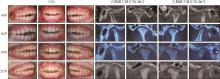
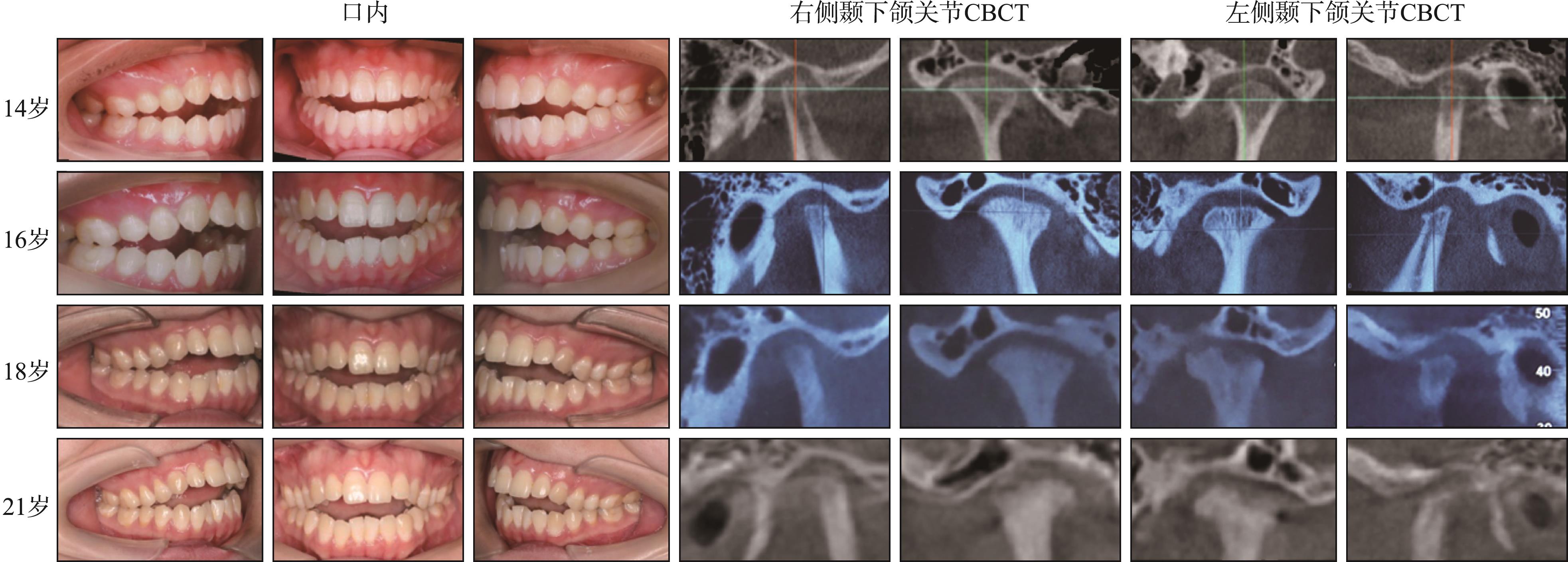
|
||