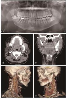
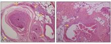
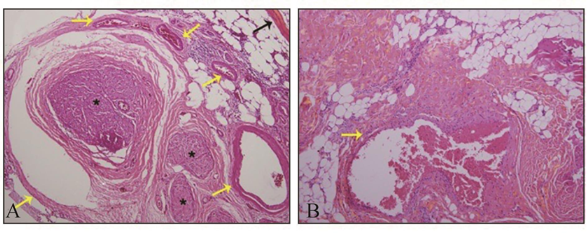
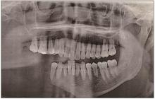
Int J Stomatol ›› 2023, Vol. 50 ›› Issue (4): 445-451.doi: 10.7518/gjkq.2023051
• Case Report • Previous Articles Next Articles
Wu Yifan( ),Lu Hao,Liu Shengwen,Xu Wanlin(),Yang Wenjun.
CLC Number:
| 1 | Saify F, Gosavi S. Gorham’s disease: a diagnostic challenge[J]. J Oral Maxillofac Pathol, 2014, 18(3): 411-414. |
| 2 | Gorham LW, Stout AP. Massive osteolysis (acute spontaneous absorption of bone, phantom bone, disappearing bone)[J]. J Bone Joint Surg Am, 1955, 37(5): 985-1004. |
| 3 | Patil DJ, Konidena A. Gorham’s disease of the maxilla-a rare case report with literature overview[J]. Radiol Case Rep, 2021, 16(7): 1754-1759. |
| 4 | Heffez L, Doku HC, Carter BL, et al. Perspectives on massive osteolysis. Report of a case and review of the literature[J]. Oral Surg Oral Med Oral Pathol, 1983, 55(4): 331-343. |
| 5 | Jackson JBS. A boneless arm[J]. Boston Med Surf, 1838, 18: 368-369. |
| 6 | Kim MK, Hong JR, Kim SG, et al. Fatal progression of gorham disease: a case report and review of the literature[J]. J Oral Maxillofac Surg, 2015, 73(12): 2352-2360. |
| 7 | Perschbacher SE, Perschbacher KA, Pharoah MJ, et al. Gorham’s disease of the maxilla: a case report[J]. Dentomaxillofac Radiol, 2010, 39(2): 119-123. |
| 8 | He J, He Y, Qiu WL, et al. Gorham disease in the maxilla[J]. J Craniofac Surg, 2012, 23(4): e293-e295. |
| 9 | Rodriguez-Vazquez JR, Chandra SR, Albertson ME, et al. Radiation-induced sarcoma on 18F-FDG PET/CT after treatment of Gorham-Stout disease of the maxilla[J]. Clin Nucl Med, 2019, 44(11): e607-e608. |
| 10 | Oujilal A, Lazrak A, Benhalima H, et al. Massive lytic osteodystrophy or Gorham-Stout disease of the craniomaxillofacial area[J]. Rev Laryngol Otol Rhinol, 2000, 121(4): 255-260. |
| 11 | Benhalima H, Lazrak A, Boulaich M, et al. Massive osteolysis of the maxillo-facial bones: case report and review of the literature[J]. Trop Dent J, 2001, 24(96): 35-40. |
| 12 | Duraisamy D, Veerasamy JS, Rajiah D, et al. Vani-shing mandible: a rare case report with accent to recent concepts on aetio-pathogenesis[J]. J Clin Diagn Res, 2015, 9(11): ZD25-ZD27. |
| 13 | Gulati U, Mohanty S, Dabas J, et al. “vanishing bone disease” in maxillofacial region: a review and our experience[J]. J Maxillofac Oral Surg, 2015, 14(3): 548-557. |
| 14 | Gataa IS, Nader NHR, Abdallah DT. Massive craniofacial gorham disease treated successfully by cisplatin and 5-fluorouracil with ten years of follow-up: a case report and literature review[J]. J Oral Maxillofac Surg, 2016, 74(9): 1774-1782. |
| 15 | Kato H, Ozeki M, Fukao T, et al. Craniofacial CT findings of Gorham-Stout disease and generalized lymphatic anomaly[J]. Neuroradiology, 2016, 58(8): 801-806. |
| 16 | Mukhopadhyay S, Chattopadhyay A, Bhattacharya R, et al. Gorham’s disease of mandible: a rare case presentation in pediatric patient[J]. J Indian Soc Pedod Prev Dent, 2016, 34(2): 180-184. |
| 17 | Zakhary I, Khanafer A. Rare incidence of Gorham disease with limited involvement of the maxilla: case report[J]. Br J Oral Maxillofac Surg, 2016, 54(7): 845-846. |
| 18 | Bocchialini G, Ferrari L, Burlini D. From tooth extraction to Gorham-Stout disease: a case report[J]. Int J Surg Case Rep, 2017, 34: 110-114. |
| 19 | Franco-Barrera MJ, Zavala-Cerna MG, Aguilar-Portillo G, et al. Gorham-stout disease: a clinical case report and immunological mechanisms in bone erosion[J]. Clin Rev Allergy Immunol, 2017, 52(1): 125-132. |
| 20 | Liu M, Liu WW, Qiao CY, et al. Mandibular Gorham-Stout disease: a case report and literature review[J]. Medicine, 2017, 96(42): e8184. |
| 21 | Mohapatra M, Jena AK, Dandapat AK, et al. Vani-shing mandible in a 7-year old child: response to radiation therapy[J]. J Clin Pediatr Dent, 2017, 41(6): 472-477. |
| 22 | Naqvi AA, Joshi SS, Bailey E. An unusual case of disappearing bone disease in the mandible and lite-rature review[J]. J Surg Case Rep, 2017, 2017(2): rjx025. |
| 23 | Bin Park S, Choi JY, Kim SJ. Gorham-stout disease affecting the mandible[J]. Clin Nucl Med, 2017, 42(10): 779-781. |
| 24 | Sinha R, Sarkar S, Khaitan T, et al. Gorham’s di-sease of the maxilla and mandible with distinctive cone beam computerized tomographic features[J]. Iran J Pathol, 2017, 12(3): 301-306. |
| 25 | Galiay L, Simon F, Lévy R, et al. Temporomandibular joint anomalies in pediatric craniofacial Gorham-Stout disease[J]. J Craniomaxillofac Surg, 2018, 46(8): 1179-1184. |
| 26 | Lova F, Vengal M, Ahsan A, et al. Gorham disease involving the maxillofacial bones: a perplexing entity[J]. Radiol Case Rep, 2018, 13(1): 96-100. |
| 27 | Mulvihill D, Kumar RS, Muzaffar J, et al. Gorham-Stout disease of the temporal bone involving the temporomandibular joint[J]. J Laryngol Otol, 2018, 132(3): 279-281. |
| 28 | Qu LY, Cai XY, Wang BL. Diagnosis and treatment of Gorham-Stout disease in maxillofacial regions[J]. J Craniofacial Surg, 2018, 29(2): 460-461. |
| 29 | Soh HY, Fauzi AA, Nazimi AJ, et al. Gorham’s di-sease of the mandible: radiological features[J]. Oral Radiol, 2018, 34(2): 179-184. |
| 30 | Zhang S, Wu DD, Shi LQ, et al. Gorham disease of the mandible: a report of two cases and a literature review[J]. Oral Surg Oral Med Oral Pathol Oral Radiol, 2019, 127(2): e71-e76. |
| 31 | Jagtap R, Gupta S, Lamfon A, et al. Gorham-Stout disease of the mandible: case report and review of literature of a rare type of osteolysis[J]. Oral Radiol, 2020, 36(4): 389-394. |
| 32 | Rahman NA, Harun MH, Rahman SA, et al. Floa-ting teeth appearance: a radiographic dilemma[J]. J Taibah Univ Med Sci, 2020, 15(2): 160-165. |
| 33 | Saify FY, Gosavi S, Jain S, et al. Vanishing bone di-sease: an enigma[J]. J Oral Maxillofac Pathol, 2021, 25(): S7-S10. |
| 34 | Aouad P, Young NM, Saratsis AM, et al. Gorham Stout disease of the temporal bone with cerebrospinal fluid leak[J]. Childs Nerv Syst, 2022, 38(2): 455-460. |
| 35 | Angelini A, Mosele N, Pagliarini E, et al. Current concepts from diagnosis to management in Gorham-Stout disease: a systematic narrative review of about 350 cases[J]. EFORT Open Rev, 2022, 7(1): 35-48. |
| 36 | Devlin RD. Interleukin-6: a potential mediator of the massive osteolysis in patients with Gorham-Stout disease[J]. J Clin Endocrinol Metab, 1996, 81(5): 1893-1897. |
| 37 | Rossi M, Buonuomo PS, Battafarano G, et al. Dissecting the mechanisms of bone loss in Gorham-Stout disease[J]. Bone, 2020, 130: 115068. |
| 38 | Rossi M, Rana I, Buonuomo PS, et al. Stimulation of treg cells to inhibit osteoclastogenesis in Gorham-Stout disease[J]. Front Cell Dev Biol, 2021, 9: 706596. |
| 39 | Dellinger MT, Garg N, Olsen BR. Viewpoints on vessels and vanishing bones in Gorham-Stout di-sease[J]. Bone, 2014, 63: 47-52. |
| 40 | Wang W, Wang H, Zhou X, et al. Lymphatic endothelial cells produce M-CSF, causing massive bone loss in mice[J]. J Bone Miner Res, 2017, 32(5): 939-950. |
| 41 | Hagendoorn J, Padera TP, Yock TI, et al. Platelet-derived growth factor receptor-beta in Gorham’s di-sease[J]. Nat Clin Pract Oncol, 2006, 3(12): 693-697. |
| 42 | Wijenayaka AR, Kogawa M, Lim HP, et al. Sclerostin stimulates osteocyte support of osteoclast activity by a RANKL-dependent pathway[J]. PLoS One, 2011, 6(10): e25900. |
| 43 | Hopman SM, van Rijn RR, Eng C, et al. PTEN ha-martoma tumor syndrome and Gorham-Stout pheno-menon[J]. Am J Med Genet A, 2012, 158A(7): 1719-1723. |
| 44 | Homayun-Sepehr N, McCarter AL, Helaers R, et al. KRAS-driven model of Gorham-Stout disease effectively treated with trametinib[J]. JCI Insight, 2021, 6(15): e149831. |
| 45 | Mowry S, Canalis R. Gorham-Stout disease of the temporal bone[J]. Laryngoscope, 2010, 120(3): 598-600. |
| 46 | Alves VM, Vieira TS, Amorim NS, et al. 99mTc(V)-DMSA SPECT-CT findings in a case of Gorham-Stout disease[J]. Nucl Med Rev, 2015, 18(2): 97-101. |
| 47 | Rossi M, Rana I, Buonuomo PS, et al. Dysregulated miRNAs in bone cells of patients with Gorham-Stout disease[J]. FASEB J, 2021, 35(3): e21424. |
| 48 | Kuriyama DK, McElligott SC, Glaser DW, et al. Treatment of Gorham-Stout disease with zoledronic acid and interferon-α: a case report and literature review[J]. J Pediatr Hematol Oncol, 2010, 32(8): 579-584. |
| [1] | Zhang Chaoying,Li Yining,Gong Jiaxing,Wang Huiming. Interpretation of the 2022 classification of head and neck tumors by the World Health Organization: odontogenic and maxillofacial bone tumors [J]. Int J Stomatol, 2023, 50(3): 263-271. |
| [2] | Zhang Xidan,Sun Jiyu,Fu Xinliang,Gan Xueqi.. Research progress on the development of mesoporous calcium silicate nanoparticles in endodontics and repairing maxillofacial bone defects [J]. Int J Stomatol, 2022, 49(4): 476-482. |
| [3] | Liu Min, Liu Weiwei, Xu Zhimin, Wang Zilin, Li Jiale, Han Bing.. Gorham-Stout syndrome in the maxillofacial region [J]. Inter J Stomatol, 2018, 45(4): 480-484. |
| [4] | Wang Yang, Shen Yuqin, Yu Wenwen, Sun Xinhua. Reasearch progress on modified mesoporous bioactive glasses for repairing maxillofacial bone defects [J]. Inter J Stomatol, 2018, 45(1): 32-35. |
| [5] | XIE Ke-xian, LIU Deng-gao, WU Yun-tang. Research progress on massive osteolysis [J]. Inter J Stomatol, 2009, 36(6): 712-715. |