Int J Stomatol ›› 2023, Vol. 50 ›› Issue (2): 186-194.doi: 10.7518/gjkq.2023035
• Reviews • Previous Articles Next Articles
Wang Mudan( ),Song Dongzhe,Huang Dingming.()
CLC Number:
| 1 | 周学东, 陈智, 岳林. 牙体牙髓病学[M]. 5版. 北京: 人民卫生出版, 2020. |
| Zhou XD, Chen Z, Yue L. Prosthodontics[M]. 5th ed. Beijing: People’s Medical Publishing House, 2020. | |
| 2 | Liao WC, Chen CH, Pan YH, et al. Vertical root fracture in non-endodontically and endodontically treated teeth: current understanding and future challenge[J]. J Pers Med, 2021, 11(12): 1375. |
| 3 | Gutmann J. Minimally invasive dentistry (Endodontics)[J]. J Conserv Dent, 2013, 16(4): 282. |
| 4 | Gluskin AH, Peters CI, Peters OA. Minimally invasive endodontics: challenging prevailing paradigms[J]. Br Dent J, 2014, 216(6): 347-353. |
| 5 | Shabbir J, Zehra T, Najmi N, et al. Access cavity preparations: classification and literature review of traditional and minimally invasive endodontic access cavity designs[J]. J Endod, 2021, 47(8): 1229-1244. |
| 6 | Silva EJNL, Pinto KP, Ferreira CM, et al. Current status on minimal access cavity preparations: a critical analysis and a proposal for a universal nomenclature[J]. Int Endod J, 2020, 53(12): 1618-1635. |
| 7 | Wang Q, Liu YX, Wang ZH, et al. Effect of access cavities and canal enlargement on biomechanics of endodontically treated teeth: a finite element analysis[J]. J Endod, 2020, 46(10): 1501-1507. |
| 8 | 高羽轩, 张岚, 周学东, 等. 直线通路微创开髓洞型对上颌第一前磨牙力学性能影响的有限元分析[J]. 中华口腔医学杂志, 2022, 57(1): 52-59. |
| Gao YX, Zhang L, Zhou XD, et al. Effect of straight-line minimally invasive access cavity on the mechanical properties of maxillary first premolars: a finite element analysis[J]. Chin J Stomatol, 2022, 57(1): 52-59. | |
| 9 | Santosh SS, Ballal S, Natanasabapathy V. Influence of minimally invasive access cavity designs on the fracture resistance of endodontically treated mandi-bular molars subjected to thermocycling and dynamic loading[J]. J Endod, 2021, 47(9): 1496-1500. |
| 10 | Silva EJNL, Lima CO, Barbosa AFA, et al. Preser-ving dentine in minimally invasive access cavities does not strengthen the fracture resistance of restored mandibular molars[J]. Int Endodontic J, 2021, 54(6): 966-974. |
| 11 | Patel S, Bhuva B, Bose R. Present status and future directions: vertical root fractures in root filled teeth[J]. Int Endod J, 2022, 55(): 804-826. |
| 12 | Silva EJNL, Versiani MA, Souza EM, et al. Minimally invasive access cavities: does size really matter[J]. Int Endod J, 2021, 54(2): 153-155. |
| 13 | Plotino G, Grande NM, Isufi A, et al. Fracture strength of endodontically treated teeth with diffe-rent access cavity designs[J]. J Endod, 2017, 43(6): 995-1000. |
| 14 | Saber SM, Hayaty DM, Nawar NN, et al. The effect of access cavity designs and sizes of root canal pre-parations on the biomechanical behavior of an en-dodontically treated mandibular first molar: a finite element analysis[J]. J Endod, 2020, 46(11): 1675-1681. |
| 15 | Jiang QZ, Huang YT, Tu XR, et al. Biomechanical properties of first maxillary molars with different endodontic cavities: a finite element analysis[J]. J Endod, 2018, 44(8): 1283-1288. |
| 16 | Zhang YY, Liu YX, She YH, et al. The effect of en-dodontic access cavities on fracture resistance of first maxillary molar using the extended finite element method[J]. J Endod, 2019, 45(3): 316-321. |
| 17 | Sabeti M, Kazem M, Dianat O, et al. Impact of access cavity design and root canal taper on fracture resistance of endodontically treated teeth: an ex vivo investigation[J]. J Endod, 2018, 44(9): 1402-1406. |
| 18 | Augusto CM, Barbosa AFA, Guimarães CC, et al. A laboratory study of the impact of ultraconservative access cavities and minimal root canal tapers on the ability to shape canals in extracted mandibular molars and their fracture resistance[J]. Int Endod J, 2020, 53(11): 1516-1529. |
| 19 | Neelakantan P, Khan K, Hei Ng GP, et al. Does the orifice-directed dentin conservation access design debride pulp chamber and mesial root canal systems of mandibular molars similar to a traditional access design[J]. J Endod, 2018, 44(2): 274-279. |
| 20 | Corsentino G, Pedullà E, Castelli L, et al. Influence of access cavity preparation and remaining tooth substance on fracture strength of endodontically treated teeth[J]. J Endod, 2018, 44(9): 1416-1421. |
| 21 | Liu YX, Liu H, Fan B. Influence of cavity designs on fracture behavior of a mandibular first premolar with a severely curved h-shaped canal[J]. J Endod, 2021, 47(6): 1000-1006. |
| 22 | Fu YJ, Zhang L, Gao Y, et al. A comparison of vo-lume of tissue removed and biomechanical analysis of different access cavity designs in 2-rooted mandibular first molars: a multisample 3-dimensional finite element analysis[J]. J Endod, 2022, 48(3): 362-369. |
| 23 | 陈新民, 赵云凤. 口腔生物力学[M]. 北京: 科学出版社, 2010. |
| Chen XM, Zhao YF. Dental biomechanics[M]. Beijing: Science Press, 2010. | |
| 24 | Welch-Phillips A, Gibbons D, Ahern DP, et al. What is finite element analysis[J]. Clin Spine Surg, 2020, 33(8): 323-324. |
| 25 | Kim SY, Kim BS, Kim H, et al. Occlusal stress distribution and remaining crack propagation of a cracked tooth treated with different materials and designs: 3D finite element analysis[J]. Dent Mater, 2021, 37(4): 731-740. |
| 26 | 刘子嫣, 赵凌, 杨丽媛, 等. 开髓方式与全冠修复对上颌中切牙应力分布影响的三维有限元分析[J]. 华西口腔医学杂志, 2019, 37(6): 642-647. |
| Liu ZY, Zhao L, Yang LY, et al. Three-dimensional finite element analysis of different endodontic access methods and full crown restoration in the ma-xillary central incisor[J]. West China J Stomatol, 2019, 37(6): 642-647. | |
| 27 | Yuan KY, Niu CG, Xie Q, et al. Comparative evaluation of the impact of minimally invasive preparation vs. conventional straight-line preparation on tooth biomechanics: a finite element analysis[J]. Eur J Oral Sci, 2016, 124(6): 591-596. |
| 28 | Jalali P, Allen C, Meyer C, et al. Stress distribution in a tooth treated through minimally invasive access compared to one treated through traditional access: a finite element analysis study[J]. J Conserv Dent, 2018, 21(5): 505. |
| 29 | Wan BY, Shahmoradi M, Zhang ZP, et al. Modelling of stress distribution and fracture in dental occlusal fissures[J]. Sci Rep, 2019, 9(1): 4682. |
| 30 | Lai H, Lin X, Zhang Y, et al. Effect of different en-dodontic access preparations on the biomechanical behavior of lithium disilicate and resin nanoceramic onlay restorations: an in vitro and 3D finite element analysis study[J]. J Prosthet Dent, 2022: S0022-3913(22)00006-3. |
| 31 | Assif D, Nissan J, Gafni Y, et al. Assessment of the resistance to fracture of endodontically treated molars restored with amalgam[J]. J Prosthet Dent, 2003, 89(5): 462-465. |
| 32 | Moore B, Verdelis K, Kishen A, et al. Impacts of contracted endodontic cavities on instrumentation efficacy and biomechanical responses in maxillary molars[J]. J Endod, 2016, 42(12): 1779-1783. |
| 33 | Rover G, Belladonna FG, Bortoluzzi EA, et al. Influence of access cavity design on root canal detection, instrumentation efficacy, and fracture resistance assessed in maxillary molars[J]. J Endod, 2017, 43(10): 1657-1662. |
| 34 | Silva AA, Belladonna FG, Rover G, et al. Does ultraconservative access affect the efficacy of root canal treatment and the fracture resistance of two-roo-ted maxillary premolars[J]. Int Endod J, 2020, 53(2): 265-275. |
| 35 | Xia J, Wang WD, Li ZM, et al. Impacts of contrac-ted endodontic cavities compared to traditional en-dodontic cavities in premolars[J]. BMC Oral Health, 2020, 20(1): 250. |
| 36 | Krishan R, Paqué F, Ossareh A, et al. Impacts of conservative endodontic cavity on root canal instrumentation efficacy and resistance to fracture assessed in incisors, premolars, and molars[J]. J Endod, 2014, 40(8): 1160-1166. |
| 37 | Silva EJNL, Rover G, Belladonna FG, et al. Impact of contracted endodontic cavities on fracture resistance of endodontically treated teeth: a systematic review of in vitro studies[J]. Clin Oral Investig, 2018, 22(1): 109-118. |
| [1] | Lu Lei,Wang Xin,Kang Zebiao,Xie Fuqiang. Research progress on the application of computer-assisted navigation surgery in complex maxillofacial fractures [J]. Int J Stomatol, 2023, 50(6): 696-703. |
| [2] | Tang Zhiwei,Gao Ying. Application and progress on targeted endodontic microsurgery techniques [J]. Int J Stomatol, 2022, 49(6): 678-683. |
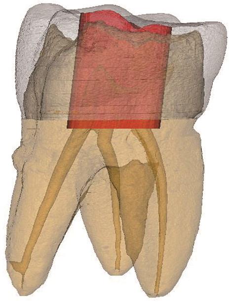
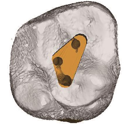
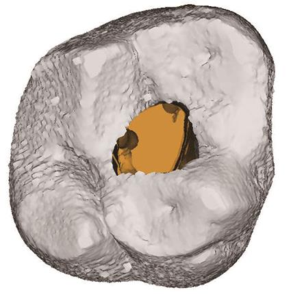
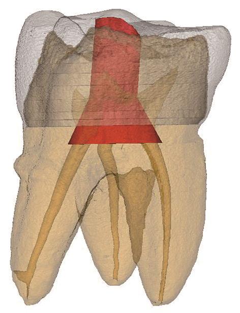
| [3] | Peng Weiqi,Gao Yuan,Xu Xin. The minimally invasive concept and research progress on access cavity design [J]. Int J Stomatol, 2021, 48(4): 433-438. |
| [4] | Ji Mengzhen,Qi Meiyao,Du Kexin,Quan Shuqi,Zhang Yuqiang,Zheng Qinghua. Three-dimensional finite element study on the effect of pulp opening cavity on the resistance of cracked teeth after full crown restoration [J]. Int J Stomatol, 2021, 48(1): 41-49. |
| [5] | Ning Chenxi,Li Xia. Research progress on the effect of root canal sealers on root fracture resistance [J]. Int J Stomatol, 2020, 47(6): 711-716. |
| [6] | Shi Haitao,Huang Jinxia,Pan Jian. Application of endoscopic technique to remove the foreign bodies in the maxillary sinus [J]. Int J Stomatol, 2020, 47(4): 452-457. |
| [7] | Tang Bei,Zhao Wenjun,Wang Hu,Zheng Guangning,You Meng. Inferior alveolar nerve injury due to apical overfilling: two cases reports [J]. Int J Stomatol, 2020, 47(3): 293-296. |
| [8] | Ding Jie, Song Guangtai.. Clinical application of minimally invasive techniques in the management of children’s dental caries [J]. Inter J Stomatol, 2018, 45(4): 473-479. |
| [9] | Ma Yanqun, Li Hong, Hou Benxiang. Research progress on the new attachment of apical periodontal ligament [J]. Inter J Stomatol, 2018, 45(3): 331-334. |
| [10] | Ma Quanquan, Cai Xiaoxiao.. Current status of immediate implant placement and provisionalization in esthetics zone [J]. Inter J Stomatol, 2017, 44(6): 731-736. |
| [11] | Huang Xiaoxiang, Zhang Ru, Hou Benxiang.. The effect of anatomy of apical zone of permanent teeth on root canal therapy [J]. Inter J Stomatol, 2017, 44(3): 261-266. |
| [12] | Li Shaorong, Zhang Ru, Hou Benxiang.. The effect of intracanal medications on mechanical properties of teeth [J]. Inter J Stomatol, 2017, 44(3): 273-278. |
| [13] | Liang Jichao, Wang Fen, Zhang Fengying, Zhang Zhenghua, Hou Meijuan, Pang Fusheng, Zhou Feng. Comparison of the accuracy of Digora and Propex on the measurement of working length of root canal [J]. Inter J Stomatol, 2016, 43(5): 515-518. |
| [14] | Ju Yingxin, Liu Luchuan. Application of erbium laser to treat periapical diseases [J]. Inter J Stomatol, 2016, 43(4): 473-476. |
| [15] | Lin Jie1, Zheng Zhifeng2, Lu Zhaojie1, Li Xiurong2, Zheng Zhiqiang1.. Design and bonding technique of posterior zirconia resin-bonded fixed partial dentures [J]. Inter J Stomatol, 2015, 42(6): 624-627. |